Introduction
It has been well established that oxidative stress plays an important role in the onset of different diseases, including atherosclerosis, rheumatoid arthritis, cancer and the degenerative diseases associated with aging (1, 2). Consequently, the supplement of dietary antioxidants will help to attenuate the damage of the body induced by oxidative stress, and can be used as potential therapeutic or preventive drugs for the risk of many free radical-mediated diseases.
In the last few decades, the natural antioxidant that may be obtained from different plant parts, flavonoids and poly phenolic compounds are paid more attention to, because phenolic compounds isolated from plants can act as free radical scavengers, metal chelators, and singlet oxygen quenchers (3).
Pistacia vera L., is a plant member of Anacardiaceae family and native to Asia. Pistachio nut is mostly produced in Iran and some other countries (4). Pistacia species have caught up the interest of researchers due to the study on different part of this plant such as leaves, kernels, hulls and gum demonstrate various biological activities such as antioxidant potential, antimicrobial, anti-inflammatory, mainly due to flavonoids and other phenolic components and anti-insect activities (5-11). It has been proved that Pistachio nuts are a rich source of phenolic compounds and have been considered because of high antioxidant potential (12).
Investigation on pistachio green hull has showed antioxidant, anti-microbial and antimutagenic activity (13). A clinical trial study on young men demonstrated that a pistachio diet improved blood glucose level, endothelial function, and some indices of inflammation and oxidative status (14). Also P. vera L. gum extract demonstrated a protective effect on oxidative damage in rat cerebral ischemia-reperfusion (15). It is also showed that a gum extract has antinocieptive and anti-inflammatory effect (16). In other study P. vera L. (Pistachio) leaves and nuts aqueous extracts showed antiemetic effect in young chicken (17).
In recent studies, the side effects of synthetic antioxidants are considered. There is an increasing interest to the use of natural substances instead of the synthetic ones. The purpose of this study was the evaluation of antioxidant effects from different parts of pistachio fruits, namely leaves and gum using three in-vitro approaches: deoxyribose assay, erythrocyte membrane peroxidation and rat liver microsomal lipid peroxidation induced by Fe2+/ascorbate.
Experimental
Ascorbic acid, Deoxyribose, Tiobarbitoric acid (TBA), ferric chloride, methanol, butylated hydroxytoluene (BHT) and trichloroacetic acid (TCA) were obtained from Merck (Germany).
Plant material and Preparation of extracts
P. vera L. was collected from (Khorasan- Gonabad region) I.R Iran. It was identified by Mr. Ahi in the Herbarium of School of Pharmacy, Mashhad University of Medical Sciences (MUMS), IR. Iran. For the decoction extract, 1 L water was added to 100 g plant material and boiled for 15 min and percolation was performed till the solvent become colorless. Afterwards, the solution was filtrated and evaporated in a water bath (maintained at 30-40°C). The extract was stored in a refrigerator at 4°C.
To obtain the ethanolic extract, the leaves and fruits of plant were prepared by defatted powder via Soxhlet with petroleum ether giving as dry residue and was macerated in ethanol 80° (v/v) for 72 h. Then, the macerated mixture was filtered and evaporated as mentioned previously.
The gum was extracted from the resin by cold maceration by hydrodistillation with ethanol. The combined hydroalcoholic extract was filtered through filter paper and evaporated to dryness on water bath.
Characterization of extract by HPLC
The separation was carried out on a millipore column (5 μm, 1.5 X 3.9 mm) using gradient elution. Gradient was performed using water-phosphoric acid (0.1 N, 99:1) and acetonitrile-phosphoric acid (0.1 N, 99:1) at a total flow rate of 1 mL/min; gradient composition (min, % acetonitrile-phosphoric acid): 0.0, 5; 5.0, 7.5; 10, 10; 15, 12.5; 20, 15; 25, 17.5; 30, 20; 35, 22.5; 40, 25). The extracts were dissolved in methanol and filtered through a membrane filter (0.45 μm). 1.0 μL sample of 10 g/L of extract was injected in to a reversed-phase column (RPC-18). The peaks were momtored at 236 nm.
Animals
Male Wistar rats weighing 200-250 g were used for the study. The animals were bred and housed in the Animal House of the Faculty of Pharmacy Mashhad University of Medical Sciences accordance with ethical committee Acts.
Deoxyribose assay
In this method, after mixing the materials, hydroxyl radicals were produced by ascorbic acid, H2O2 and Fe3+-EDTA thus deoxyribose degradation take placed and produced malondialdhyde. The reaction mixture contained 100 μL of 28 mM 2-deoxy- 2-ribose 500 μL solution of various concentration of the material test (aqueous and ethanolic extracts of pistachio fruits and leaves and hydroalchoholic extracts of gum in buffer), 200 μL of 200 μM FeCl3 and 1.04 mM EDTA (1:1 v/v), 100 μL H2O2 (1 mM) and 100 μL ascorbic acid (1 mM). All solutions were prepared freshly. After an incubation period of 1 h at 37°C, 1 mL of TBA (1%in 50 mM NaOH) and 1 mL of TCA were added to the reaction Mixture and the tubes were heated at 100°C for 20 min. The degree of deoxyribose degradation was measured by the TBA reaction. Absorbance was read at 532 nm (18, 19). The percentage Absorbance was read at 532 nm. The percentage of inhibition of deoxyribose degradation was calculated using the following equation:
%Inhibition = (A0 - A1) / A0 × 100
Here, A0 is the absorbance of the control in the absence of samples; A1 is the absorbance in the presence of samples. DMSO was used as a positive control (20) and the negative control was all of the reaction mixture without extracts.
Site-specific reactions assay
This assay was prepared in the following three ways:
This test was done as mentioned previous to except that 100 mL of FeCl3 instead of 200 mL of Fe3+-EDTA Solution was extra, for evaluated potency of sample radical scavenging and Fe chelating.
The evaluation was performed without ascorbic acid as a starter for oxidation.
Deoxyribose itself was omitted from the test (18).
Erythrocyte membrane peroxidation
According to the method that was described before (19, 21), the evaluation of protective effects of pistachio extracts, were done with modification. After anesthizing the Wistar rats (200-250 g) with chloroform, whole blood were collected via a cardiac puncture to heparinized tubes. The RBC was separated from plasma by centrifugation at 1500 g for 15 min. Packed RBC was washed twice with NaCl 0.15 M, and preincubated with phosphate buffer (pH = 7.4) containing sodium azide (1 mM) to inhibit catalase. Then H2O2 (10 mM) was added, peroxidation was initiated H2O2 (10 mM). 100 μL solutions of various concentrations of the material test were added and the mixture was incubated at 37°C for 60 min. The addition of 28% (w/v) trichloroacetic acid terminated reaction. Lipid peroxidation was measured using thiobarbitoric method. The quantity of MDA was determined by measuring the absorbance at 532 nm BHT was used as a positive control (21, 22).
Liver microsomal preparation and lipid peroxidation induced by Fe2+/ascorbat
This test was carried out according to the method that was described previously (23). Wistar rats were anesthetized and liver was perfused with ice-cold saline through the portal vein until getting uniformly pale and were immediately removed. Then, pieces of liver were homogenized with 4 volume of ice-cold 0.1 M potassium phosphate buffer (pH = 7.4) containing 1.15% (w/v) KCl. The homogenate was centrifuged at 10000 g for 60 min. The supernatant was used for the study. According to the mentioned previous method, (23) for measuring antioxidant activity, rat liver microsome (2 mg/ mL) was mixed with 0.1 mL of FeSO4 (26% mM), 0.1 mL of ascorbate (0.13 mM), 0.1 mL of the sample in 150 mM KCl/Tris-HCl buffer solution (pH = 7.4). The mixture was incubated at 37°C for 60 min in a water bath; 0.75 mL of 2 M trichloroacetic acid/1.7 M HCl was added to stop trhe reaction, then tubes were centrifuged (4000 rpm, 10 min) and 0.5 mL of the supernatant was mixed with 0.15 mL TBA and was heated at 95°C for 10 min. The level of malondialdehyde was determined by measuring the absorbance at 532 nm. The percent of lipid peroxidation inhibition was calculated by following Equation:
%I = (A0 - A1 / A0) × 100
Here, A0 is the absorbance of the control reaction; A1 is the absorbance in the presence of the agents. BHT was used as a positive control (20).
Phytochemical test
Phytochemical screening of the extract was performed using the following reagents and chemicals (24): Alkaloids with Dragendorff’s reagent, flavonoids by the use of Mg and HCl; tannins with 1% gelatin and 10% NaCl solutions and saponins with ability to produce hemolysis of RBC.
Statistical analysis
Statistical analysis was performed using one-way ANOVA followed by Tukey-Kramer post hoc test for multiple comparisons. The p-value less than p < 0.05 were considered to be statistically significant. PCS software was used to calculate IC50 value.
Results and Discussion
Yield of extraction
Yield of extraction was 4.22% for leaves and 10% for fruits. In ehanolic extraction method, yield of extraction for leaves and fruits were 26% and 6.9% respectively. For Pistachio gum, the yield was 6.9%.
Deoxyribose degradation assay
In deoxyribose degradation method, absorption spectra at 532 nm were evaluated for extracts in various concentration (25, 100, 250, 500 and 1000 μg/mL) and blank was used. All of tested compound showed antioxidant activity and OH radical scavenging effect (p < 0.001 VS control). The IC50 values of aqueous and ethanolic extracts of fruits were149.2 μg/mL and 64.7 μg/mL and for leaves 105.4 μg/mL and 84.8 μg/mL, respectively and for gum extract was 285.5 μg/mL. The results are shown in Table 1. In deoxyribose assay ability of radical scavenging and inhibition of deoxyribose degradation (18), all exracts inhibited MDA production in a dose-dependent manner. The effect of fruit ethanolic extract (1000 μg/mL) was similar to positive control.
Table1
Free radical scavenging activity of Pistacia vera extracts in deoxyribose assay
| Concentr Concentration | %Inhibition | ||||
|---|---|---|---|---|---|
| Ethanolic fruit extract | Aqueous fruit extract | Ethanolic Leaf extract | Aqueous Leaf extract | Hydroalcholic Gum extract | |
| Control | - | - | - | - | - |
| 25 (μg/mL) | 38.6*** | 24.2*** | 34.3*** | 27.4*** | 14.7*** |
| 100 | 53*** | 37.5*** | 54.9*** | 52.7*** | 34.1*** |
| 250 | 64.6*** | 63*** | 56.6*** | 59.1*** | 46.2*** |
| 500 | 85.3*** | 70.7*** | 72.8*** | 73*** | 63.5*** |
| 1000 | 90.3*** | 77.1*** | 88.5*** | 84.5*** | 67.4*** |
| DMSO(20 mM) | 91.5*** | 91.8*** | 90.9*** | 91.3*** | 91.3*** |
Table 2
Results of the assay for site-specific reactions of the ethanol and aqueous extracts of fruit, leaf and gum of of Pistacia vera
| Sample | Concentration | Omit EDTA | Omit Vit C | Omit deoxyribose |
|---|---|---|---|---|
| Ethanolic | Negative control | 0.617 | 0.156 | 0.084 |
| 1000μg/mL | 0.099 | 0.210 | 0.037 | |
| 500 μg/mL | 0.163 | 0.170 | 0.030 | |
| 250 μg/mL | 0.256 | 0.144 | 0.024 | |
| 100 μg/mL | 0.340 | 0.127 | 0.019 | |
| 25 μg/mL | 0.426 | 0.102 | 0.009 | |
| Aqueous | Negative control | 0.617 | 0.156 | 0.082 |
| 1000μg/mL | 0.114 | 0.143 | 0.064 | |
| 500 μg/mL | 0.167 | 0.141 | 0.056 | |
| 250 μg/mL | 0.297 | 0.129 | 0.027 | |
| 100 μg/mL | 0.470 | 0.108 | 0.009 | |
| 25 μg/mL | 0.326 | 0.077 | 0.002 | |
| Ethanolic | Negative control | 0.609 | 0.159 | 0.079 |
| 1000μg/mL | 0.111 | 0.197 | 0.069 | |
| 500 μg/mL | 0.178 | 0.149 | 0.061 | |
| 250 μg/mL | 0.262 | 0.152 | 0.032 | |
| 100 μg/mL | 0.419 | 0.116 | 0.027 | |
| 25 μg/mL | 0.487 | 0.103 | 0.015 | |
| Aqueous | Negative control | 0.609 | 0.159 | 0.079 |
| 1000μg/mL | 0.171 | 0.168 | 0.058 | |
| 500 μg/mL | 0.210 | 0.140 | 0.037 | |
| 250 μg/mL | 0.293 | 0.112 | 0.019 | |
| 100 μg/mL | 0.327 | 0.098 | 0.016 | |
| 25 μg/mL | 0.402 | 0.061 | 0.007 | |
| Hydroalcholic | Negative control | 0.609 | 0.159 | 0.083 |
| 1000μg/mL | 0.236 | 0.131 | 0.056 | |
| 500 μg/mL | 0.280 | 0.119 | 0.045 | |
| 250 μg/mL | 0.341 | 0.117 | 0.027 | |
| 100 μg/mL | 0.399 | 0.081 | 0.021 | |
| 25 μg/mL | 0.419 | 0.042 | 0.010 |
RBC lipid peroxidation
High polyunsaturated fatty acid contents cause membrane lipids particularly susceptible to oxidative damage (21, 25). Lipid peroxidation is one of the important reactions that induced by oxidative stress (26, 27).
TBARS were expressed with Pmol MDA produced in the presence of different concentrations of extracts. The IC50 values of the aqueous and ethanolic extracts of fruits were 768.3 μg/mL and 325.1 μg/mL and for leaves were 314.5 μg/mL and 231.4 μg/mL, respectively and gum extract showed antioxidant effect less than 50%. Results are shown in Tables 3-5.
All extracts prevented oxyradical generation. The fruits ethanolic extract was more effective than aqueous extract probably due to existence of linoleaic and linolenic fatty acids. The antioxidant effect of pistachio fruits (28, 29) was due to antioxidant effects of phenolic compounds (flavonoids and tannin) (30). It was shown that Pistachio skins has better antioxidant activity i compared with seeds in different tests such as (DPPH assay, Folin-Ciocalteau colorimetric method and TEAC assay, SOD-mimetic assay). The excellent antioxidant activity of pistachio skins can be due to its higher content of antioxidant phenolic compounds (31). In another study P. lentiscus resin showed antioxidant activity, Fe2+ chelating activity and it inhibited inflammation (32).
Table 3
Effects of ethanolic and aqueous extracts of Pistacia vera fruits in RBC lipid peroxidation assay
| Concentration | Aqueous extract | Ethanolic extract | ||||
|---|---|---|---|---|---|---|
| MDA | p | Inhibition% | MDA | p | Inhibition% | |
| Control | 365.38 ± 9.26 | - | - | 385.79 ± 6.52 | - | - |
| 25 (μg/mL) | 360.9 ± 4.13 | Ns | 1.2 | 366.62 ± 9.26 | Ns | 5 |
| 100 | 339.35 ± 4.65 | Ns | 7.1 | 339.34 ± 5.72 | 0.001 | 12 |
| 250 | 255.48 ± 6.27 | 0.001 | 30 | 192.56 ± 7.26 | 0.001 | 50 |
| 500 | 211.94 ± 3.64 | 0.001 | 42 | 125.91 ± 9.38 | 0.001 | 67.4 |
| 1000 | 178.8 ± 9.56 | 0.001 | 51 | 96.42 ± 3.64 | 0.001 | 75 |
| BHT(0.04) mM | 74.34 ± 2.15 | 0.001 | 79.6 | 78.18 ± 2.25 | 0.001 | 80 |
Table 4
Effects of ethanolic and aqueous extracts of Pistacia vera leaves in RBC lipid peroxidation assay
| Concentration | Aqueous extract | Ethanolic extract | ||||
|---|---|---|---|---|---|---|
| MDA | p | Inhibition% | MDA | p | Inhibition% | |
| Control | 377.15 ± 9.33 | - | - | 402.06 ± 3.4 | - | - |
| 25 (μg/mL) | 356.9 ± 6.22 | Ns | 5.4 | 351.41 ± 10.51 | Ns | 12.6 |
| 100 | 298.81 ± 5.38 | 0.001 | 20.8 | 291.01 ± 6.82 | 0.001 | 28 |
| 250 | 214.23 ± 9.15 | 0.001 | 43.2 | 233.3 ± 5.24 | 0.001 | 42 |
| 500 | 109.22 ± 5 | 0.001 | 71 | 92.601 ± 3.29 | 0.001 | 77 |
| 1000 | 106.23 ± 3.65 | 0.001 | 71.8 | 88.93 ± 4.97 | 0.001 | 77.9 |
| BHT (0.04) mM | 80.7 ± 1.91 | 0.001 | 78.6 | 77.25 ± 2.75 | 0.001 | 80.8 |
Table 5
Effects of hydroalcoholic extracts of Pistacia vera gum in RBC lipid peroxidation assay
| Concentration | MDA | p | Inhibition% |
|---|---|---|---|
| Control | 371.6 ± 6.49 | - | - |
| 25 (μg/mL) | 363.51 ± 4.06 | Ns | 2.1 |
| 100 | 349.86 ± 8.22 | Ns | 5.8 |
| 250 | 316.31 ± 5.78 | 0.001 | 14.9 |
| 500 | 203.49 ± 7.42 | 0.001 | 45.2 |
| 1000 | 196.74 ± 7.56 | 0.001 | 47 |
| BHT (0.04) mM | 75.78 ± 2.66 | 0.001 | 79.6 |
Liver microsomal lipid peroxidation
Treatment of liver microsomal with extracts was produced a significant decrease of MDA generation as compared with control treatment. The IC50 values of aqueous and ethanolic extracts of fruits 1441.5 μg/mL and 648.7 μg/mL, and leaves were 1101.1 μg/mL and 700.1 μg/mL, respectively and gum extract showed antioxidant effect less than 50%. Results are shown in Tables 6-8.
Table 6
Effects of ethanolic and aqueous extracts of Pistacia vera fruits in microsomal lipid peroxidation assay
| Aqueous extract | Ethanolic extract | ||||||
|---|---|---|---|---|---|---|---|
| Concentration | MDA | p | Inhibition% | MDA | p | Inhibition% | |
| Control | 1.98 ± 0.083 | - | - | 1.89 ± 0.073 | - | - | |
| 25 (μg/mL) | 1.84 ± 0.074 | Ns | 7.2 | 1.66 ± 0.044 | Ns | 12.1 | |
| 100 | 1.53 ± 0.027 | 0.001 | 22.7 | 1.37 ± 0.049 | 0.001 | 27.6 | |
| 250 | 1.49 ± 0.043 | 0.001 | 24.7 | 1.29 ± 0.034 | 0.001 | 31.4 | |
| 500 | 1.34 ± 0.074 | 0.001 | 32.6 | 1.04 ± 0.063 | 0.001 | 44.9 | |
| 1000 | 1.051 ± 0.076 | 0.001 | 47.1 | 0.77 ± 0.096 | 0.001 | 59.5 | |
| BHT (0.04) mM | 0.55 ± 0.02 | 0.001 | 72.2 | 0.53 ± 0.052 | 0.001 | 71.8 | |
Table 7
Effects of ethanolic and aqueous extracts of Pistacia vera leaves in microsomal lipid peroxidation assay
| Concentration | Aqueous extract | Ethanolic extract | ||||
|---|---|---|---|---|---|---|
| MDA | p | Inhibition% | MDA | p | Inhibition% | |
| Control | 1.98 ± 0.08 | - | - | 1.89 ± 0.073 | - | - |
| 25 (μg/mL) | 1.80 ± 0.06 | Ns | 9.1 | 1.79 ± 0.063 | Ns | 5.4 |
| 100 | 1.67 ± 0.05 | 0.05 | 15.9 | 1.57 ± 0.08 | 0.05 | 16.9 |
| 250 | 1.61 ± 0.06 | 0.01 | 18.7 | 1.32 ± 0.056 | 0.001 | 30.7 |
| 500 | 1.07 ± 0.07 | 0.001 | 45.8 | 1.02 ± 0.043 | 0.001 | 46.1 |
| 1000 | 0.97 ± 0.06 | 0.001 | 51 | 0.84 ± 0.094 | 0.001 | 55.2 |
| BHT (0.04) mM | 0.55 ± 0.02 | 0.001 | 72.2 | 0.53 ± 0.052 | 0.001 | 71.8 |
Table 8
Effects of hydroalcoholic extracts of Pistacia vera gum in microsomal lipid peroxidation assay
| Concentration | MDA | p | Inhibition% |
|---|---|---|---|
| Control | 1.89 ± 0.05 | - | - |
| 25 (μg/mL) | 1.78 ± 0.04 | Ns | 6 |
| 100 | 1.74 ± 0.02 | Ns | 8.1 |
| 250 | 1.66 ± 0.07 | 0.05 | 12.6 |
| 500 | 1.37 ± 0.06 | 0.001 | 27.4 |
| 1000 | 1.24 ± 0.05 | 0.001 | 34.5 |
| BHT (0.04) mM | 0.53 ± 0.04 | 0.001 | 72 |
Phytochemical tests
All extracts were negative as for the existence of alkaloid. The aqueous and ethanolic extracts of fruits were negative for tannin content but both extracts of leaves and gum were positive in tannin test. The aqueous and methanolic extracts of fruits had considerable amount of flavonoids but leaves ethanolic extracts and gum extracts had fewer amount. Saponin was not found in the extracts. According to the result of phytochemical tests in this research, the protective effect of extract is probably due to the presence of tannin in gum and leaves extract and flavonoids in fruit extract. HPLC fingerprints of the extract indicated five peaks (Figure 1).
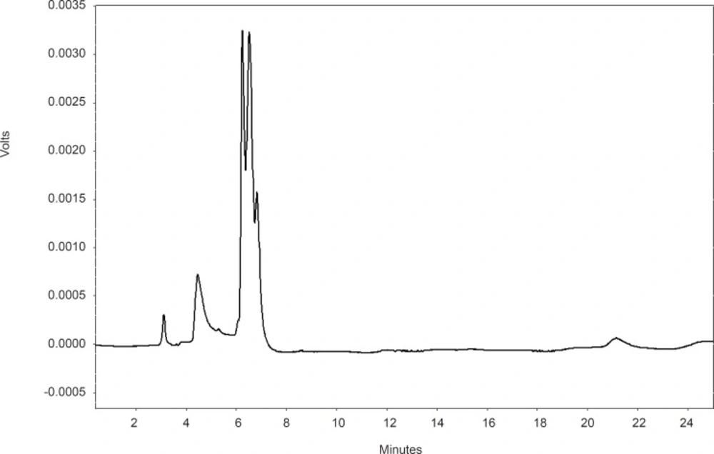
Figure 1
HPLC fingerprint of P. vera ethanolic gum extract
Conclusion
The present study showed that extracts of different part of P. vera have antioxidant activity in different in vitro methods. The ethanolic extracts of leaves and fruits showed more roles for antioxidant properties and gum hydroalcoholic extract demonstrated less antioxidant effect.