Plant material
A. buettneri leaves were collected in April 2006 from the University of Lomé’s botanical garden located in the Faculty of Science department in Togo. These were then subsequently analysed and verified by the Laboratory of Botany. A reference sample was deposited in the Laboratory of Botany and Plant Ecology’s Herbarium, part of the Faculty of Science, “University of Lomé” (UL-MET 001)
Extract preparation
The selected leaves were washed, then dried under air-conditioning and finally ground to attain a powder. The powder was subsequently extracted with a mixture of water : ethanol (1:1, v/v) for 72 h and after that filtered. The filtrate was evaporated and a dark hydro-alcohol extract (yield: 24.25%) was obtained. Phytochemical screening of the extract revealed the presence of tannins, flavonoids and alkaloids.
Animals
Wistar rats, of both sexes weighing 150-200 g were the subject of testing. They were placed in an Animal house within the Faculty of Science’s Laboratory of Physiology/Pharmacology. The animals were kept under conditions of ambient temperature, humidity and dark-light cycle (12 h–12 h). The animals had free access to food and water.
Ethanol gastric ulcer induction
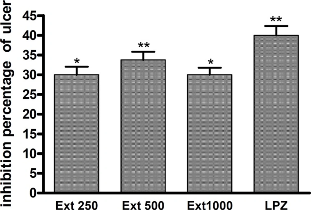
A gastric ulcer was induced by an oral administration of 1 mL/100 g body weight of ethanol 95°. Rats were subjected to fasting for 24 h preceding their ulcer induction. The control group received distilled water and the treated groups received 250; 500 or 1000 mg/kg of A. buettneri extract (Ext) 30 min prior to ulcer induction. Lansoprazole (LPZ) 30 mg/kg was used as a reference drug. Two hours following ulcer induction the subjected rats were then sacrificed under ether anaesthesia. Their stomachs were removed and opened along the greater curvature. Following this the dimension of the ulcer was evaluated by means of planimetry, using 0.25 mm2 ulcer area by unit. The percentage of ulcer inhibition was then calculated using the following formula: (the dimension of the control rat’s ulcer-the dimension of the treated rat’s ulcer) the dimension of the control rat’s ulcer.
Evaluation of gastric pH measurement
Extract (250, 500 mg/kg; p.o.) was administered to rats and two hours later they were sacrificed under the same afore mentioned conditions. The stomach of each rat was removed, opened and the gastric pH was measured.
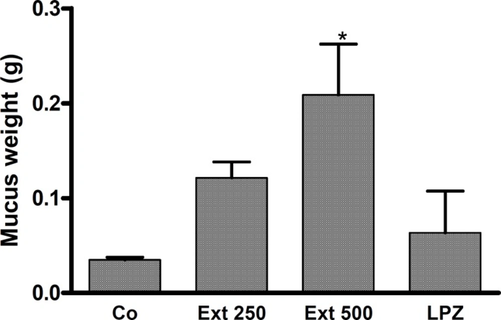
Evaluation of mucus production
Gastric mucus production was measured according to the method used by Salehi
et al. (
7). Two hours after the extract was administered (250, 500 mg/kg) the rats were sacrificed. Using a glass slide the gastric mucosa of each rat was gently scraped then, subsequently weighed using a precision electronic balance.
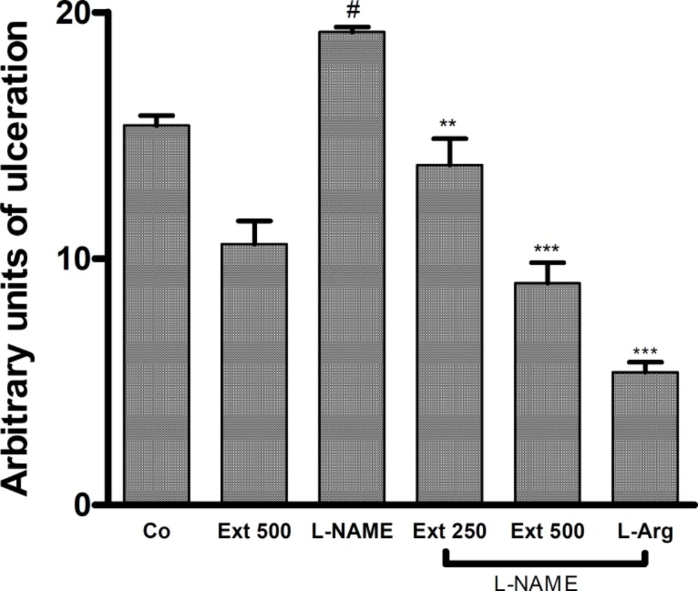
Ethanol induced gastric mucosal lesion in L-NAME pre-treated rats
The method used by Maria
et al. (
8) was implemented. Animals were subjected to fasting for 24 h and deprived of water for 19 h prior to commencing the experiment. The animals were divided into groups each consisting of 5 rats. Ethanol 95° was administered orally at a dose of 1 mL/100g body weight. Prior to ethanol administration, the rats orally received either distilled water or extract as follows:
Group I, control group, received saline solution (IV) and distilled water (orally), 45 and 30 min prior to ulcer induction.
Group II were administered saline solution (IV) and a 500 mg/kg extract of A. buettneri (orally), 45 and 30 min before ulcer induction.
Group III were given L-NAME 40 mg/kg (IV) and distilled water (orally), 45 and 30 min before ulcer induction.
Groups IV and V received L-NAME 40 mg/kg (IV) and extracts of A. buettneri 250 and 500 mg/kg (orally) 30 min before ethanol administration.
Group VI was treated with L-arginine (IV) 400 mg/kg immediately, prior to being injected with L-NAME.
Two hours after the ulcers were induced, the rats were sacrificed. The stomach of each rat was removed, opened along the greater curvature and the dimension of the induced ulcer was evaluated by planimetry.
The effect of A. Buettneri hydro-alcohol extract on gastric mucosal damage induced by ethanol 95°. The extract was administered 30 min before ulcer induction. Results are mean ± SEM for 5 rats. *p < 0.05; **p < 0.01 (control vs treated).
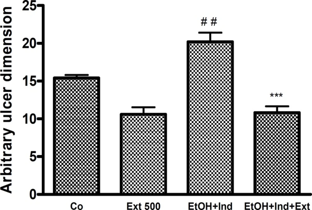
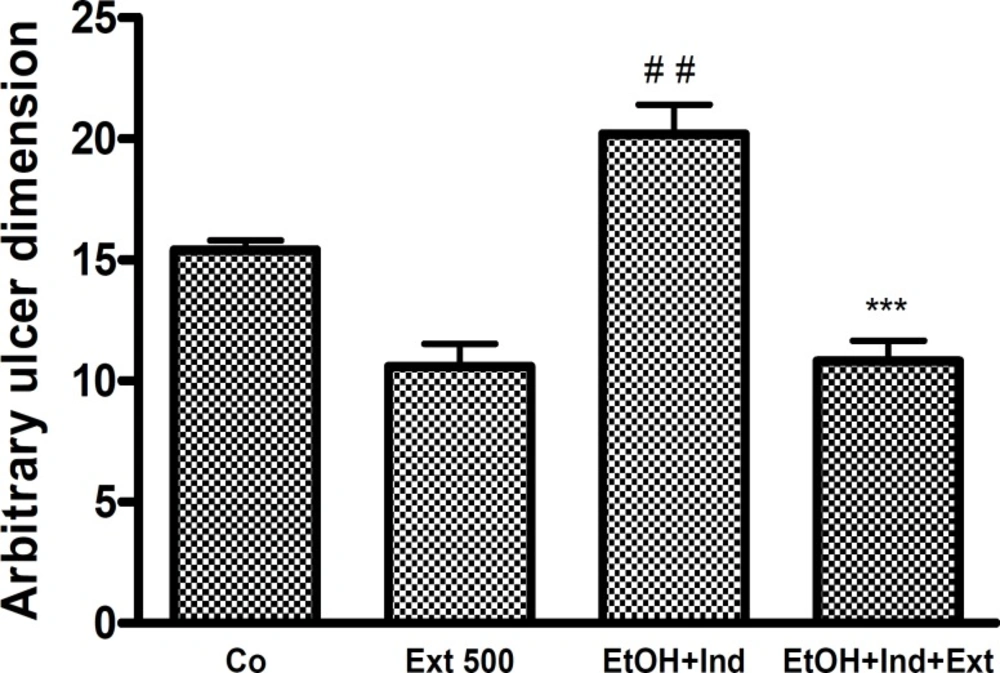
Ethanol induced gastric mucosal lesion in indomethacin pre-treated rats
Ulcer inducement in indomethacin pre-treated rats was divided into three groups (I, III, and IV). Group I was used as a control (Co) and group II was not pre-treated with indomethacin instead receiving Nacl solution (IP). 30 min before indomethacin administration the rats were given distilled water or an extract of Indomethacin 300 mg/kg was administered (IP) to rats that had been subjected to fasting overnight. Four hours subsequent to the administration of indomethacin, the rats received ethanol 95° orally. Two hours later the rats were then sacrificed and treated as indicated above.
Statistical analysis
Data obtained from the animal experiments was expressed as mean ± SEM statistical tests including a one-way analysis of variance (ANOVA) followed by bonferroni’s significant difference test were used to analyze any differences between the groups that were subjected to testing. A p-value of less than 0.05 was considered as being statistically significant.