Chemicals
Fetal bovine serum (FBS) was purchased from Invitrogen/Life Technologies (Carlsbad, CA, USA). Dimethyl sulfoxide (DMSO) was the products of Sigma-Aldrich (St. Louis, USA). Hygromycin B was purchased from Sigma-Aldrich (St. Louis, USA). Lipofectamine 2000 reagent was purchased from Invitrogen/Life Technologies (CA, USA).
Generation of recombinant vector
Primers for the enhancer sequence of IE (immediate-early) genes of human cytomegalovirus (hCMV) were designed in order to proliferate a fragment of about 420 bp length including KpnI-HindIII recognition sites. The amplified fragments were purified on a 1.5% agarose gel and cloned at the KpnI-HindIII sites of pGL4.26 using standard protocols. The orientation and sequences of this element were confirmed by sequencing of the plasmids.
Development of a toxicity sensitive luciferase reporter plasmid
The toxicity reporter plasmids were generated using the pGL4.26-minimal promoter vector (Promega, UK, Southampton, United Kingdom) containing a minimal TATA promoter upstream of the firefly luciferase gene (luc). Enhancer element of CMV IE genes was inserted into the pGL4.26 [minP] multiple cloning site upstream of the luc gene. Consequently, TOP10 competent cells were transformed with the recombinant DNA for amplification. Engineered vector contains one copy of enhancer sequence of HCMV immediate-early genes that have been inserted through KpnI-HindIII restriction sites upstream of the promoter-luc + transcriptional unit. Five positive clones were sequenced using RV3 primer (Rhinovirus universal primer).
Cell viability assay
Cell viability was assessed by methyl thiazol tetrazolium (MTT) assay (Sigma-Aldrich, St Louis, USA) according to the manufacturer’s instruction. Briefly, the cells (1 × 10⁴) were cultured overnight in a 96-well plate. Afterwards, the medium of each well was replaced by 200 μL fresh medium plus 50 μL of the MTT solution (5 mg/mL in PBS). The plates were incubated at 37 °C for 4 h. The absorbance being proportional to cell was subsequently measured at 570 nm in each well using an enzyme linked immunosorbent assay plate reader (BioRad 680, USA). All experiments were performed in triplicate, and the relative cell viability (%) was calculated as a percentage relative to the untreated control cells.
Generation of stable cell line
The Huh7-CMV-luc containing the Hygromycin B selectable marker was stably transfected into the Huh7 cells using the Lipofectamine® 2000 reagent. According to the LD50 value, the transfected cells were selected using 450 μM Hygromycin B (Invitrogen/Life Technologies, CA, USA) in the media for 21 days. The Hygromycin B-resistant clones were isolated and screened by measuring their basal and inducible luciferase activity at different concentration of toxins. Positive clones showed high luciferase activity. The cells were maintained in growth medium containing 450 μM hygromycin for further analysis.
Cell culture conditions
The human hepatocarcinoma cell line (Huh-7) was purchased from National Cell Bank, Pasteur Institute of Iran and were grown in Dulbecco′s Modified Eagle′s Medium (DMEM) (Invitrogen/Life Technologies, Carlsbad, CA, USA). The cells were su lemented with 10 % fetal bovine serum, 100 U/mL penicillin, and 100 mg/mL streptomycin (Invitrogen/Life Technologies, Carlsbad, CA, USA). The cells were grown at 37 °C in a humidified atmosphere of 5% carbon dioxide. the cells were tripsinized/sub cultured every 2 to 3 days.
Chemical exposure
Sterile stock solutions of chemical food toxins were prepared in DMEM just before use. Briefly, the cells were seeded at a density of 2 × 10⁵ per well in 24-well microtiter plates, and incubated until the cells reached 70–80% confluence. Following overnight recovery, the culture medium was replaced by the fresh DMEM su lemented with antibiotics along with a range of chemical concentrations in triplicate for 4 h, 6 h, 12 h and 24 h to estimate the luciferase shortest induction time.
Huh7-CMV-luc reporter-gene assay
Huh7 cells were seeded in 24-well plates at a density of 2 × 10⁵ cells per well and grown overnight. Consequently, the cells were transiently transfected with the recombinant reporter plasmids. The plasmid pGL4.26 without the CMV enhancer fragment was considered to control the performance of the construction. Transfection was done by Lipofectamine 2000® (Invitrogen, Carlsbad, CA, USA) reagent in triplicate according to the manufacturer’s instructions. Following transfection the culture medium was replaced 24 h later with the fresh growth medium containing 0.1 to 1o μmol/L Nitrite and lead which was prepared immediately before each experiment. The cells were left for 24 h to respond to the toxins, and then the firefly luciferase activities in their lysates were monitored.
Luciferase assay
Luciferase assays were performed following the manufacturer’s instruction (Promega). The cells were washed twice with phosphate buffered saline (PBS). Each well received 50 μL lysis buffer (Promega, UK, Southampton, United Kingdom) after removing PBS. The cell lysates were harvested and spin for 5 min. The cell lysates (50 μL) were added to 10 μL luciferase assay reagent (Promega, UK, Southampton, United Kingdom). Luciferase bioluminescence measurements were performed at room temperature using a luminometer (Sirius tube Luminometer, Berthold Detection System, Germany). The activity was expressed as relative light units (RLU) emitted from total assays and it was calculated versus background activity.
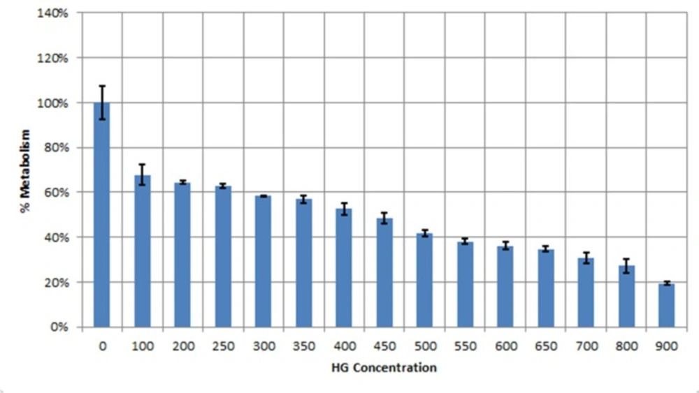
MTT assay indicating the inhibitory effect of Hygromycin B on human hepatoma cells (Huh7). Data showed LD50 value of450 μM Hygromycin B for Huh7 cell line
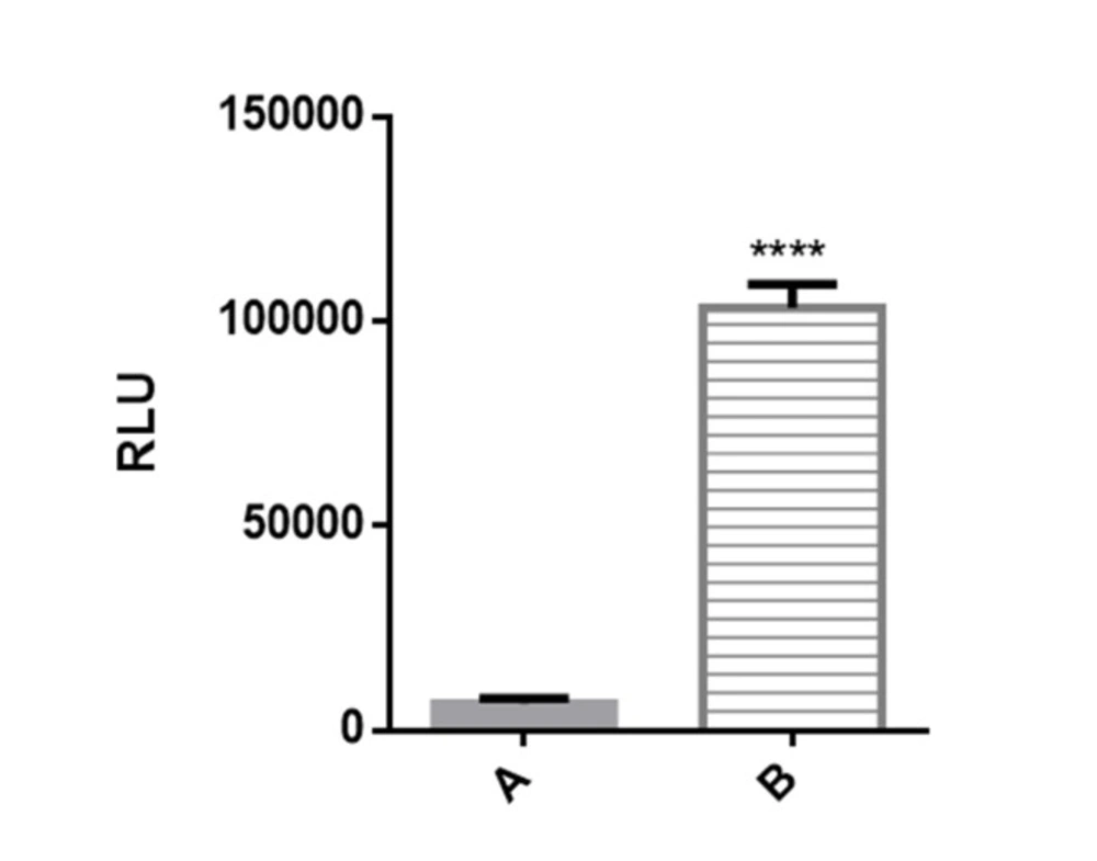
Figure 2. Luciferase reporter activity in transiently transfected Huh7-CMV-luc cells. Recombinant cells were seeded overnight in 24 well plates at 2 × 10⁵ cells per well. After 24 h luciferase activity was assessed by measuring luciferase activity in cell lysates. Transfected cells with recombinant vector including CMV enhancer element (column B) showed significant increase in luciferase activity compared to control cells which have PGL4.26 plasmid with minimal promoter lacking CMV fragment (column A).
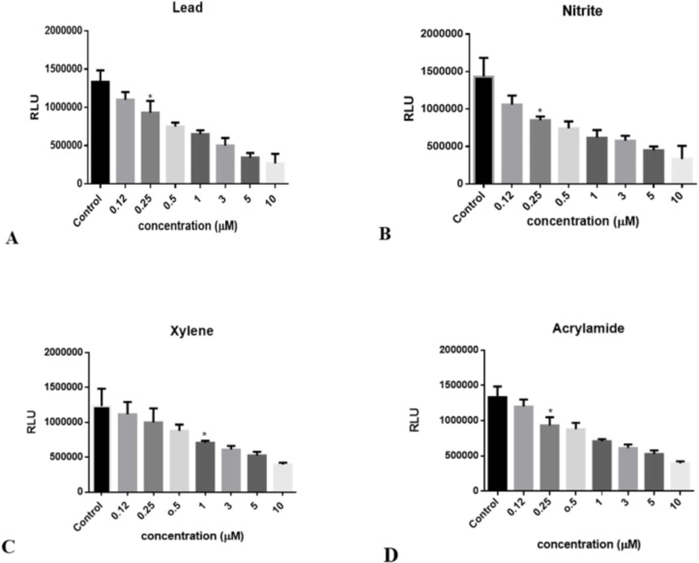
Decrement in luciferase activity following to ATP reduction in Huh7-CMV-luc stable cell line exposed to: (A) lead concentrations (0.1 to 10 μM), (B) Nitrite concentrations (0.1 to 10 μM), (C) Xylene concentrations (0.1 to 10 μM), and (D) Acrylamide concentrations (0.1 to 10 μM). Luciferase activity was measured after 24 hours. The numbers in the left column represent the relative luciferase activities. Luciferase activity was decreased following treatment with increasing concentrations of toxins. Each bar shows the mean ± SD; *, p = 0.0327 for (A), p = 0.0170 for (B), p = 0.0228 for (C), and p = 0.0224 for (D) compared to control
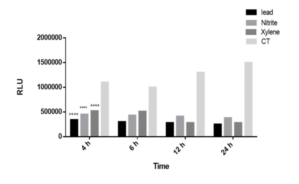
The time course of luciferase activity induction. The Recombinant cell line was incubated with Lead, Nitrite and Xylene (5 μM) at 37 °C for the indicated time after which luciferase activity in cell lysates was measured as described under the “Methods” section. Luciferase activity was expressed as RLUs in recombinant Huh7 cell line. The luciferase induced significantly after 4 h as the shortest induction time. Each bar shows the mean ± SD; ****,P < 0.0001 compared to control
Statistical analysis
All experiments were conducted in triplicate, and the results were expressed as mean ± SD (standard deviation). One-way analysis of variance (ANOVA) was used to assess the statistical analysis, and p < 0.05 was considered to be significant. The data were analyzed using Microsoft Excel software and GraphPad Prism software.