


1. Background
2. Objectives
3. Methods
3.1. Cell Culture and Reagents
3.2. Herpes Simplex Virus Type 1 Infection and Time-Course Sampling
3.3. Western Blotting
3.4. Cell Viability (MTT) Assay
3.5. Quantitative Real-time PCR
| Genes and Primer Type | Sequence (5′→3′) | Amplicon Size (bp) |
|---|---|---|
| HSV-1 ICP0 | 143 | |
| Forward | CGCTGTTCTCCTTGGACTTG | |
| Reverse | TTGTTGGGCGTGTTTCTTGT | |
| HSV-1 ICP4 | 175 | |
| Forward | GCTCCGAAGAACTGGACTGA | |
| Reverse | CTTGGTGACGTGGTTGTTGT | |
| HSV-1 gB | 118 | |
| Forward | GGACGAGGCGCCGTTTACGA | |
| Reverse | AGCAGGGTGCTCGTGTTTGG | |
| HSV-1 gC | 132 | |
| Forward | CTTGACGACGATGACGACGA | |
| Reverse | GTTGTCGTCGTCGTAGTCGT | |
| IL-6 (human) | 149 | |
| Forward | ACTCACCTCTTCAGAACGAATTG | |
| Reverse | CCATCTTTGGAAGGTTCAGGTTG | |
| TNF-α (human) | 136 | |
| Forward | CCTCTCTCTAATCAGCCCTCTG | |
| Reverse | GAGGACCTGGGAGTAGATGAG | |
| GAPDH (human, control) | 121 | |
| Forward | GAAGGTGAAGGTCGGAGTC | |
| Reverse | GAAGATGGTGATGGGATTTC |
Abbreviations: HSV-1, Herpes simplex virus type 1; TNF-α, tumor necrosis factor-alpha; IL-6, interleukin-6.
3.6. Immunofluorescence and Confocal Microscopy
3.7. Plaque Assay
3.8. Enzyme-Linked Immunosorbent Assay
3.9. Statistical Analysis
4. Results
4.1. Herpes Simplex Virus Type 1 Infection Induces extracellular Signal-Regulated Kinase 1/2 and Activation of Protein Kinase B with a Concurrent Decline in HaCaT Cell Viability
![Herpes simplex virus type 1 (HSV-1) infection activates extracellular signal-regulated kinase 1/2 (ERK1/2) and activation of protein kinase B (AKT) signaling and decreases cell viability in HaCaT cells: A, Western blot analysis showing time-dependent phosphorylation of ERK1/2 following HSV-1 infection [multiplicity of infection (MOI) = 1]; B, densitometric quantification of phosphorylated extracellular signal-regulated kinase 1/2 (p-ERK1/2) normalized to total ERK1/2; C, Western blot showing phosphorylation of AKT at corresponding time points; D, densitometric quantification of phosphorylated activation of protein kinase B (p-AKT) normalized to total AKT; E, MTT assay showing progressive decline in cell viability from 0 to 24-hours post-infection (hpi). GAPDH served as the loading control [data are expressed as mean ± standard deviation (SD); n = 3; ** P < 0.01 and *** P < 0.001 vs. control].](https://brieflands.com/journals/ijpr/articles/164639/figures/ijpr-24-1-164639-i001-preview.webp "Herpes simplex virus type 1 (HSV-1) infection activates extracellular signal-regulated kinase 1/2 (ERK1/2) and activation of protein kinase B (AKT) signaling and decreases cell viability in HaCaT cells: A, Western blot analysis showing time-dependent phosphorylation of ERK1/2 following HSV-1 infection [multiplicity of infection (MOI) = 1]; B, densitometric quantification of phosphorylated extracellular signal-regulated kinase 1/2 (p-ERK1/2) normalized to total ERK1/2; C, Western blot showing phosphorylation of AKT at corresponding time points; D, densitometric quantification of phosphorylated activation of protein kinase B (p-AKT) normalized to total AKT; E, MTT assay showing progressive decline in cell viability from 0 to 24-hours post-infection (hpi). GAPDH served as the loading control [data are expressed as mean ± standard deviation (SD); n = 3; ** P < 0.01 and *** P < 0.001 vs. control].")
Herpes simplex virus type 1 (HSV-1) infection activates extracellular signal-regulated kinase 1/2 (ERK1/2) and activation of protein kinase B (AKT) signaling and decreases cell viability in HaCaT cells: A, Western blot analysis showing time-dependent phosphorylation of ERK1/2 following HSV-1 infection [multiplicity of infection (MOI) = 1]; B, densitometric quantification of phosphorylated extracellular signal-regulated kinase 1/2 (p-ERK1/2) normalized to total ERK1/2; C, Western blot showing phosphorylation of AKT at corresponding time points; D, densitometric quantification of phosphorylated activation of protein kinase B (p-AKT) normalized to total AKT; E, MTT assay showing progressive decline in cell viability from 0 to 24-hours post-infection (hpi). GAPDH served as the loading control [data are expressed as mean ± standard deviation (SD); n = 3; ** P < 0.01 and *** P < 0.001 vs. control].
4.2. Upregulation of Viral and Host Inflammatory Genes in Response to Herpes Simplex Virus Type 1 Infection
![Herpes simplex virus type 1 (HSV-1) infection upregulates viral and inflammatory gene expression in HaCaT keratinocytes: A, qRT-PCR analysis showing time-dependent increases in viral transcripts (ICP0, ICP4, gB, and gC); B, qRT-PCR of host cytokines [interleukin-6 (IL-6) and tumor necrosis factor-alpha (TNF-α)] demonstrating parallel induction of inflammatory genes; C, enzyme-linked immunosorbent assay (ELISA) confirming increased secretion of IL-6 and TNF-α in culture supernatants. Results are normalized to GAPDH [data are expressed as mean ± standard deviation (SD); n = 3; ** P < 0.01, *** P < 0.001 vs. control].](https://brieflands.com/journals/ijpr/articles/164639/figures/ijpr-24-1-164639-i002-preview.webp "Herpes simplex virus type 1 (HSV-1) infection upregulates viral and inflammatory gene expression in HaCaT keratinocytes: A, qRT-PCR analysis showing time-dependent increases in viral transcripts (ICP0, ICP4, gB, and gC); B, qRT-PCR of host cytokines [interleukin-6 (IL-6) and tumor necrosis factor-alpha (TNF-α)] demonstrating parallel induction of inflammatory genes; C, enzyme-linked immunosorbent assay (ELISA) confirming increased secretion of IL-6 and TNF-α in culture supernatants. Results are normalized to GAPDH [data are expressed as mean ± standard deviation (SD); n = 3; ** P < 0.01, *** P < 0.001 vs. control].")
Herpes simplex virus type 1 (HSV-1) infection upregulates viral and inflammatory gene expression in HaCaT keratinocytes: A, qRT-PCR analysis showing time-dependent increases in viral transcripts (ICP0, ICP4, gB, and gC); B, qRT-PCR of host cytokines [interleukin-6 (IL-6) and tumor necrosis factor-alpha (TNF-α)] demonstrating parallel induction of inflammatory genes; C, enzyme-linked immunosorbent assay (ELISA) confirming increased secretion of IL-6 and TNF-α in culture supernatants. Results are normalized to GAPDH [data are expressed as mean ± standard deviation (SD); n = 3; ** P < 0.01, *** P < 0.001 vs. control].
4.3. Pharmacological Inhibition of Mitogen-Activated Protein Kinase and Phosphatidylinositol 3-Kinase/Protein Kinase B Pathways Attenuates Signaling Activation and Restores Cell Viability in Herpes Simplex Virus Type 1-Infected HaCaT Cells
![U0126 and LY294002 inhibit extracellular signal-regulated kinase 1/2 (ERK1/2) and phosphatidylinositol 3-kinase/protein kinase B (PI3K/AKT) signaling and restore cell viability in Herpes simplex virus type 1 (HSV-1)-infected HaCaT cells: A, Western blot analysis showing reduced phosphorylated extracellular signal-regulated kinase 1/2 (p-ERK1/2) levels following U0126 treatment and reduced phosphorylated activation of protein kinase B (p-AKT) after LY294002 treatment and densitometric quantification confirming pathway-specific inhibition; B, MTT assay indicating partial recovery of viability in inhibitor-treated cells relative to untreated infection. GAPDH served as the internal control [data are expressed as mean ± standard deviation (SD); n = 3; ** P < 0.01, *** P < 0.001 vs. control].](https://brieflands.com/journals/ijpr/articles/164639/figures/ijpr-24-1-164639-i003-preview.webp "U0126 and LY294002 inhibit extracellular signal-regulated kinase 1/2 (ERK1/2) and phosphatidylinositol 3-kinase/protein kinase B (PI3K/AKT) signaling and restore cell viability in Herpes simplex virus type 1 (HSV-1)-infected HaCaT cells: A, Western blot analysis showing reduced phosphorylated extracellular signal-regulated kinase 1/2 (p-ERK1/2) levels following U0126 treatment and reduced phosphorylated activation of protein kinase B (p-AKT) after LY294002 treatment and densitometric quantification confirming pathway-specific inhibition; B, MTT assay indicating partial recovery of viability in inhibitor-treated cells relative to untreated infection. GAPDH served as the internal control [data are expressed as mean ± standard deviation (SD); n = 3; ** P < 0.01, *** P < 0.001 vs. control].")
U0126 and LY294002 inhibit extracellular signal-regulated kinase 1/2 (ERK1/2) and phosphatidylinositol 3-kinase/protein kinase B (PI3K/AKT) signaling and restore cell viability in Herpes simplex virus type 1 (HSV-1)-infected HaCaT cells: A, Western blot analysis showing reduced phosphorylated extracellular signal-regulated kinase 1/2 (p-ERK1/2) levels following U0126 treatment and reduced phosphorylated activation of protein kinase B (p-AKT) after LY294002 treatment and densitometric quantification confirming pathway-specific inhibition; B, MTT assay indicating partial recovery of viability in inhibitor-treated cells relative to untreated infection. GAPDH served as the internal control [data are expressed as mean ± standard deviation (SD); n = 3; ** P < 0.01, *** P < 0.001 vs. control].
4.4. Confocal Microscopy Reveals Distinct Subcellular Localization Patterns of Phosphorylated Extracellular Signal-Regulated Kinase 1/2, Phosphorylated Activation of Protein Kinase B, and Herpes Simplex Virus Type 1 Following Pathway Inhibition
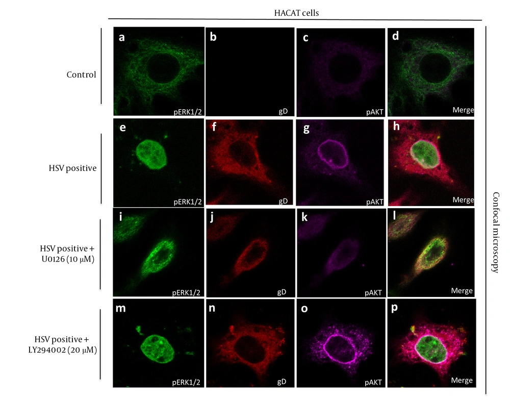
Pathway inhibition alters nuclear localization of extracellular signal-regulated kinase 1/2 (ERK1/2) and activation of protein kinase B (AKT) in herpes simplex virus type 1 (HSV-1)-infected keratinocytes: A - D, confocal images showing nuclear accumulation of phosphorylated extracellular signal-regulated kinase 1/2 (p-ERK1/2) and phosphorylated activation of protein kinase B (p-AKT) in control cells; E - H, confocal images showing nuclear accumulation of p-ERK1/2 and p-AKT in HSV-1-infected cells; I - L, U0126 treatment restricts p-ERK1/2 to the cytoplasm, while LY294002 prevents p-AKT nuclear localization; M - P, HSV-1 glycoprotein D (gD) remains cytoplasmic across conditions, confirming that inhibitors affect post-entry signaling. Representative images from three independent experiments are shown.
4.5. Pharmacological Inhibition of Extracellular Signal-Regulated Kinase 1/2 and Phosphatidylinositol 3-Kinase/Protein Kinase B Reduces Herpes Simplex Virus Type 1 Replication in HaCaT Cells
![Pharmacological inhibition of extracellular signal-regulated kinase 1/2 (ERK1/2) and phosphatidylinositol 3-kinase/protein kinase B (PI3K/AKT) signaling suppresses herpes simplex virus type 1 (HSV-1) replication: A, plaque formation is markedly reduced in cells treated with U0126 or LY294002 compared with untreated infection; B, quantification of plaque-forming units (PFU) showing significant declines in infectious viral titers in inhibitor-treated groups [data are expressed as mean ± standard deviation (SD); n = 3; ** P < 0.01 vs. control].](https://brieflands.com/journals/ijpr/articles/164639/figures/ijpr-24-1-164639-i005-preview.webp "Pharmacological inhibition of extracellular signal-regulated kinase 1/2 (ERK1/2) and phosphatidylinositol 3-kinase/protein kinase B (PI3K/AKT) signaling suppresses herpes simplex virus type 1 (HSV-1) replication: A, plaque formation is markedly reduced in cells treated with U0126 or LY294002 compared with untreated infection; B, quantification of plaque-forming units (PFU) showing significant declines in infectious viral titers in inhibitor-treated groups [data are expressed as mean ± standard deviation (SD); n = 3; ** P < 0.01 vs. control].")
Pharmacological inhibition of extracellular signal-regulated kinase 1/2 (ERK1/2) and phosphatidylinositol 3-kinase/protein kinase B (PI3K/AKT) signaling suppresses herpes simplex virus type 1 (HSV-1) replication: A, plaque formation is markedly reduced in cells treated with U0126 or LY294002 compared with untreated infection; B, quantification of plaque-forming units (PFU) showing significant declines in infectious viral titers in inhibitor-treated groups [data are expressed as mean ± standard deviation (SD); n = 3; ** P < 0.01 vs. control].
5. Discussion
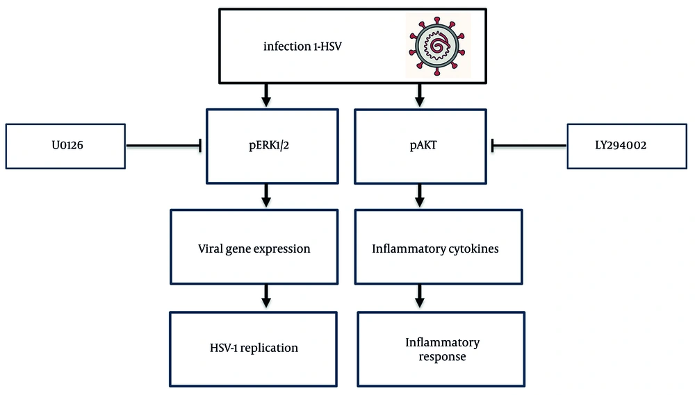
Schematic model summarizing herpes simplex virus type 1 (HSV-1)-induced signaling and the effects of pathway inhibition; HSV-1 activates mitogen-activated protein kinase/extracellular signal-regulated kinase 1/2 (MAPK/ERK1/2) and phosphatidylinositol 3-kinase/protein kinase B (PI3K/AKT) pathways, promoting viral replication and inflammatory gene expression. U0126 and LY294002 disrupt pathway activation, inhibit nuclear translocation of phosphorylated extracellular signal-regulated kinase 1/2 (p-ERK1/2) and phosphorylated activation of protein kinase B (p-AKT), and reduce viral replication and cytokine release.




