


1. Background
2. Objectives
3. Methods
3.1. Plant Material
3.2. Chemicals
3.3. Analysis of the Structure
3.4. Extraction and Isolation
3.5. Cell Culture
3.6. Hispidulin Cytotoxicity Analysis
3.7. Enzyme-Linked Immunosorbent Assay
3.8. Western Blotting Analysis
3.9. Reactive Oxygen Species Analysis
3.10. Assessment of NADPH Oxidase Activity
3.11. Assessment of Glucose Uptake Induced by Inactivated Insulin
3.12. Statistical Method
4. Result
4.1. Structure of Compound 1
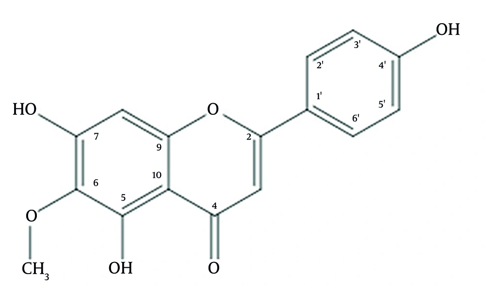
The structure of hispidulin purified from Vietnamese Clerodendrum chinense leaves
4.2. Analysis of the Cytotoxicity of Hispidulin
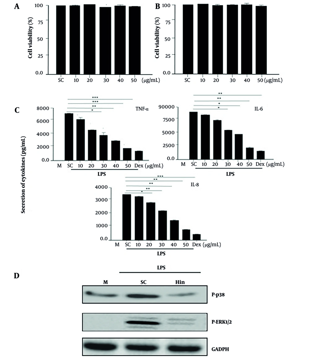
The viability and anti-inflammatory activity of hispidulin in macrophages stimulated by LPS. A, hispidulin concentrations from 10 to 50 µg/mL did not affect the viability of Raw 246.7; B, hispidulin concentrations from 10 to 50 µg/mL did not affect the viability of 3T3-L; C, the secretion of inflammatory cytokines stimulated by LPS was inhibited by hispidulin in macrophages; D, the phosphorylation of ERK1/2 and p38 activated by LPS was inhibited by hispidulin in macrophages, LPS (1µg/mL) was incubated for 30 minutes after cells were pre-treated with either the control solvent or hispidulin (50 µg/mL) for 45 minutes. The experiment was repeated five times (n = 5). LPS, lipopolysaccharide; M, medium; Hin, hispidulin; SC, solvent control (0.1% DMSO). *** P < 0.001 is a significant difference.
4.3. Evaluation of LPS-Induced Inflammatory Inhibition
4.4. Determination of Reactive Oxygen Species
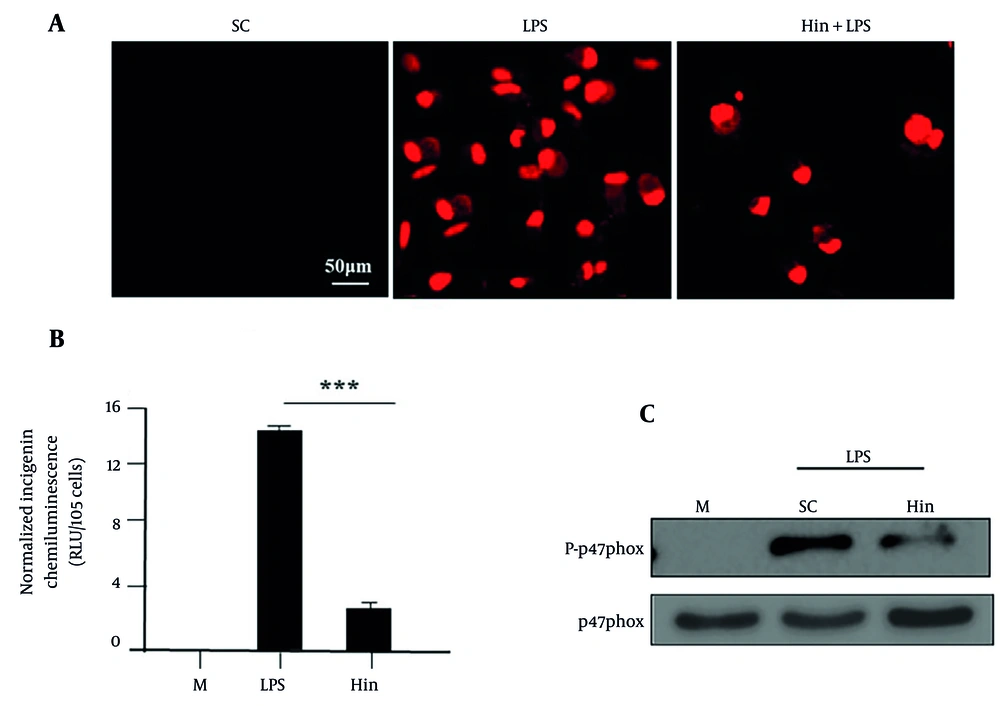
The production of reactive oxygen species (ROS) by LPS is disrupted by hispidulin in macrophages. A, ROS in macrophages were inhibited by hispidulin, LPS (1 µg/mL) was added for 30 minutes after cells were pre-treated with hispidulin (50 µg/mL) for 45 minutes; B, the activity of NADPH oxidase induced by LPS was inhibited by hispidulin in macrophages, LPS (1 µg/mL) was incubated for 30 minutes after cells were pre-treated with hispidulin (50 µg/mL) for 45 minutes; C, the phosphorylation of p47phox was inhibited by hispidulin, LPS (1 µg/mL) was added 15 minutes after macrophages were incubated with hispidulin (50 µg/mL) for 45 minutes, the cells were lysed and approximately 50 µg of protein was added to each well, the phosphorylation of p47phox was analyzed by Western blot. The experiment was repeated five times (n=5). LPS, lipopolysaccharide; M, medium; SC, solvent control (0.1% DMSO). *** P < 0.001 is a significant difference.
4.5. Analysis of the Reactivation of Insulin Resistance
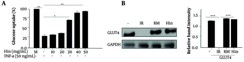
Effect of hispidulin on insulin resistance. A, glucose uptake effect of hispidulin; B, effect of hispidulin on Glucose transporter type 4, the insulin-resistant cells were supplemented with either a control solvent or hispidulin (50 µg/mL) or rosiglitazone maleate (90 μM). The cells were analyzed by Western blotting. -, solvent control (0.1% DMSO); IR, insulin-resistant; RM, rosiglitazone maleate; Hin, hispidulin. The experiment was repeated thrice (n = 5). *** P < 0.001 is a significant difference.

