


1. Background
2. Objectives
3. Patients and Methods
3.1. Patients
3.2. FDG-PET/CT Protocol
3.3. FDG-PET/CT Scan Interpretation
3.4. Histopathological Analysis
3.5. Statistical Analysis
4. Results
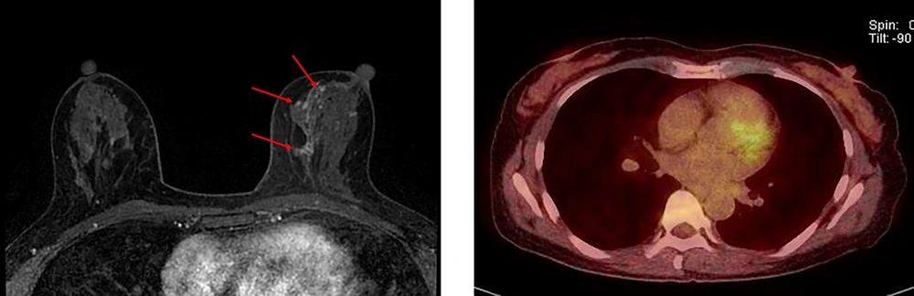
A 52-year-old woman with pure ductal carcinoma in situ (DCIS) in the left breast. A, Axial dynamic enhanced T1-weighted MRI demonstrates a segmental clumped non-mass enhancement (3.5 cm) (arrows) in the left middle inner part of the breast. B, The FDG-PET/CT scan shows no discernible FDG uptake in the left middle inner part of the breast.
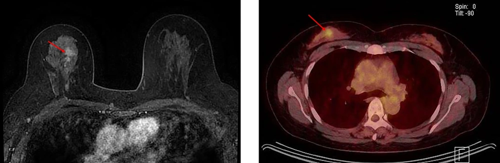
A 49-year-old woman with ductal carcinoma in situ (DCIS) upgraded to invasive ductal carcinoma (IDC) in the right breast. A, Axial dynamic enhanced T1-weighted MRI demonstrates a heterogeneously enhanced mass (2.1 cm) with an irregular shape and margins (arrow) in the right middle upper part of the breast. B, The FDG-PET/CT scan shows focal FDG uptake (arrow) in the right middle upper part of the breast with a maximum standardized uptake value (SUVmax) of 2.9.
| DCIS (n = 119) | DCIS with invasion (n = 46) | P-value | |
|---|---|---|---|
| Age (y) | |||
| Mean ± SD | 54.0 ± 10.5 | 49.3 ± 8.8 | 0.008 a |
| Median (range) | 52.5 (26.0 - 82.0) | 49.0 (28.0 - 69.0) | |
| Biopsy method | |||
| Core needle biopsy | 96 (80.7) | 42 (91.3) | 0.098 |
| Vacuum-assisted biopsy | 23 (19.3) | 4 (8.7) | |
| Tumor grade | |||
| 1 & 2 | 55 (46.2) | 14 (30.4) | 0.065 |
| 3 | 64 (53.8) | 32 (69.6) | |
| SUVmax | |||
| Mean ± SD | 1.9 ± 1.1 | 3.2 ± 2.5 | < 0.001 a |
| Median (range) | 1.6 (0.7 - 5.6) | 2.5 (0.7 - 13.5) | |
| < 1.9 (cutoff point) | 67 (56.3) | 10 (21.7) | < 0.001 a |
| ≥ 1.9 (cutoff point) | 52 (43.7) | 36 (78.3) | |
| Visual analysis of FDG-PET/CT | |||
| Negative | 39 (32.8) | 4 (8.7) | 0.002 a |
| Positive | 80 (67.2) | 42 (91.3) | |
| FDG uptake of axillary LNs | |||
| Negative | 112 (94.1) | 42 (91.3) | 0.502 |
| Positive | 7 (5.9) | 4 (8.7) | |
| Pathological size (DCIS) (cm) | |||
| Mean ± SD | 2.8 ± 2.3 | 4.1 ± 2.3 | < 0.001 a |
| Median (range) | 2.0 (0.2 - 11.3) | 3.9 (0.5 - 10.0) | |
| LN metastasis in pathology | |||
| Negative | 118 (99.2) | 44 (95.7) | 0.188 |
| Positive | 1 (0.8) | 2 (4.3) | |
| ER | |||
| Negative | 42 (35.3) | 15 (32.6) | 0.745 |
| Positive | 77 (64.7) | 31 (67.4) | |
| PR | |||
| Negative | 49 (41.2) | 17 (37.0) | 0.620 |
| Positive | 70 (58.8) | 29 (63.0) | |
| HER-2 | |||
| Negative | 63 (52.9) | 21 (45.7) | 0.401 |
| Positive | 56 (47.1) | 25 (54.3) | |
| Ki-67 | |||
| < 14% | 62 (52.1) | 18 (39.1) | 0.135 |
| ≥ 14% | 57 (47.9) | 28 (60.9) | |
| EGFR | |||
| Negative | 86 (72.3) | 33 (71.7) | 0.946 |
| Positive | 33 (27.7) | 13 (28.3) |
Abbreviations: DCIS, ductal carcinoma in situ; EGFR, epidermal growth factor receptor; ER, estrogen receptor; FDG-PET/CT, 18F-fluorodeoxyglucose-positron emission tomography/computed tomography; HER-2, human epidermal growth factor receptor type 2; LN, lymph node; PR, progesterone receptor; SD, standard deviation; SUVmax, maximum standardized uptake value.
aP-value shows a significant difference between DCIS and IDC, based on Chi-square or Fisher's exact test and independent t-test or Mann-Whitney U test.
| Univariate analysis | Multivariate analysis | |||||
|---|---|---|---|---|---|---|
| Crude OR (95% CI) | P-value | Model 1: SUVmax + age + tumor size | Model 2: Visual detection by PET/CT + age + tumor size | |||
| Adjusted OR (95% CI) | P-value | Adjusted OR (95% CI) | P-value | |||
| Age (y) | 0.95 (0.92 - 0.99) | 0.011 a | 0.97 (0.93 - 1.00) | 0.069 | 0.96 (0.92 - 1.00) | 0.032 a |
| Biopsy method | ||||||
| VAB | 1.0 (Reference) | |||||
| CNB | 0.43 (0.15 - 1.29) | 0.134 | ||||
| Tumor grade of biopsy specimen | ||||||
| 1 & 2 | 1.0 (Reference) | |||||
| 3 | 1.93 (0.94 - 3.96) | 0.074 | ||||
| SUVmax (≥ 1.9 as the cutoff point) | 4.47 (2.05 - 9.75) | < 0.001 a | 3.53 (1.58 - 7.89) | 0.002 a | ||
| Visual analysis of FDG-PET/CT | ||||||
| Negative | 1.0 (Reference) | 1.0 (Reference) | ||||
| Positive | 4.64 (1.61 - 13.31) | 0.004 a | 4.67 (1.55 - 14.08) | 0.006 a | ||
| FDG uptake of axillary LNs | ||||||
| Negative | 1.0 (Reference) | |||||
| Positive | 1.59 (0.45 - 5.66) | 0.476 | ||||
| Pathological size (DCIS) (cm) | 1.25 (1.08 - 1.44) | 0.003 a | 1.20 (1.03 - 1.40) | 0.022 a | 1.25 (1.07 - 1.47) | 0.005 a |
| LN metastasis in pathology | ||||||
| Negative | 1.0 (Reference) | |||||
| Positive | 4.44 (0.42 - 47.09) | 0.217 | ||||
| ER | ||||||
| Negative | 1.0 (Reference) | |||||
| Positive | 1.11 (0.54 - 2.29) | 0.768 | ||||
| PR | ||||||
| Negative | 1.0 (Reference) | z | ||||
| Positive | 1.18 (0.59 - 2.38) | 0.636 | ||||
| HER-2 | ||||||
| Negative | 1.0 (Reference) | |||||
| Positive | 1.33 (0.67 - 2.63) | 0.408 | ||||
| Ki-67 | ||||||
| < 14% | 1.0 (Reference) | |||||
| ≥ 14% | 1.67 (0.84 - 3.34) | 0.143 | ||||
| EGFR | ||||||
| Negative | 1.0 (Reference) | |||||
| Positive | 1.04 (0.49 - 2.21) | 0.918 | ||||
Abbreviations: CI, confidence interval; CNB, core needle biopsy; DCIS, ductal carcinoma in situ; EGFR, epidermal growth factor receptor; ER, estrogen receptor; FDG-PET/CT, 18F-fluorodeoxyglucose-positron emission tomography/computed tomography; HER-2, human epidermal growth factor receptor type 2; LN, lymph node; OR, odds ratio; PR, progesterone receptor; SUVmax, maximum standardized uptake value; VAB, vacuum-assisted biopsy.
aStatistically significant.
| Sensitivity (95% CI) | Specificity (95% CI) | PPV (95% CI) | NPV (95% CI) | Accuracy (95% CI) | |
|---|---|---|---|---|---|
| Semi-quantitative analysis with SUVmax (≥ 1.9, cutoff point) | 0.78 (0.64-0.89) | 0.56 (0.47-0.65) | 0.41 (0.31-0.52) | 0.87 (0.77-0.94) | 0.62 (0.55-0.70) |
| Visual analysis | 0.91 (0.79-0.98) | 0.33 (0.24-0.42) | 0.34 (0.26-0.44) | 0.91 (0.78-0.97) | 0.49 (0.41-0.57) |
| P-value (visual vs. semi-quantitative) | 0.014 a | < 0.001 a | 0.012 a | 0.297 | <0.001 a |
Abbreviations: FDG-PET/CT, 18F-fluorodeoxyglucose-positron emission tomography/computed tomography; SUVmax, maximum standardized uptake value; CI, confidence interval; PPV, positive predictive value; NPV, negative predictive value.
aP-value for differences was determined using McNemar test (sensitivity, specificity, and accuracy) or generalized score statistic (PPV and NPV).


