


1. Background
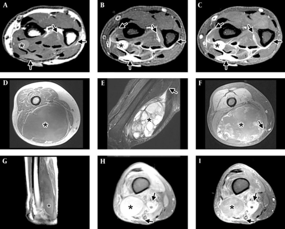
Imaging patterns of myxofibrosarcoma (MFS). A-C, Infiltrative pattern in the distal forearm, with tail-like multidirectional spread along fascial planes (arrows) beyond the tiny nodule (*). A, Short-axis T1-WI shows the mass has intermediate T1 signal, hyperintense to muscle. Tail-like extensions (arrows) can be faintly seen. B, Short-axis T2-WI with fat suppression shows the lesion to have high signal, close to that expected for fluid. The tail-like extensions can be seen more clearly. C, Short-axis post-contrast T1-WI with fat suppression shows the mass and the tail-like extensions to enhance avidly. D-F, Focal mass (*) in the distal thigh, with minimal to no spread along fascial planes (arrow) and good demarcation of the tumor by a thin rim. D, Axial T1-WI shows an intermediate signal mass (*) with areas of subtle increased signal compared to muscle located in the posterior compartment of the thigh. Tail-like extension is not well seen. E, Sagittal T2-WI with fat suppression shows the dominant multiloculated mass (*) with high signal internally and multiple low-signal septations. A long tail (arrow) can be seen extending proximally. F, Axial post-contrast T1-WI with fat suppression shows a mass (*) with peripheral nodular and central feather-like pattern of enhancement typically seen with myxoid neoplasms. A small lateral tail can be seen laterally. G-I, Mixed pattern, with a dominant mass (*) and extensive surrounding infiltration (arrows). G, Sagittal T1-WI shows a mass in the posterior compartment of the thigh with a higher signal component distally (*). H, Axial PD fat suppressed image at the distal thigh shows a high-signal mass (*) and areas of extension (arrows) in the adjacent tissues. I, Axial T1-WI with fat suppression shows a mass (*) with central feather-like pattern of enhancement typically seen with myxoid neoplasms and enhancing infiltration (arrows) of the adjacent tissues.
2. Objectives
3. Patients and Methods
3.1. Data Collection
3.2. Image Acquisition and Analysis
Abbreviations: M, male F, female MRI, magnetic resonance imaging.
aPatients in group 1 had at least one MRI (at recurrence), with or without MRI prior to surgery.
bPatients in group 2 had 2 MRIs: at presentation (prior to surgery) and at recurrence.
