


1. Background
2. Objectives
3. Materials and Methods
3.1. Study Subjects
3.2. MRI Protocol
3.3. Post-Processing
| Direction No. | X | Y | Z |
|---|---|---|---|
| 0 | 0.000 | 0.000 | 0.000 |
| 1 | 1.000 | 0.000 | 0.000 |
| 2 | 0.849 | 0.528 | 0.000 |
| 3 | -0.108 | 0.565 | 0.818 |
| 4 | 0.884 | -0.345 | -0.315 |
| 5 | -0.003 | -0.736 | 0.667 |
| 6 | -0.868 | -0.238 | 0.436 |
| 7 | 0.799 | 0.370 | 0.475 |
| 8 | -0.162 | 0.987 | 0.000 |
| 9 | 0.866 | -0.129 | 0.438 |
| 10 | -0.212 | -0.936 | 0.281 |
| 11 | 0.068 | -0.892 | -0.446 |
| 12 | 0.550 | -0.544 | -0.634 |
| 13 | -0.435 | -0.422 | 0.795 |
| 14 | -0.599 | 0.780 | 0.182 |
| 15 | -0.525 | 0.030 | -0.851 |
| 16 | -0.600 | -0.688 | 0.409 |
| 17 | 0.653 | -0.060 | -0.755 |
| 18 | 0.207 | -0.076 | -0.975 |
| 19 | -0.413 | -0.699 | -0.584 |
| 20 | -0.436 | 0.822 | -0.366 |
| 21 | 0.462 | 0.874 | 0.148 |
| 22 | -0.503 | 0.488 | -0.713 |
| 23 | 0.824 | -0.530 | 0.202 |
| 24 | 0.297 | 0.349 | 0.889 |
| 25 | -0.040 | 0.318 | -0.947 |
Abbreviations: DTI, diffusion tensor imaging.
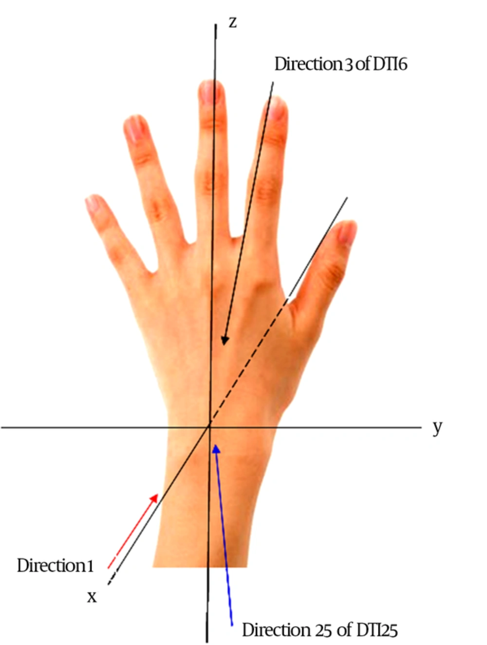
Scheme of the hand showing the X, Y, and Z axes of the wrist and directions selected in the gradient tabulations
3.4. Statistical Analyses
| Direction No. | X | Y | Z |
|---|---|---|---|
| 0 | 0.000 | 0.000 | 0.000 |
| 1 | 1.000 | 0.000 | 0.000 |
| 2 | 0.446 | 0.895 | 0.000 |
| 3 | 0.447 | 0.275 | 0.851 |
| 4 | 0.448 | -0.723 | -0.525 |
| 5 | 0.447 | -0.724 | 0.526 |
| 6 | -0.449 | -0.277 | 0.850 |
Abbreviations: DTI, diffusion tensor imaging.
4. Results
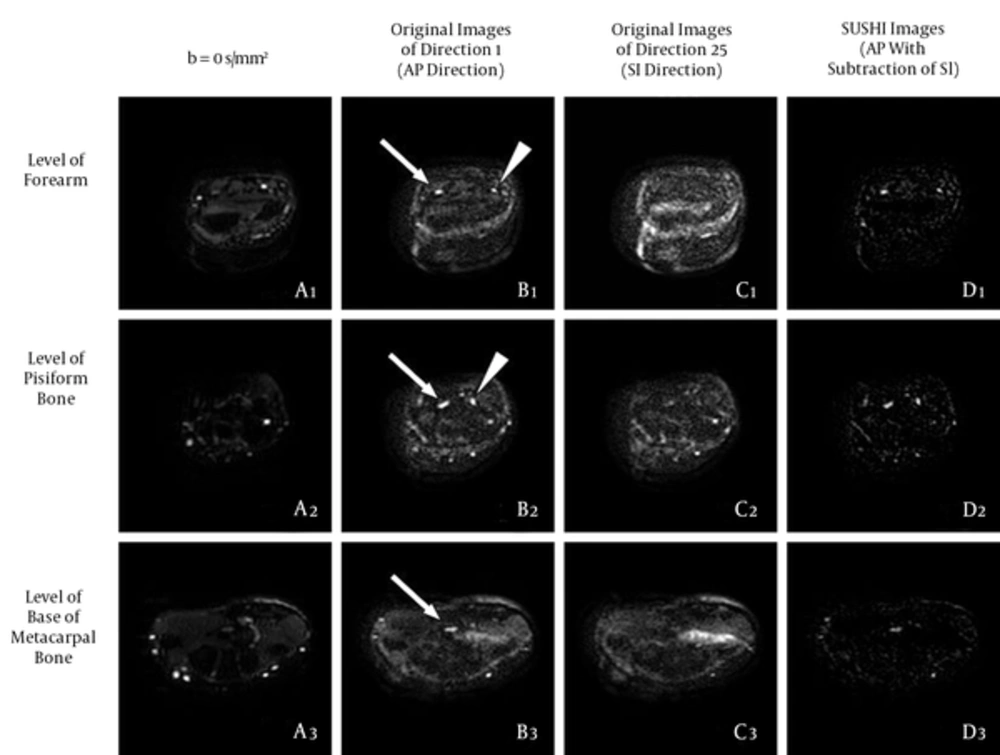
The original DTI25 images at all levels before and after SUSHI post-processing. Column A: Series of b = 0 s/mm2 images for providing the anatomical reference; these were similar to the T2-weighted images. Column B: series of original images in the AP direction, in which the nerves showed high signal intensity; most of the bone and vascular signals were suppressed, but some noise was apparent. Column C, Series of original images in the SI direction, in which only noise was apparent; the scope of the noise was similar to that of Column B, column D: Series of original images after SUSHI post-processing (AP direction with the subtraction of the SI direction), in which the nerve kept a high signal; most of the noise was subtracted, and the remnant noise was not connected with the nerve. Arrows, median nerve; arrowheads, ulnar nerve (AP, anterior-posterior; DTI, diffusion tensor imaging; SI, superior-inferior; SUSHI, suppression of heavily isotropic objects).
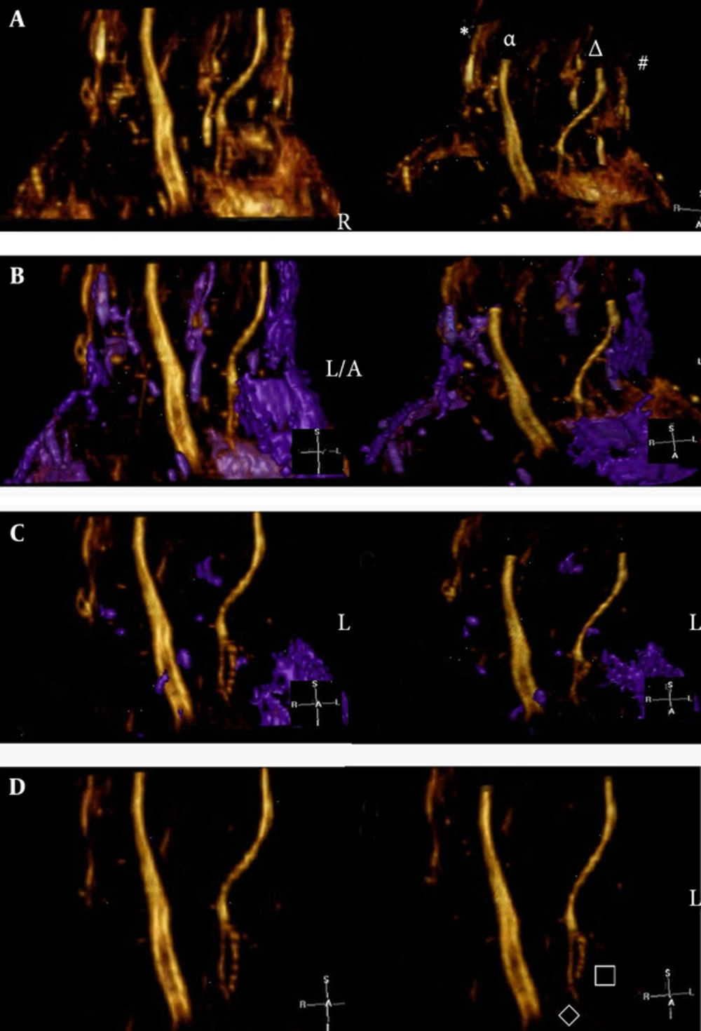
The effect of single-direction (AP direction) VR reconstruction by using the Voxar workstation software. A, Before denoising, the nerves were displayed clearly but were surrounded by noise. The symbols in the illustration are indicative as follows: (*) is the superficial branch of the radial nerve, (α) is the median nerve, (Δ) is the ulnar nerve, and (#) is the dorsal branch of the ulnar nerve. B, Denoising process. The noise region (i.e., the purple area) was selected and removed. The noise could be selected and removed completely by adjusting the window width and level over the course of multiple procedures. Note: In this example, the dorsal branch of the ulnar nerve was connected with noise; these were selected together for subsequent removal. C, Second denoising procedure. D, After denoising, the median nerve, ulnar nerve, and superficial branch of the radial nerve were clearly visible; moreover, the deep branch (□) and superficial branch (◊) of the ulnar nerve were clearly visible (AP, anterior-posterior; VR, volume rendering).
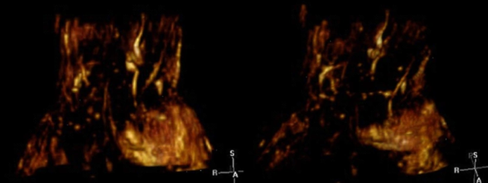
The effect of VR reconstruction for the SI direction. Only noise was visible, and the nerve could not be observed (VR, volume rendering; SI, superior-inferior).
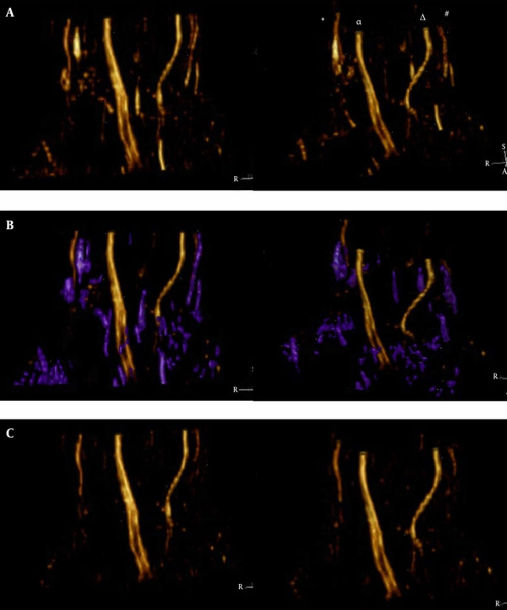
The effect of VR rfigure 5ction for SUSHI using Voxar workstation software. A, The nerves were displayed clearly before denoising, and the noise was lower than in Figure 3. The symbols are indicative as follows: (*) is the superficial branch of the radial nerve, (α) is the median nerve, (Δ) is the ulnar nerve, and (#) is the dorsal branch of the ulnar nerve. B, Denoising process. The noise region (i.e., the purple area) was selected and was removed easily in a single procedure. Note: In this example, the dorsal branch of the ulnar nerve was not connected with noise and was preserved. C, After denoising, the median nerve, ulnar nerve, superficial branch of the radial nerve, and the dorsal, deep, and superficial branches of the ulnar nerve were clearly shown (VR, volume rendering; SUSHI, suppression of heavily isotropic objects)
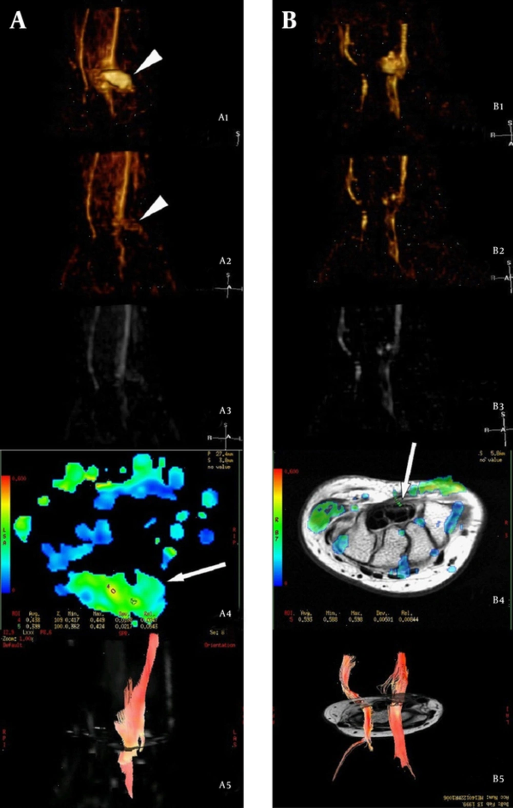
Manifestations in a subject with median nerve laceration (12-year-old female) followed up at 3 months (column A) and 18 months (column B) after repair. A1 and B1: VR reconstruction for the AP single-direction, in which the median nerve appeared intumescent with scar formation at the transection level (arrowheads). The regenerated nerve within the scar seemed undetectable. A2, B2, A3 and B3: VR reconstruction and MIP reconstruction of SUSHI, respectively, which showed that the scar (arrowheads) had been subtracted because of low anisotropy. By comparing A2 with B2, we found that the diameter of the injured nerve had increased and the signal was enhanced at the level distal to nerve injury at 18 months compared to 3 months post-injury, which were in accordance with the order of nerve regeneration. A4: FA map at the level of transection 3 months after repair, which showed that the FA value was unevenly distributed within the scar (arrows), and the highest FA value was lower than the normal value. B4: FA map at the level of the hamate bone 18 months after repair; the FA value had recovered to normal in a partial area, but the resumptive area (arrows) was smaller than normal, suggesting that nerve regeneration had not restored the nerves to their pre-injury status. A5 and B5: Fiber-tracking images, showing that more prolonged fibers were traced and the signal returned to normal at a later follow up; this was in line with ongoing nerve regeneration (VR, volume rendering; AP, anterior-posterior; MIP, maximum intensity projection; SUSHI, suppression of heavily isotropic objects; FA, fractional anisotropy).
| Single-Direction | SUSHI | P Value | |
|---|---|---|---|
| Median-DTI6 | 4 (3 - 4) | 4 (2 - 4) | 0.157 |
| Median-DTI25 | 4 (3 - 4) | 4 (3 - 4) | 1.000 |
| Ulnar-DTI6 | 3 (2 - 4) | 3 (1 - 4) | 0.039 |
| Ulnar-DTI25 | 4 (2 - 4) | 4 (1 - 4) | 0.218 |
Abbreviations: SUSHI, suppression of heavily isotropic objects; DTI, diffusion tensor imaging.
adata are mentioned as Median (Range).
| Scores | Noise Level | Noise-Removal Difficulty | ||||
|---|---|---|---|---|---|---|
| Single-direction | SUSHI | P Value | Single-direction | SUSHI | P Value | |
| DTI6 | 2 (1 - 4) | 3 (2 - 4) | < 0.001 | 3 (2 - 4) | 4 (2 - 4) | 0.002 |
| DTI25 | 2 (2 - 4) | 3 (2 - 4) | 0.005 | 3 (2 - 4) | 4 (3 - 4) | 0.002 |
Abbreviations: SUSHI, suppression of heavily isotropic objects; DTI, diffusion tensor imaging.
aThe data are mentioned as Median (Range).
