


1. Context
2. Objectives
3. Methods
3.1. Study Protocol and Registration
3.2. Selection Criteria
3.3. Literature Search Strategy
3.4. Screening and Data Extraction
3.5. Quality Appraisal
4. Results
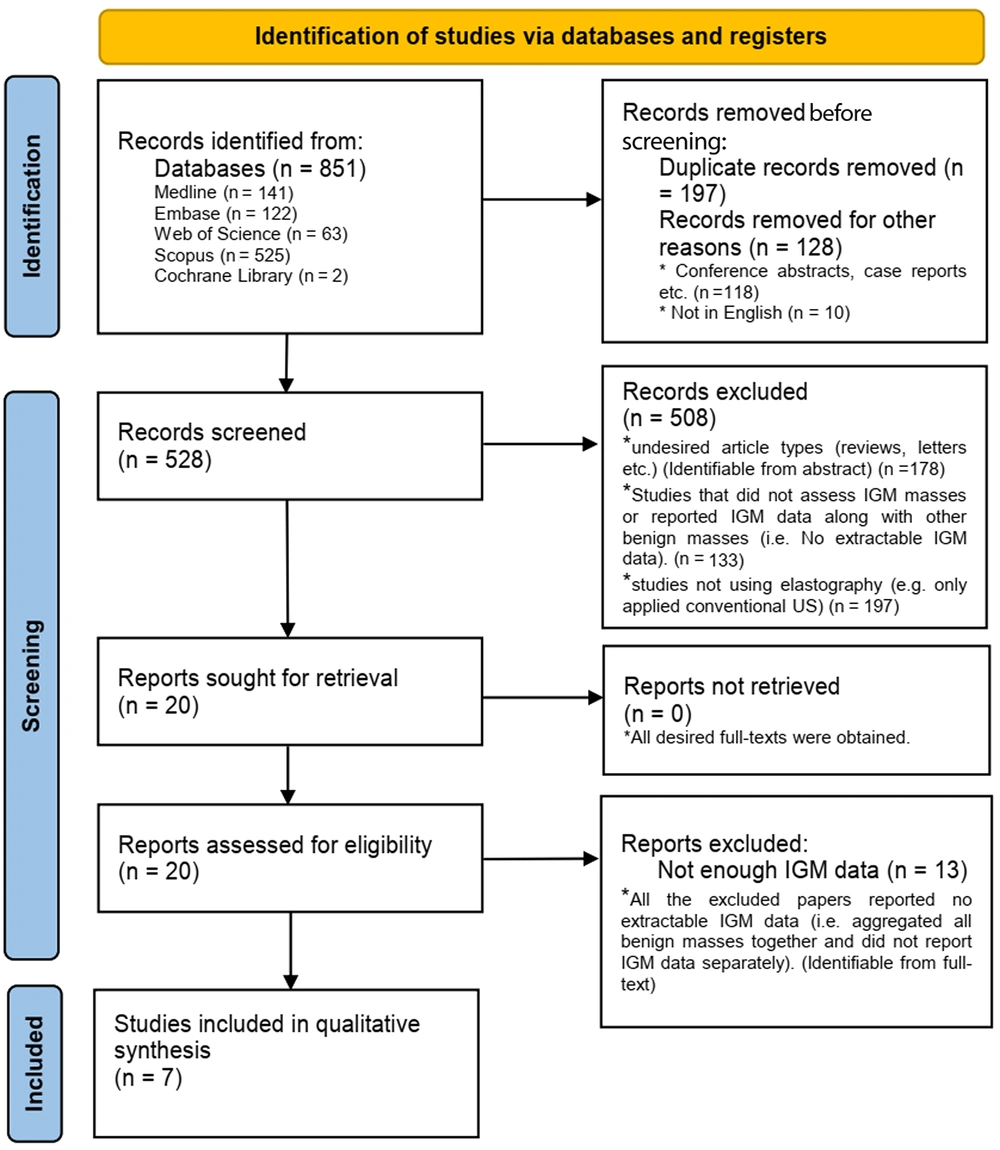
The PRISMA flow diagram of the study
| Authors | Year of publication | Number of participants | Age of participants | Objectives | Presenting symptoms | Type of modality | Device | Region of interest | Conventional US evaluation method | Quantitative USE parameter | Qualitative USE method |
|---|---|---|---|---|---|---|---|---|---|---|---|
| Durur‑Karakaya et al. (10) | 2014 | IGM=27 | IGM (37.8 ± 7.1 y) | Description of elastography findings for IGM | Palpable mass, breast pain, erythema, fistula formation, axillary lymphadenopathy, and failure to respond to treatment | Conventional US and strain elastography | EUB-6500; Hitachi® Medical, Tokyo, Japan | Breast and axilla | Maximum lesion diameter and lesion type (diffuse, tubular, mass, and cystic) | SR | Tsukuba classification |
| Teke et al. (11) | 2016 | IGM=4; Malignancy=122 (DCIS=9, IDC=98, ILC=10, malignant epithelial tumor=9) | IGM (39.5 ± 6.3 y); Malignancy (50.1 ± 9.7 y) | Differentiation between IGM and malignant breast masses | N/A | Conventional US and SWE | Acuson S2000 US system; Siemens Medical Solutions, Mountain View, CA, USA | Breast and axilla | BI-RADS score, tumor margin, shape, size, echo pattern, posterior acoustic features, and distribution | SWV | Tozaki classification |
| Yagci et al. (12) | 2017 | IGM=23; Malignancy=45 (IDC=42, ILC=2, malignant fibroepithelial tumor=1) | IGM (37.9 ± 6.6 y); Malignancy (52.8 ± 12.0 y) | Differentiation between IGM and malignant breast masses | N/A | Conventional US and strain elastography | Hi-Vision Preirus; Hitachi Medical Systems, Japan | Breast and axilla | Size, shape, margin, echogenicity, and posterior acoustic features | SR | N/A |
| Arslan et al. (13) | 2018 | IGM=77; Malignancy=36 (IDC=64, DCIS=5, ILC=4, mucinous carcinoma=2, medullary carcinoma=2) | IGM (35.6 ± 8.65 y); Malignancy (54.8 ± 11.8 y) | Differentiation between IGM and malignant breast masses | Palpable mass, breast pain, erythema, and nipple change | Conventional US and strain elastography | Aplio 500; Toshiba Medical Systems Corporation, Tokyo, Japan | Breast and axilla | BI-RADS score, tumor smargin, size, shape, echo pattern, posterior acoustic features, and distribution | SR | Tsukuba classification |
| Aslan et al. (14) | 2018 | IGM=39 | Group 1 (conservative treatment): 38.44 ± 9.6 y; Group 2 (surgery): 36.05 ± 7.44 y | Analysis of the correlation between the severity of IGM and the pretreatment SWE findings | Palpable mass, erythema, nipple retraction, and sinus formation | Conventional US and SWE | Acuson S 2000; Siemens Medical Solutions, Mountain View, CA, USA | Breast | BI-RADS score | SWV | Tozaki classification |
| Makal and Guvenc (15) | 2020 | IGM=88; Malignancy=80 | IGM (37 ± 9 y); Malignancy (49 ± 13 y) | Differentiation between IGM and malignant breast masses | Palpable mass, breast pain, erythema, nipple change, and abscess formation | Conventional US and SWE | Acuson S2000 Ultrasound System with Color Doppler Imaging; Siemens Healthcare, Erlangen, Germany | Breast | BI-RADS score, lesion location, and size | SWV | Tsukuba classification |
| Toprak et al. (16) | 2020 | IGM=39; Malignancy (IDC)=94 | IGM (33.94 ± 6.29 y); Malignancy (50.58 ± 11.55 y) | Differentiation between IGM and malignant breast masses | N/A | Conventional US and SWE | Acuson S2000 US System; Siemens Medical Solutions, Mountain View, CA, USA | Breast | BI-RADS score, lesion size, shape, orientation, margin, echo pattern, posterior acoustic features, and calcifications | SWV | Tozaki classification |
Abbreviations: IGM, idiopathic granulomatous mastitis; SR, strain ratio; ED, elastic diameter; DCIS, ductal carcinoma in situ; IDC, invasive ductal carcinoma; ILC, invasive lobular carcinoma; US, ultrasound; USE, ultrasound elastography; SWV, shear wave velocity; BI-RADS, breast imaging-reporting and data system; VTI, velocity time integral; SWE, shear wave elastography; y, year; N/A, not applicable (or available).
4.1. Quality Appraisal
4.2. Elastography Technique and Parameters
| Authors | Conventional USE findings | Quantitative USE findings | Qualitative USE findings |
|---|---|---|---|
| Durur‑Karakaya et al. (10) | Most common pattern: Diffuse. Mean maximum diameter of lesions: 24.74 ± 17.83 mm. | SR: 1.10 ± 0.79 (0.29 - 4.00). | Tsukuba score: 1.66 ± 0.55 (1.00 - 3.00) |
| Teke et al. (11) | Most common pattern: IGM: Irregular heterogeneous hypoechoic mass with tubular extensions and axillary adenopathy. Malignancy: Spiculated contours and posterior acoustic shadowing. IGM: BI-RADS 3 (n=18) and BI-RADS 4 (n=30). Malignancy: BI-RADS 3 (n=39) and BI-RADS 5 (n=83). | SWV: IGM: Internal SWV [2.76 (1.14 – 4.12)] (n=27); marginal SWV [3.19 (2.49 – 5.82)] (n=48); size (mm) [36 (7 – 135)] (n=48). Malignancy: Internal SWV [4.79 (2.12 – 8.02)] (n=73); marginal SWV [5.05 (2.09 – 8.46)] (n=122); size (mm) [25 (8 – 62)] (n=122). | Tozaki classification: IGM: Pattern 2 (n=2), pattern 3 (n=31), and pattern 4a (n=15). Malignancy: Pattern 3 (n=7), pattern 4a (n=34), and pattern 4b (n=81). |
| Yagci et al. (12) | Most common pattern: IGM: Irregular microlobulated contours and a heterogeneous hypoechoic structure. Malignancy: A heterogeneous hypoechoic structure with posterior acoustic shadowing. | SR: IGM: 1.5 ± 0.8 (0.2 - 4); Malignancy: 5.3 ± 5.2 (1.4 - 33). | N/A |
| Arslan et al. (13) | Most common pattern: IGM: An irregular heterogeneous hypoechoic mass with tubular extensions and unilateral axillary adenopathy. Malignancy: N/A. IGM: BI-RADS 3 (n=24) and BI-RADS 4 or 5 (n=12). | SR: IGM: 1.08 ± 0.58 (0.32–2.70); Malignancy: 4.71 ± 1.56 (1.18–7.53). | Tsukuba score: IGM: 1.36 ± 0.54 (1 – 3); Malignancy, 4.28 ± 1.01 (2 – 5). |
| Aslan et al. (14) | Group 1: Tubular hypoechoic structures (66.7%). Group 2: Tubular hypoechoic structures (57.2%). Group 1: BI-RADS 3 (27.8%), BI-RADS 4 (50%), and BI-RADS 5 (22.2%). Group 2: BI-RADS 3 (28.6%), BI-RADS 4 (47.6%), and BI-RADS 5 (23.8%). | SWV: Group 1, 1.98 ± 1.02 m/s; group 2, 2.82 ± 1.66 m/s. | Tozaki classification: Group 1: Pattern 1 (n=3), pattern 2 (n=5), pattern 3 (n=6), and pattern 4b (n=4); group 2: Pattern 1 (n=2), pattern 2 (n=3), pattern 3 (n=9), pattern 4a (n=1), and pattern 4b (n=6). |
| Makal and Guvenc (15) | Most common pattern: IGM: An irregular heterogeneous hypoechoic mass with tubular extensions. Malignancy: N/A. BI-RADS: IGM: 3.61 ± 0.65; Malignancy, 4.62 ± 0.49. | SWV: IGM: 2.5 ± 1.17 m/s; Malignancy: >5 m/s. | SWE score: IGM: 3.07 ± 0.54; Malignancy, 4.62 ± 0.49. |
| Toprak et al. (16) | Most common pattern: IGM: Angular contours. Malignancy: Spiculated contours and posterior acoustic shadowing. All lesions: BI-RADS≥4. | SWV: IGM: 3.78 ± 1.26 m/s; Malignancy, 5.34 ± 1.43 m/s. | Tozaki classification: IGM: Pattern 1 (n=9), pattern 2 (n=11), pattern 3 (n=17), and pattern 4a (n=2). Malignancy: Pattern 3 (n=15), pattern 4a (n=12), and pattern 4b (n=67). |
Abbreviations: SR, strain ratio; IGM, idiopathic granulomatous mastitis; US, ultrasound; USE, ultrasound elastography; BI-RADS, breast imaging-reporting and data system; SWV, shear wave velocity; SWE, shear wave elastography; N/A, not applicable (or available).
4.3. Exclusive Studies on IGM
4.4. USE Application for Distinguishing IGM from Cancerous Lesions
| Authors | Diagnostic parameter | Diagnostic cut-off point | Sensitivity (%) | Specificity (%) | PPV | NPV | Accuracy (%) | AUC | Confounding factors | Number of operators and setting |
|---|---|---|---|---|---|---|---|---|---|---|
| Teke et al. (11) | SWV | 4.07 m/s | 91 | 91.7 | 96.5 | 80 | 91.2 | N/A | N/A | 2, N/A |
| Yagci et al. (12) | SR | 2.5 | 87 | 96 | N/A | N/A | N/A | 0.939 | Operator dependent | 1, Blinded to the results |
| Arslan et al. (13) | Conventional US, ES, and US+ES | Conventional US: Category 3; ES: Score 3; SR: 2.71 | Conventional US: 94.8; ES: 83.1; SR: 87; US+SR: 96.1; US+ES: 96.1 | Conventional US: 66.7; ES: 100; SR: 100; US+SR: 100; US+ES: 100 | Conventional US: 85.9; ES: 100; SR: 100; US+SR: 100; US+ES: 100 | US: 85.7; ES: 73.5; SR: 78.3; US+SR: 92.3; US+ES: 92.3 | Conventional US: 85.8; ES: 88.5; SR: 91.1; US+SR: 97.3; US+ES: 97.3 | Conventional US: 0.80; ES: 0.91; SR: 0.97; US+SR: 0.98; US+ES: 0.98 | Operator dependent | 2, Blinded to the results |
| Makal and Guvenc (15) | SWV | 4.1 m/s | 97.5 | 93 | 92.6 | 97.6 | 95.2 | 0.94 | Abscess in IGM may cause SWV to be lower. | 1, N/A |
| Toprak et al. (16) | SWV | 4.34 m/s | 74 | 72 | 86 | 51 | 70 | 0.796 | The size and central necrosis of malignant lesions may explain the incidence of pattern 3. | 1, Blinded to the results |
Abbreviations: AUC, area under the curve; PPV, positive predictive value; NPV, negative predictive value; SWV, shear wave velocity; SR, strain ratio; ES, elasticity score; US, ultrasound; IGM, idiopathic granulomatous mastitis; N/A, not applicable (or available).
5. Discussion
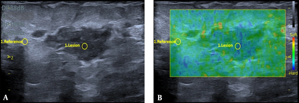
Grey-scale US (A) exhibits an irregular parallel mass with mild peripheral edema in a 29-year-old woman with a history of mastitis in the same breast over the past year. The strain elastography (B) indicates a strain ratio (SR) of 2.68, and the biopsy findings indicate an idiopathic granulomatous mastitis (IGM) lesion.
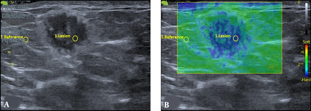
Grey-scale US (A) displays a round microlobulated mass with a thick halo in a 41-year-old woman. The mass appears stiff on strain elastography (B). The strain ratio (SR) is 4.76, and the lesion is shown to be invasive ductal carcinoma following core needle biopsy.


