


1. Background
2. Objectives
3. Patients and Methods
3.1. Selection Criteria
3.2. Sample Size Justification
3.3. Clinical and Laboratory Evaluation
| Variables | Recovery | Test Results | |
|---|---|---|---|
| No | Yes | ||
| Gender | χ2 = 0.00; P = 0.99 | ||
| Female | 4 (57.10) | 4 (57.10) | |
| Male | 3 (42.90) | 3 (42.90) | |
| Exposure | χ2 = 1.07; P = 0.29 | ||
| No | 0 (0.00) | 1 (14.30) | |
| Yes | 7 (100.00) | 6 (85.70) | |
| Treatment type | χ2 = 6.53; P = 0.11 | ||
| Heart failure treatment+ inotropic agents +IVIg + pulse methylprednisolone | 2 (28.60) | 3 (42.90) | |
| Heart failure treatment + inotropic agents + IVIg + pulse methylprednisolone | 0 (0.00) | 2 (28.60) | |
| IVIg + pulse methylprednisolone | 1 (14.30) | 2 (28.60) | |
| Heart failure treatment | 4 (57.10) | 0 (0.00) | |
| Chest X-ray | χ2 = 7.77; P = 0.02 c | ||
| No cardiomegaly | 0 (0.00) | 5 (71.40) | |
| Cardiomegaly | 7 (100.00) | 2 (28.60) | |
| Age, y | 6.49 ± 4.33 | 5.57 ± 3.25 | Z = - 0.38; P = 0.70 |
| Weight, kg | 21.00 ± 10.01 | 25.00 ± 12.07 | Z = - 0.70; P = 0.48 |
| Body surface area (BSA) | 0.84 ± 0.30 | 0.92 ± 0.29 | Z = - 0.70; P = 0.48 |
| Pulse rate | 117.86 ± 22.70 | 138.57 ± 31.71 | Z = - 1.28; P = 0.19 |
| Respiratory rate | 30.00 ± 8.02 | 32.86 ± 10.52 | Z = - 0.51; P = 0.60 |
| Systolic blood pressure (SBP) | 88.57 ± 6.26 | 90.29 ± 11.57 | Z = - 0.78; P = 0.43 |
| Diastolic blood pressure (DBP) | 62.14 ± 6.36 | 57.57 ± 9.37 | Z = - 1.11; P = 0.26 |
Abbreviation: IVIg, intravenous immune globulin.
a Values are reported as mean ± SD or No. (%).
b Inotropic agents: Milrinone, epinephrine, dopamine, and dobutamine.
c Significant at the level of 0.05.
| Variables | Recovery | Test Results | |
|---|---|---|---|
| No | Yes | ||
| NTproBNP | χ2 = 2.77; P = 0.28 | ||
| < 1000 | 0 (0.00) | 1 (14.30) | |
| 1000 - 5000 | 2 (28.60) | 4 (57.10) | |
| > 5000 | 5 (71.40) | 2 (28.60) | |
| D-dimer | χ2 = 1.50; P = 0.63 | ||
| < 1000 | 3 (42.90) | 1 (14.30) | |
| 1000 - 2000 | 1 (14.30) | 2 (28.60) | |
| > 2000 | 3 (42.90) | 4 (57.10) | |
| TPI | χ2 = 2.80; P = 0.26 | ||
| Negative | 3 (42.90) | 6 (85.70) | |
| Positive | 4 (57.10) | 1 (14.30) | |
| SPO2 | 96.00 ± 3.16 | 96.43 ± 2.57 | Z = - 0.06; P = 0.94 |
| WBC | 10.67 ± 6.65 | 12.18 ± 7.31 | Z = - 0.83; P = 0.40 |
| PMN | 37.71 ± 11.75 | 81.44 ± 13.06 | Z = - 3.00; P = 0.003 b |
| Lymph | 48.71 ± 20.08 | 12.51 ± 7.26 | Z = - 2.74; P = 0.006 b |
| Hb | 12.60 ± 1.55 | 10.10 ± 1.62 | Z = - 2.36; P = 0.01 b |
| MCV | 85.90 ± 5.67 | 79.37 ± 5.23 | Z = - 2.37; P = 0.01 b |
| Plt | 226428.57 ± 62406.88 | 185857.14 ± 96340.41 | Z = - 0.19; P = 0.56 |
| ESR | 8.86 ± 13.60 | 30.29 ± 21.33 | Z = - 2.49; P = 0.01 b |
| CRP | 18.91 ± 27.25 | 100.57 ± 85.67 | Z = - 2.23; P = 0.02 b |
| PT | 15.05 ± 3.99 | 13.85 ± 0.89 | Z = - 0.19; P = 0.84 |
| PTT | 27.86 ± 2.41 | 32.86 ± 7.17 | Z = - 1.28; P = 0.19 |
| INR | 1.31 ± 0.46 | 1.11 ± 0.12 | Z = - 0.98; P = 0.32 |
| AST | 593.57 ± 1392.35 | 79.14 ± 51.79 | Z = - 0.12; P = 0.89 |
| ALT | 910.29 ± 2240.41 | 63.57 ± 36.74 | Z = - 0.12; P = 0.89 |
| ALP | 441.43 ± 160.22 | 435.14 ± 71.92 | Z = - 0.97; P = 0.33 |
| BUN | 37.43 ± 18.66 | 36.57 ± 15.36 | Z = 0.00; P = 0.99 |
| Cr | 0.69 ± 0.22 | 0.59 ± 0.08 | Z = - 0.78; P = 0.43 |
| LDH | 983.71 ± 1163.67 | 600.71 ± 150.05 | Z = - 0.25; P = 0.79 |
| Alb | 3.48 ± 0.80 | 2.94 ± 0.47 | Z = - 1.66; P = 0.09 |
| D-dimer | 1848.00 ± 1714.84 | 2619.43 ± 1562.89 | Z = - 0.83; P = 0.40 |
Abbreviations: NTproBNP, N-terminal pro–B-type natriuretic peptide; TPI, Troponin I; SPO2, Oxygen saturation; PMN, polymorphonuclear neutrophil; MCV, mean corpuscular volume; ESR, erythrocyte sedimentation rate; Hb, hemoglobin; CRP, C-reactive protein; LDH, lactate dehydrogenase; ALT, alanine aminotransferase; AST, aspartate transaminase; BUN, blood urea nitrogen; Cr, creatinine; PT, prothrombin time; PTT, partialthromboplastin time; INR, international normalizedratio; ALP, alkaline phosphatase; Alb, albumin.
a Values are reported as mean ± SD or No. (%).
b Significant at the level of 0.05.
3.4. Follow-Up
| Variables | Echocardiography Findings on Admission | Echocardiography Findings in Follow-up | ||||
|---|---|---|---|---|---|---|
| Recovery | Test Results | Recovery | Test Results | |||
| No | Yes | No | Yes | |||
| Aortic regurgitation | χ2 = 3.81; P = 0.19 | - | - | - | ||
| No or trivial | 7 (100.00) | 4 (57.10) | ||||
| Mild | 0 (0.00) | 3 (42.90) | ||||
| Pericardia effusion | χ2 = 0.31; P = 0.99 | - | - | - | ||
| No | 4 (57.10) | 5 (71.40) | ||||
| Mild | 3 (42.90) | 2 (28.60) | ||||
| Pleural effusion | χ2 = 0.66; P = 0.99 | - | - | - | ||
| No | 3 (42.90) | 4 (57.10) | ||||
| Mild | 3 (42.90) | 2 (28.60) | ||||
| Severe | 1 (14.30) | 1 (14.30) | ||||
| Electrocardiogram | χ2 = 0.28; P = 0.99 | - | - | - | ||
| Normal | 3 (42.90) | 4 (57.10) | ||||
| Abnormal | 4 (57.10) | 3 (42.90) | ||||
| TR | χ2 = 7.66; P = 0.02 b | χ2 = 4.56; P = 0.13 | ||||
| No or trivial | 1 (14.30) | 0 (0.00) | 0 (0.00) | 2 (28.60) | ||
| Mild | 0 (0.00) | 5 (71.40) | 1 (33.30) | 5 (71.40) | ||
| Moderate | 6 (85.70) | 2 (28.60) | 2 (66.70) | 0 (0.00) | ||
| MR | χ2 = 1.72; P = 0.56 | χ2 = 4.70; P = 0.07 | ||||
| No or trivial | 0 (0.00) | 1 (14.30) | 0 (0.00) | 3 (42.90) | ||
| Mild | 1 (14.30) | 2 (28.60) | 1 (33.30) | 4 (57.10) | ||
| Moderate | 6 (85.70) | 4 (57.10) | 2 (66.70) | 0 (0.00) | ||
| EF (M-mode), % | 21.71 ± 14.62 | 56.14 ± 14.17 | Z = - 2.82; P = 0.005 b | 33.33 ± 17.00 | 64.42 ± 2.63 | Z = - 2.42; P = 0.01 b |
| TAPSE | 1.10 ± 0.24 | 1.93 ± 0.36 | Z = - 2.82; P = 0.005 b | 1.36 ± 0.32 | 2.03 ± 0.13 | Z = - 2.43; P = 0.01 b |
| Ao_VTI | 10.57 ± 2.24 | 19.00 ± 5.41 | Z = - 2.43; P = 0.01 b | 15.16 ± 6.60 | 20.92 ± 2.09 | Z = - 1.38; P = 0.16 |
| E_A | 2.06 ± 0.86 | 1.73 ± 0.46 | Z = - 0.51; P = 0.60 | 1.50 ± 0.56 | 1.81 ± 0.45 | Z = - 0.58; P = 0.55 |
| E_Em | 13.10 ± 8.66 | 10.94 ± 5.96 | Z = - 1.08; P = 0.27 | 9.66 ± 1.52 | 9.18 ± 1.60 | Z = - 0.23; P = 0.81 |
| LVEDd_cm | 5.37 ± 1.01 | 4.59 ± 0.36 | Z = - 1.15; P = 0.24 | - | - | - |
| EF_sympson | 13.86 ± 14.17 | 50.14 ± 9.33 | Z= - 3.01; P = 0.003 b | - | - | - |
| Em_cm_s | 5.80 ± 2.35 | 9.50 ± 2.81 | Z = - 2.23; P = 0.02 b | - | - | - |
| E_cm_s | 80.92 ± 19.36 | 92.00 ± 12.76 | Z = - 1.28; P = 0.20 | - | - | - |
Abbreviations: MR, mitral regurgitation; TR, tricuspid regurgitation; AO_VTI, aortic velocity time integral; E_A, mitral EA-wave velocity; E_EM, mitral E- Em-wave velocity; TAPSE, tricuspid annular plane systolic excursion; EF, ejection fraction; LVEDd, left ventricular end-diastolic diameter.
a Values are reported as mean ± SD or No. (%).
b Significant at the level of 0.05.
| Echocardiography Findings and Admission | Follow-up | Test Results | ||
|---|---|---|---|---|
| No or Trivial | Mild | Moderate | ||
| TR | χ2 = 3.00; P = 0.39 | |||
| No or trivial | 0 (0.00) | 0 (0.00) | 1 (10.00) | |
| Mild | 1 (10.00) | 4 (40.00) | 0 (0.00) | |
| Moderate | 1 (10.00) | 2 (20.00) | 1 (10.00) | |
| MR | χ2 = 6.00; P = 0.11 | |||
| No or trivial | 0 (0.00) | 1 (10.00) | 0 (0.00) | |
| Mild | 0 (0.00) | 2 (20.00) | 0 (0.00) | |
| Moderate | 3 (30.00) | 2 (20.00) | 2 (20.00) | |
Abbreviations: MR, mitral regurgitation; TR, tricuspid regurgitation.
a Values are reported as No. (%).
3.5. Echocardiography
3.6. Cardiac Magnetic Resonance
3.7. CMR Imaging Post-processing
3.8. Ethics
3.9. Statistical Analysis
4. Results
4.1. Patients’ Demographics
4.2. Treatment Outcomes
4.3. Laboratory Findings
4.4. Echocardiography Findings on Admission
4.5. Echocardiography Findings at Follow-up
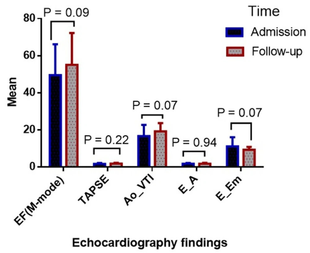
Comparing the quantitative echocardiography findings at the time of admission with the follow-up time. AO_VTI, aortic velocity time integral; E_A, mitral EA-wave velocity; E_EM, mitral E-Em-wave velocity; TAPSE, tricuspid annular plane systolic excursion; EF, ejection fraction.
4.6. Cardiac Magnetic Resonance Imaging Findings
| ID | Indexed LV End-Diastolic Volume (ILVEDV, mL/m2) | Indexed LV End-Systolic Volume (ILVESV, mL/m2) | Indexed LV Systolic Volume ILVSV, mL/m2) | LVEF (%) | Peak Longitudinal strain (%) | Peak Circumferential strain (%) | Peak Radial strain (%) | LGE | Pericardial Effusion | Time to CMR Imaging |
|---|---|---|---|---|---|---|---|---|---|---|
| 1 | 163.00 | 150.00 | 13.00 | 8.00 | 3.00 | 4.00 | 3.00 | LGE | No | 3 mo |
| 2 | 45.80 | 42.50 | 32.80 | 7.10 | 3.00 | 4.00 | 3.00 | LGE | No | 3 mo |
| 3 | 211.00 | 186.00 | 25.40 | 12.00 | 1.00 | 3.00 | 4.00 | LGE | No | 2 mo |
| 4 | 62.70 | 25.90 | 36.70 | 58.60 | 15.00 | 17.00 | 27.00 | No LGE | No | 8 mo |
| 5 | 65.00 | 23.60 | 36.30 | 64.00 | 15.00 | 17.00 | 27.00 | No LGE | No | 2 w |
| 6 | 44.60 | 15.50 | 29.00 | 65.00 | 14.00 | 16.00 | 20.00 | No LGE | No | 2 w |
| 7 | 83.00 | 39.00 | 44.00 | 53.00 | 1.00 | 3.00 | 4.00 | LGE | No | 1 mo |
| 8 | 61.60 | 21.10 | 40.50 | 65.70 | 14.50 | 18.00 | 14.50 | LGE | No | 2 w |
| 9 | 68.00 | 32.00 | 45.90 | 52.00 | 11.00 | 16.00 | 14.00 | No LGE | Yes | 2 w |
| 10 | 68.10 | 14.90 | 53.20 | 78.00 | 14.00 | 18.00 | 26.00 | LGE | Yes | 2 w |
| 11 | 99.00 | 77.00 | 22.00 | 22.00 | 1.00 | 4.00 | 3.00 | LGE | No | 1 mo |
| 12 | 158.00 | 115.00 | 42.60 | 27.00 | 1.00 | 3.00 | 4.00 | LGE | No | 4 mo |
| 13 | 225.10 | 218.00 | 6.60 | 3.00 | 1.00 | 3.00 | 4.00 | LGE | Yes | 2 mo |
| 14 | 59.90 | 22.60 | 32.20 | 53.00 | 11.00 | 14.00 | 16.00 | LGE | No | 2 w |
| Mean ± SD | 101.05 ± 61.59 | 70.22 ± 68.91 | 32.87 ± 12.89 | 40.60 ± 26.13 | 7.53 ± 6.33 | 10.00 ± 6.89 | 12.10 ± 9.74 | - | - |
Abbreviations: LVEF, left ventricular ejection fraction, LGE, late gadolinium enhancement, CMR, cardiac magnetic resonance, SD, standard deviation.
4.7. Global Function Measurement Using LVEF
4.8. Specific Case Findings
4.9. Patients Outcomes
5. Discussion
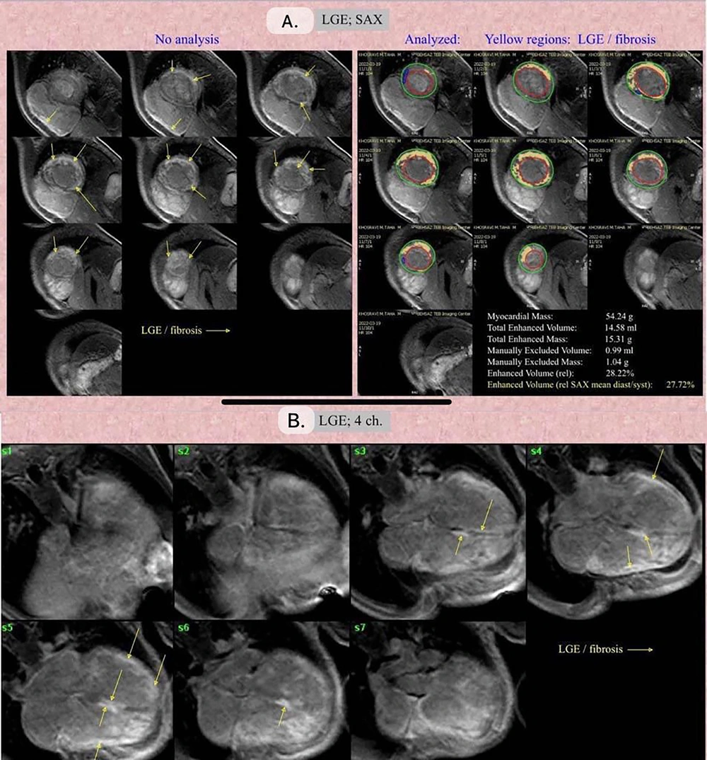
A, late gadolinium enhancement (LGE), Short Axis View (SAX) view: The yellow arrows point towards the LGE/fibrosis regions. The yellow regions highlight the areas of LGE and fibrosis. The green and red contours represent the Epicardial and Endocardial contouring, respectively. B, LGE; 4-chamber view: The yellow arrows indicate the LGE/fibrosis regions in a four-chamber view of the heart.




