


1. Background
2. Objectives
3. Patients and Methods
3.1. Statistical Analysis
4. Results
| Location | No. (%) |
|---|---|
| Upper/outer | 30 (34.1) |
| Upper/inner | 2 (2.3) |
| Lower/inner | 4 (4.5) |
| Lower/outer | 6 (6.8) |
| Upper | 18 (20.5) |
| Medial | 2 (2.3) |
| Lower | 8 (9.1) |
| Lateral | 5 (5.7) |
| Retroareolar/Central | 13 (14.8) |
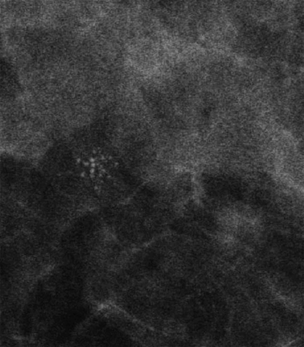
Calcification groups that have amorphous morphology as seen here on magnification view. They were suspicious, which led to the diagnosis of fibrocystic changes after histopathological assessment.
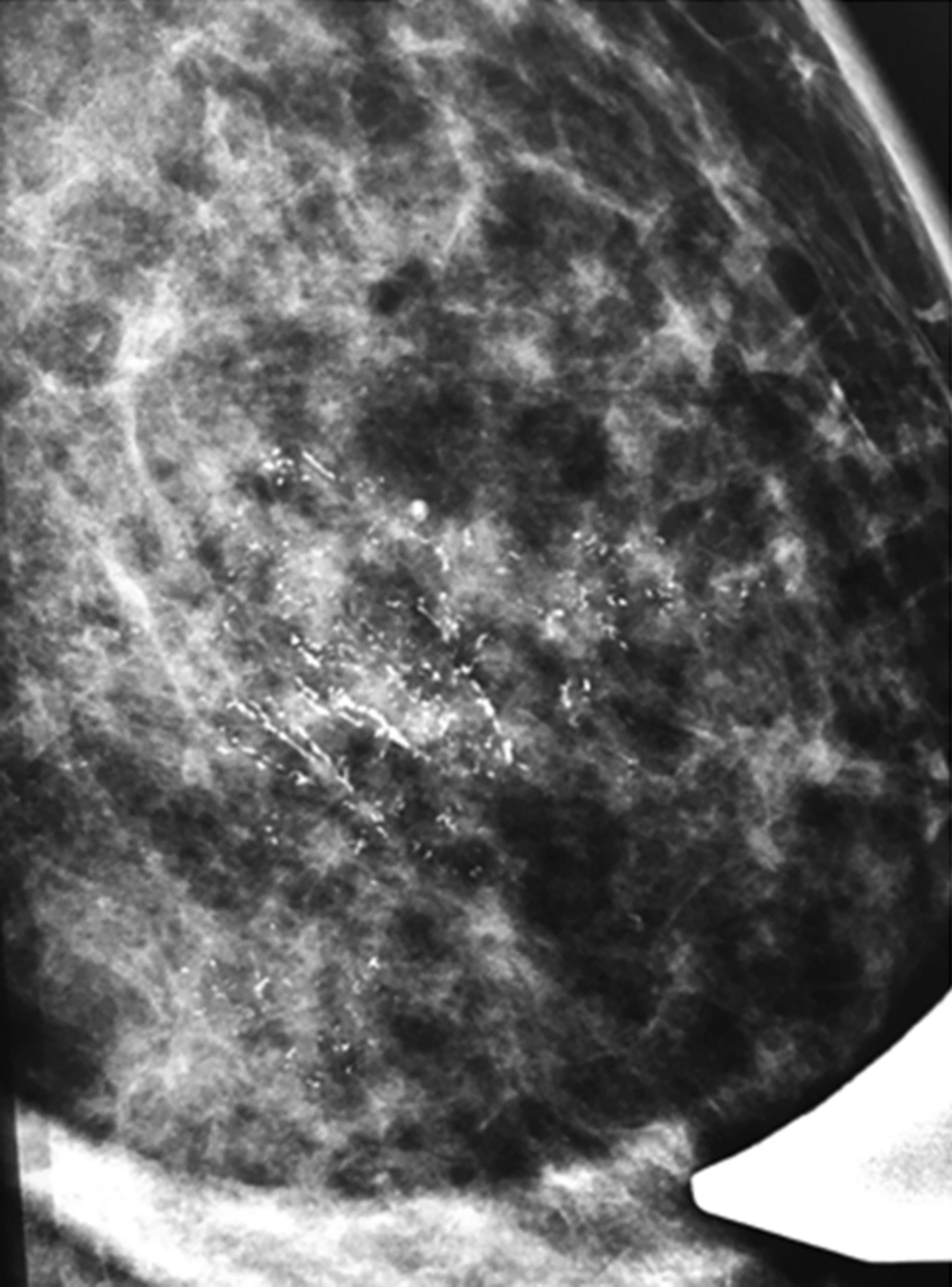
There are calcifications with fine linear morphology and fine-linear branching morphology on this magnification view. As we know, they should be judged by those with the worse morphology. Core biopsy showed invasive ductal carcinoma.
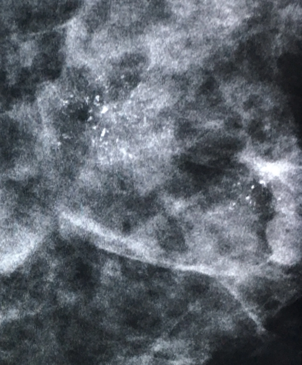
In this mammogram, groups of coarse heterogeneous morphology with tissue distortion are due to invasive ductal carcinoma.
| Morphology descriptor | Probability of malignancy | Positive predictive value |
|---|---|---|
| Amorphous | 7/26 (29) | 7.9 |
| Coarse heterogeneous | 8/19 (42.1) | 17.8 |
| Fine linear or fine-linear branching | 23/27 (85.2) | 63.2 |
| Fine pleomorphic | 15/16 (93) | 100 |
aValues are expressed as No.(%)
| Variable | TP | FN | TN | FP | Sen %95CI | Spec %95CI | PPV %95CI | NPV %95CI | PLR %95CI | NLR %95CI | Accuracy %95CI |
|---|---|---|---|---|---|---|---|---|---|---|---|
| Number (<10and >10) | 47 | 6 | 6 | 30 | 0.89 (0.77-0.96) | 0.17 (0.06-0.33) | 0.61 (0.49-0.72) | 0.5 (0.21-0.79) | 1.1 (0.89-1.3) | 1.47 (0.5-4.1) | 0.6 (0.49-0.7) |
| Size heterogeneity | 51 | 2 | 6 | 30 | 0.96 (0.87-0.99) | 0.17 (0.06-0.33) | 0.63 (0.52-0.73) | 0.75 (0.35-0.17) | 1.2 (0.99-1.3) | 4.4 (0.94-20.7) | 0.64 (0.53-0.74) |
| Density heterogeneity | 45 | 8 | 12 | 24 | 0.85 (0.72-0.93) | 0.33 (0.19-0.51) | 0.65 (0.53-0.76) | 0.6 (0.36-0.81) | 1.3 (0.98-1.6) | 2.2 (1-4.9) | 0.63 (0.5-0.74) |
| Morphology heterogeneity | 53 | 0 | 2 | 34 | 1 (0.93-1) | 0.06 (0.01-0.19) | 0.61 (0.5-0.71) | 1 (0.16-1) | 1.1 (0.98-1.1) | - | 0.62 (0.51-0.72) |
| Distribution (1 vs. >1) | 13 | 40 | 28 | 8 | 0.25 (0.14-0.38) | 0.78 (0.61-0.9) | 0.62 (0.38-0.82) | 0.41 (0.29-0.54) | 1.1 (0.51-2.4) | 1.3 (0.82-1.3) | 0.46 (0.35-0.57) |
| Associated (yes vs. no) | 27 | 26 | 23 | 13 | 0.51 (0.37-0.65) | 0.64 (0.46-0.79) | 0.68 (0.51-0.81) | 0.47 (0.33-0.62) | 1.4 (0.85-2.3) | 1.3 (0.9-1.9) | 0.56 (0.45-0.67) |
Abbreviations: CI, confidence interval; FN, false negative; FP, false positive; NLR, negative likelihood ratio; NPV, negative predictive value; PLR, positive likelihood ratio; PPV, positive predictive value; Sen, sensitivity; spec, specificity; TN, true negative; TP, true positive.
| BI-RADS Version | Cutt-Off Point | TP | FN | TN | FP | Sen [95%CI] | Spec [95%CI] | PPV [95%CI] | NPV [95%CI] | PLR [95%CI] | NLR [95%CI] | Accuracy [95%CI] |
|---|---|---|---|---|---|---|---|---|---|---|---|---|
| 4th | 4a versus others | 58 | 1 | 5 | 24 | 98 [91-99] | 17 [6-36] | 71 [60-80] | 83 [36-99] | 1.2 [1-1.4] | 10.2 [1.2-83] | 72 [61-81] |
| (4a and 4b) versus others | 41 | 18 | 22 | 7 | 69 [56-81] | 76 [56-90] | 85 [72-94] | 55 [38-71] | 2.9 [1.5-5.6] | 2.5 [1.6-3.8] | 72 [61-81] | |
| 4 versus 5 | 37 | 22 | 24 | 5 | 63 [49-75] | 83 [64-94] | 88 [74-96] | 52 [37-67] | 3.6 [1.6-8.3] | 2.2 [1.5-3.2] | 69 [59-79] | |
| 5th | 4b versus others | 41 | 18 | 22 | 7 | 69 [56-81] | 76 [56-90] | 85 [72-94] | 55 [38-71] | 2.9 [1.5-5.6] | 2.5 [1.6-3.8] | 72 [61-81] |
| 4 versus 5 | 7 | 52 | 29 | 0 | 12 [5-23] | 100 [88-100] | 100 [59-100] | 36 [25-47] | - | 1.1 [1-1.2] | 41 [31-52] |
Abbreviations: BI-RADS, breast imaging reporting and data system; CI, confidence interval; FN, false negative; FP, false positive; NLR, negative likelihood ratio; NPV, negative predictive value; PLR, positive likelihood ratio; PPV, positive predictive value; Sen, sensitivity; spec, specificity; TN, true negative; TP, true positive.


