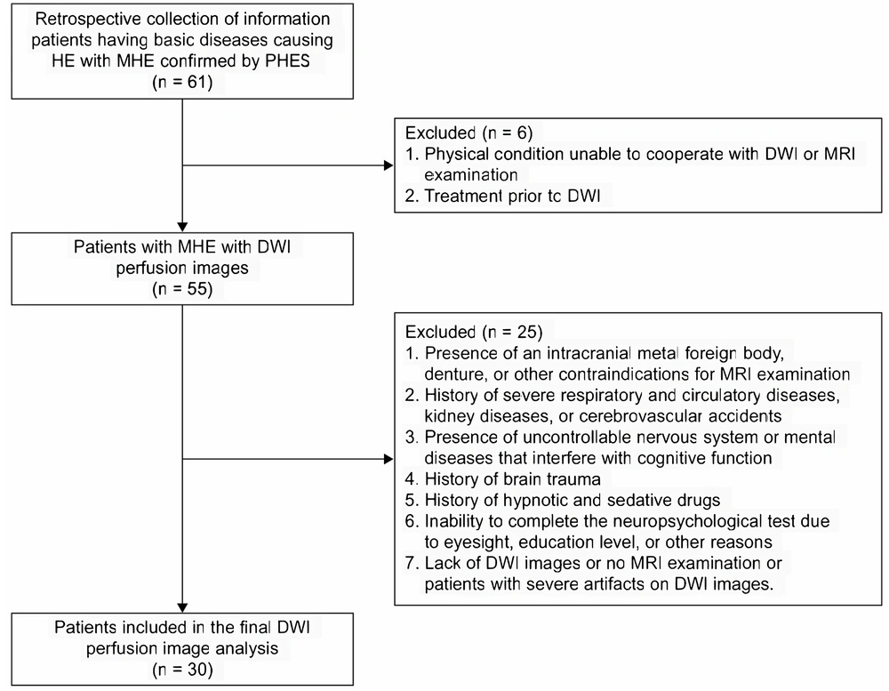
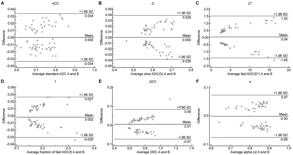
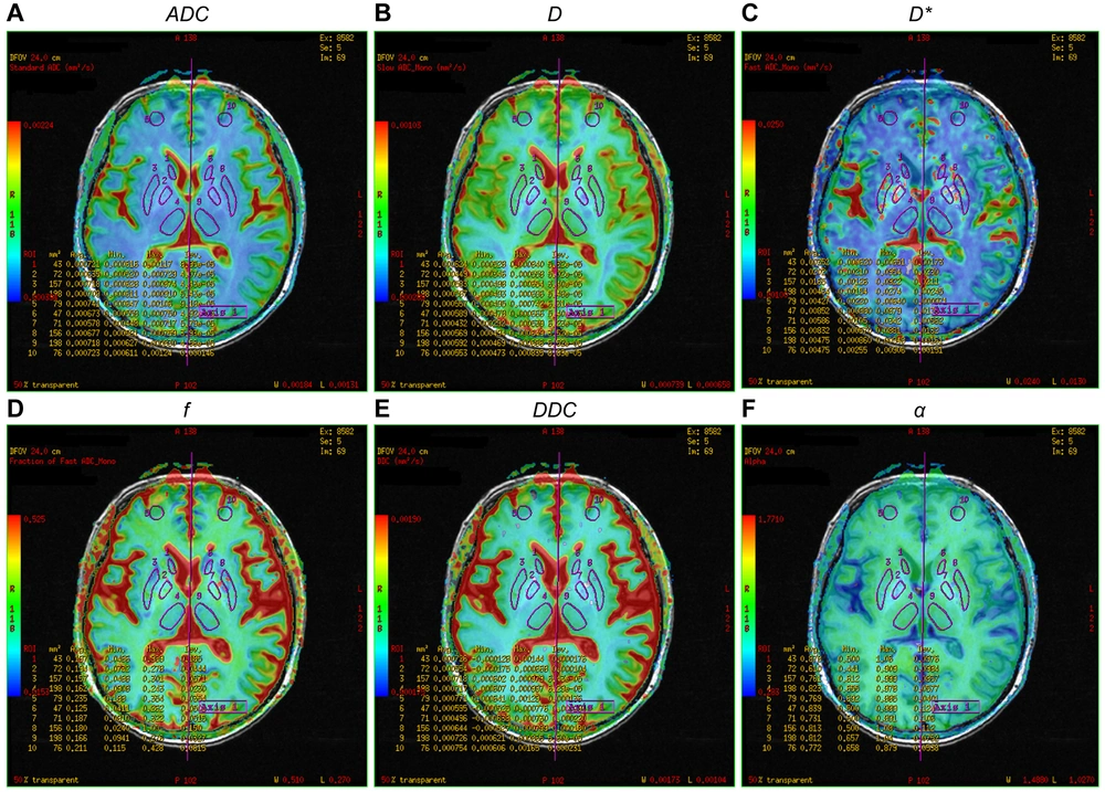
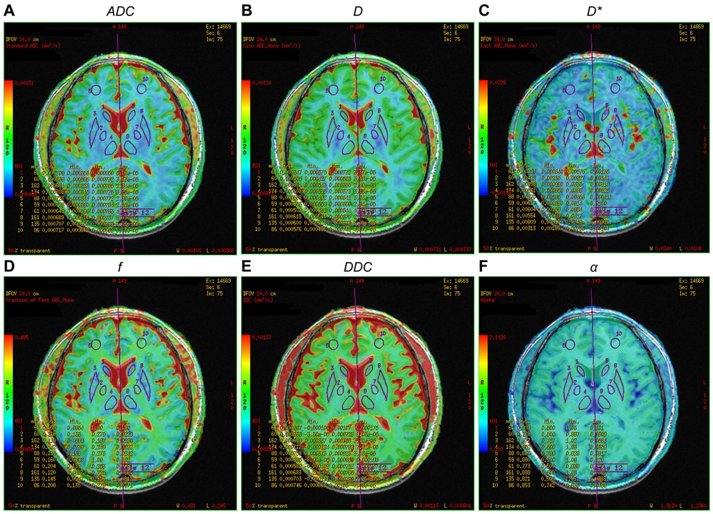
MRI has unique advantages as a non-invasive method to detect MHE. Advanced MRI techniques have been intensively used to reveal aberrant features of cerebral structure, metabolism, and function, providing more objective and reliable information than neuropsychological testing in identifying MHE. However, there is no recognized diagnostic standard for MHE currently (
2). Diffusion-weighted imaging is an MRI sequence that quantifies subtle changes in the Brownian motion of water molecules. Intravoxel incoherent motion is used to estimate the random flow perfusion of blood flow in capillaries and simulate diffusion. Furthermore, it can simultaneously measure tissue microstructure and microcirculation (
13,
14). Improvements in the scan sequences of IVIM have met the signal-to-noise ratio requirement, allowing IVIM perfusion images to be obtained simultaneously with a DWI scan (
15). Intravoxel incoherent motion DWI is widely used in the brain, mammary gland, abdomen, bone, and joints (
16-
19).
In this study, the parameters of the mono-, bi-, and stretched exponential models of the bilateral temporal lobe, occipital lobe, parietal lobe, and cingulate gyrus significantly differed between the MHE and control groups. The values of the multiple model parameters (
ADC,
D,
D*,
f,
DDC, and
α) in the 8 ROIs in the MHE group were higher than those in the control group, consistent with the results of Abdelhamid et al. (
8). Matsusue et al. reported that the cingulate gyrus is symmetrically involved (
20), and our study confirms this. The early appearance of mild cognitive dysfunction, sleep disorders, and personality changes in patients with MHE may be related to the high perfusion of blood flow in these brain regions. Moreover, this study revealed that
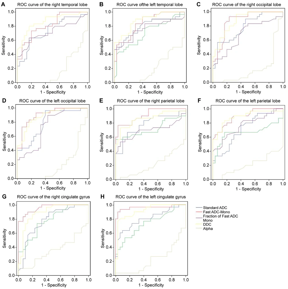
DDC had the highest AUC in the bilateral temporal and right occipital lobes. These regions can be focused on when screening for MHE, as abnormalities in these areas can provide clues to the presence of MHE.
In this study, ROC curve analysis was used to evaluate the diagnostic efficacy of the parameters from the three models for MHE. Notably, D* and DDC might be effective parameters for diagnosing MHE. The double mono-exponential model is more accurate than the mono-exponential model, which is influenced by both diffusion and perfusion.
In this study, the MHE group had higher
D* values in the bilateral frontal lobe, temporal lobe, occipital lobe, parietal lobe, and cingulate gyrus than the control group, except for the bilateral thalamus. The increased microcirculation blood perfusion in each ROI suggests widespread low-level cerebral edema in MHE (
21). High ammonia levels can lead to mitochondrial edema. Extensive low-level edema may result in astrocyte dysfunction, inhibiting their energy metabolism and compromising the integrity of the brain microstructure, leading to brain function disorders in MHE. Local cortical atrophy, thinning, and edema in patients with cirrhosis and cognitive impairment may be associated with early manifestations of MHE, such as attention deficit (
22). Among the eight ROIs with significant multi-model parameters,
D* had the highest AUC in the bilateral parietal lobe, bilateral cingulate gyrus, and left occipital lobe, suggesting that
D* may be a sensitive indicator for detecting MHE. Hyperammonia leads to vasodilation and hyperperfusion. Dysperfusion in the cerebral microcirculation—for example, due to shunting—might explain the increased D values (
23). Some studies have reported that
D* can evaluate the progression of cerebrovascular disease (
24,
25) and possess diagnostic efficacy for nasopharyngeal carcinoma (
26). However, other studies have found no significant difference in
D* values between patients with lung cancer and lymph node metastasis and those with pleural invasion (
27). Therefore, the reproducibility of
D* in MHE diagnosis should be further studied due to differences in sample size, severity of liver disease, and degree of brain injury.
The
DDC value of the stretched exponential model reflects the movement of water molecules in tissues and the complexity of lesions.
DDC has been used for diagnosing and classifying prostate cancer, hepatocellular carcinoma, and endometrial cancer (
28-
30). The thalamic functional connection network (cortico-thalamic-cortical circuit) is crucial for information processing, integration, and transmission. Cognitive dysfunction in patients with MHE may be related to thalamic abnormalities (
31). In this study, the
DDC values of all ROIs (bilateral frontal lobe, temporal lobe, occipital lobe, parietal lobe, cingulate gyrus, and thalamus) in the MHE group were higher than those in the control group, and the difference was significant, which is consistent with the results of Pigoni et al. (
32). The onset of MHE may be associated with diffuse gray matter degeneration and extensive cerebral edema of white matter (
33,
34). Increased thalamic volume may be due to hypertrophy or hyperplasia of neurons or glial cells and be considered a compensatory effect of basal ganglia (BG) dysfunction (
35). In this study, the increased
DDC value may compensate for the osmotic imbalance caused by astrocyte accumulation. Fluid moves from the outside of astrocytes to the inside of the cells, impairing the movement of Brownian molecules in the extracellular space. Moreover, angiogenic and cytotoxic edema can occur in MHE. Hyperammonemia leads to mild oxidative stress and autophagic degradation of mitochondria. Ammonia induces the formation of reactive oxygen radicals in astrocytes, affecting protein homeostasis, such as proteasome degradation, and ultimately leading to autophagy of astrocytes (
36). Astrocytes are the main cells responsible for clearing ammonia. Edema and autophagy of astrocytes hinder neurotransmitter production and ammonia clearance, potentially leading to cytotoxic edema, mild cognitive function or behavioral abnormalities, and personality changes in MHE (
1).
In the early stage of MHE, the brain responds to pathophysiological changes through its own humoral neuromodulation. This response includes autophagy and mild oxidative stress in astrocytes, cytotoxic edema, increased neuronal blood flow, and a compensatory increase in gray matter perfusion. A low
ADC value in the affected area reflects cytotoxic edema (
37). The biexponential model
D was significantly lower than the monoexponential model
ADC, which may be associated with cerebral vasodilatation in HE patients. The biexponential model eliminates the influence of microcirculation perfusion on the true diffusion of water molecules. The
F value represents the local volumetric ratio of microcirculatory perfusion effects to overall diffusion effects, including microcirculatory perfusion, fluid flow in the microstructure of glandular ducts, and glandular secretion. The
f value itself cannot distinguish between these physiological activities. The α value was lower than in the control group and tended to be 0, which may be associated with the reduced differentiation of brain tissue and increased tissue heterogeneity in MHE patients.
In contrast to previous works, a quantitative assessment was made for the cerebral blood flow of MHE using 3D-ASL. While minimum CBF accurately reflected local minimum perfusion, the mean CBF tended to vary widely (
38). Intravoxel incoherent motion DWI may be more effective for detecting underlying pathologic injury in early-stage patients (
39).
This study had some limitations. First, the sample size was small. Future studies must increase the sample size and grade patients according to Child-Pugh to reduce possible result deviations caused by differences in liver decompensation. Second, the ROIs were placed manually; two observers recorded the mean values to reduce sampling error, and the delineated area was at least 15 mm². Computer automatic partitioning and measurement or whole-brain comparison and voxel-based false discovery rate (FDR) correction are required to further reduce errors (
40).
In conclusion, MHE is a clinically significant problem that can impact the progression of cirrhosis and overall survival. No scientifically proven imaging method unequivocally diagnoses MHE. Intravoxel incoherent motion DWI is an imaging method that can detect MHE, and D* and DDC parameters might have higher diagnostic values. The bilateral temporal lobe, occipital lobe, parietal lobe, and cingulate gyrus regions should be focused on when evaluating MHE edema. This study found substantial heterogeneity and was conducted with a relatively small sample size. There is a lack of practical clinical application of IVIM-DWI in diagnosing and predicting MHE. Further research should focus on expanding the sample size, standardization, and validation of imaging biomarkers of IVIM-DWI to ultimately improve the management of MHE.