Over the past decades, CPSS in children has been increasingly recognized due to the rapid development of imaging technologies, but it is still insufficiently understood. The incomplete involution of the embryogenic veins, including the vitelline, cardinal, and umbilical vein systems, affects the development of hepatic sinusoids and leads to abnormal communications between any vein of the portal system and any vein of the inferior vena cava system, based on the anatomical site (right or left, proximal or distal). This allows intestinal blood to bypass the liver, potentially leading to multisystem morbidity. Some small intrahepatic shunts located between the portal branches and hepatic veins may regress and close spontaneously by the age of 1 to 3 years (
13-
15). However, most shunts, such as persisting intrahepatic shunts, PDV, or extrahepatic shunts, rarely involute and can remain asymptomatic until adulthood (
16). In this study, four cases were under 3 years old, including three cases of EHPS and one case of IHPS. Based on the anatomical classifications and the evident clinical manifestations such as hepatopulmonary syndrome (HPS), pulmonary hypertension (PHTN), and liver dysfunction, intervention therapy was adopted.
In early CPSS cases, some atypical symptoms or complications such as HPS, PHTN, and PAVF were noted instead of liver lesions or portal hypertension, which are typically not liver-related. Moreover, under most conditions, asymptomatic shunts were detected fortuitously on an abdominal ultrasound or abnormal liver function tests performed for other purposes (
17), often delaying referral and diagnosis (
18).
This case series revealed that hyperammonemia with or without symptoms of portosystemic shunt-related encephalopathy (100%) was the most common presentation. Pulmonary involvement, including HPS (44%), PHTN (31%), and pulmonary arteriovenous fistula (PAVF, 22%), was the main complication.
Neurological abnormalities in CPSS differ from hepatic encephalopathy in adults, which is secondary to portosystemic shunts related to liver cirrhosis or portal vein occlusion. Therefore, "portosystemic encephalopathy" is a more appropriate term to describe the neurological abnormalities and/or hyperammonemia caused by CPSS in childhood or adolescence. Portosystemic encephalopathy is a complex neuropsychiatric syndrome that includes not only asymptomatic/subtle hyperammonemia encephalopathy but also unexplained cognitive impairment, speech delay, behavioral problems, and learning difficulties, as reported in other studies (
19,
20). In the early stages of CPSS, it always exhibits subtle neurological abnormalities but may lead to irreversible brain damage. Studies have revealed that remittent and progressive blood ammonia levels are characteristic of CPSS-related hyperammonemia (
16,
21), but they do not reliably correlate with the degree of encephalopathy; rather, the peak and duration of hyperammonemia are more relevant (
22,
23). In our study, all 32 cases were accompanied by hyperammonemia, but only one showed portosystemic encephalopathy. In this case, venous ammonia was moderately elevated, and both the GFI and API were not higher than the mean index values.
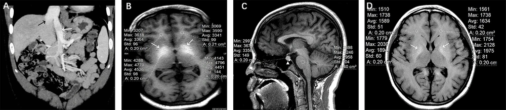
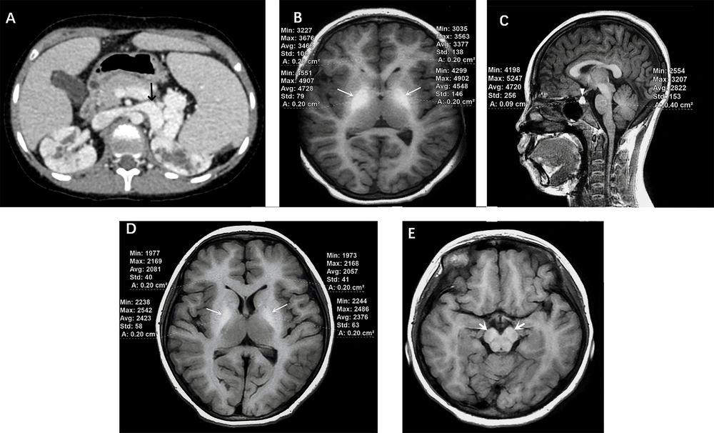
Due to the augmented accumulation of manganese, which escapes hepatic clearance in CPSS patients, different degrees of T1-weighted signal intensity can be observed on brain MRI. Commonly involved regions include the globus pallidus, putamen, caudate in the basal ganglia, subthalamic region, dentate nuclei, and cerebral peduncle (
Figure 2E). When the disease is extensive, white matter atrophy and anterior pituitary (AP) involvement can also be observed, representing subclinical brain damage (
24). Research has shown that long-term involvement can lead to irreversible brain injury, although the incidence of portosystemic encephalopathy is only about 10%. Abnormal MR signal intensities are (at least partially) reversible in the majority of patients with appropriate management of the shunts and treatment. Therefore, we consider it necessary to have routine brain MRI screening for portosystemic shunt patients, whether or not they exhibit neurological symptoms.
Plasma catecholamines are nervous mediators that include norepinephrine (NE), epinephrine (E), and dopamine (DA). The basal ganglia, particularly the globus pallidus, is the richest area of type 1 and type 2 DA receptors and is the earliest region affected by Mn toxicity, which can lead to extrapyramidal motor dysfunction. Meanwhile, type 2 DA receptors exist on the lactotrophs of the anterior pituitary (AP). As this region lacks a blood-brain barrier, it allows for unrestricted exchange activities, causing instability in the chemical environment. In this study, we selected the globus pallidus and AP to observe the brain involvement of CPSS in children. It showed that both the globus pallidus and AP could be involved in young CPSS patients. Studies in rats with portosystemic hepatic encephalopathy showed that although plasma catecholamines such as E and NE contents were significantly increased, there was no significant change in hypothalamic-AP-dopaminergic activity (
25).
The 3D gradient-echo (GE) sequence with high spatial resolution of the brain is often employed in neuroscience and clinical research to obtain structural T1-weighted images. Moreover, quantitative assessment of brain tissues and the volume of individual brain structures have become important tools for more research-oriented applications (
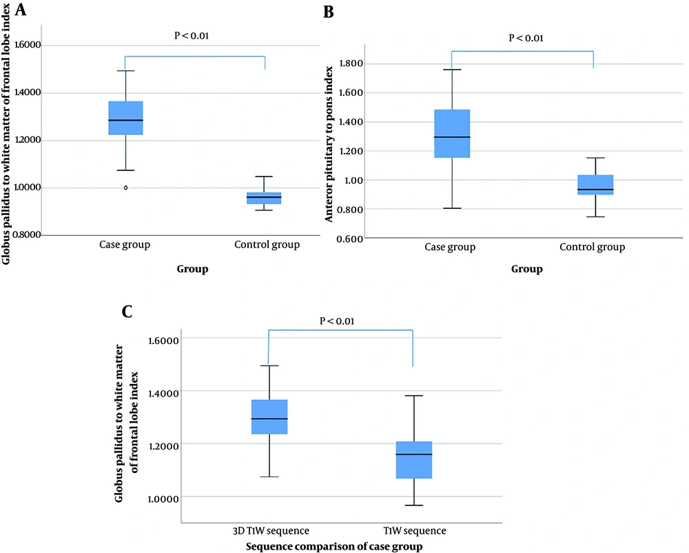
26). The typical clinical routine 3D T1W protocols have an isotropic resolution of 1 mm and require a data acquisition time ranging from 4 to 5 minutes. In this study, we measured the signals in the globus pallidus using both the axial 3D T1W sequence and the standard T1W sequence to verify if there was a difference between the ratios. Statistical analysis showed a significant difference in GFI between the 3D T1W sequence and the standard T1W sequence (P < 0.01). This difference could be attributed to the thin slice thickness avoiding partial volume issues, excellent tissue contrast, and isotropic resolution, all contributing to a better quantitative assessment.
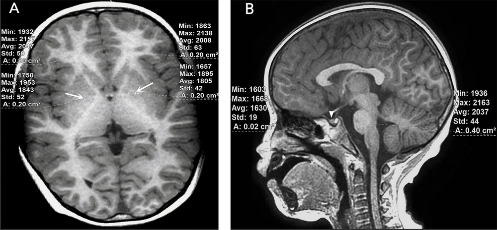
Because the globus pallidus, frontal lobe, AP, and pons are different sizes, we used an ROI measurement tool with average areas ranging from 0.02 cm² to 0.40 cm² to measure these regions. As the globus pallidus and frontal lobes are bilateral structures, we measured the values on both sides, calculated the average values of the globus pallidus and frontal lobes, and then calculated the GFI. Additional sequences such as T2-weighted images and diffusion-weighted images may also show signal differences, but to a much lesser extent and are often reported as normal (
27,
28).
In this study, we provided a comprehensive description of the characteristics of both the cases and controls, including relevant demographic and clinical information, which helps enhance the understanding of the findings and the P-values between the two groups. However, there were some limitations. This was a retrospective case-control design rather than a prospective study, providing only a snapshot view rather than signal differences over time. This design may potentially impact the results and lead to deviations in the conclusions. To further and accurately assess such differences, a longitudinal study with multiple measurements would be necessary. Additionally, the relatively small patient population (n = 32) limited the results of this study. The wide age range also influenced the ratios of the results, although age-matched controls were used. Inadequate brain MRI data after DSA or surgery made it incapable of statistically comparing the singular differences before and after therapy.
Subsequently, more adequately powered controlled studies are required for future investigations, including a more detailed natural history or clinical manifestations delineation, exploring the correlation between the complications and the CPSS classifications, and comparing the intracranial signal differences pre- and post-therapy. Unfortunately, due to the rarity of this disease, a long-term and challenging multidisciplinary team collaborative study is necessary for future medical progress.
In conclusion, the T1W signal intensity of the globus pallidus and anterior pituitary showed varying degrees of difference on brain MRI in children with CPSS compared to controls. Quantitative MRI assessment based on the 3D T1-weighted sequence could be used to evaluate portosystemic shunt-related brain signal differences. A longitudinal study with multiple measurements would be necessary to more accurately assess these differences, take timely interventions, reduce complications, and avoid long-term drug therapy.