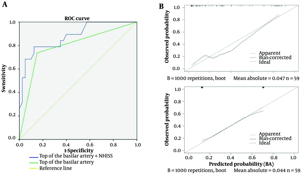
In our study, we identified a simple indicator (available image of the basilar artery apex) for assessing the favorable outcome of patients with BAO. We found that admission NIHSS, blood glucose, pc-CT, and smoking are potential predictors of favorable outcomes for BAO patients treated with EVT. By using LASSO/logistic regression to minimize confounding factors as much as possible, we concluded that basilar artery opacification has good performance (AUC, 0.799; 95% CI, 0.66-0.927) and calibration ability (mean squared error = 0.044) for favorable outcomes after EVT. In sensitivity analyses, when combined with admission NIHSS, the predictive value is further enhanced (AUC 0.877; 95% CI, 0.781-0.913; mean squared error = 0.047). This indicates that the association between the available image of the basilar artery apex and favorable outcome remained robust. These results suggest that our primary findings are unlikely to be substantially influenced by potential biases or methodological choices.
In the present study, endovascular mechanical thrombectomy techniques have made significant advances in the treatment of acute ischemic stroke. Current research indicates that selected anterior circulation large vessel occlusive acute ischemic strokes benefit significantly from EVT (
6). Additionally, the results of the Basilar Artery International Cooperation Study (BASILAR) demonstrate the safety and efficacy of EVT for acute BAO (
7). Studies now recognize that favorable outcomes are associated with treatment timing, successful recanalization, NIHSS score on admission, stroke etiology, and collateral circulation (
8,
9).
Currently, the prognosis prediction of clinical outcomes primarily relies on imaging assessments, such as pc-CS, pc-CTA, and BATMAN scoring systems for BAO (
10-
12). These classifications score the main vessels, branches, and compensatory mechanisms of the posterior circulation. Among them, pc-CS and BATMAN scores indicate better compensation with higher scores, while pc-CTA scores indicate poorer compensation with higher scores. Recently, Gao et al. (
13) reported a new collateral circulation grading system (ACGS-BAO), which consists of four levels with higher grades indicating better compensation. Pc-CTA scoring primarily assesses thrombus burden in BAO, BATMAN scoring evaluates thrombus location and collateral circulation in the basilar artery. ACGS-BAO grading and pc-CS primarily target posterior circulation after BAO. These four scoring systems have certain limitations and are cumbersome in clinical applications, especially when patients score high in pc-CS and pc-CTA but low in BATMAN scoring (
14). Among these systems, BATMAN scoring is more accurate. ACGS-BAO grading has not yet shown positive results in influencing prognosis. However, all these scoring systems underscore the importance of collateral circulation in the prognosis of BAO.
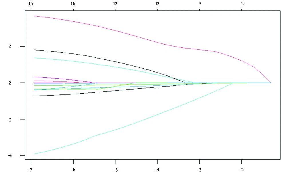
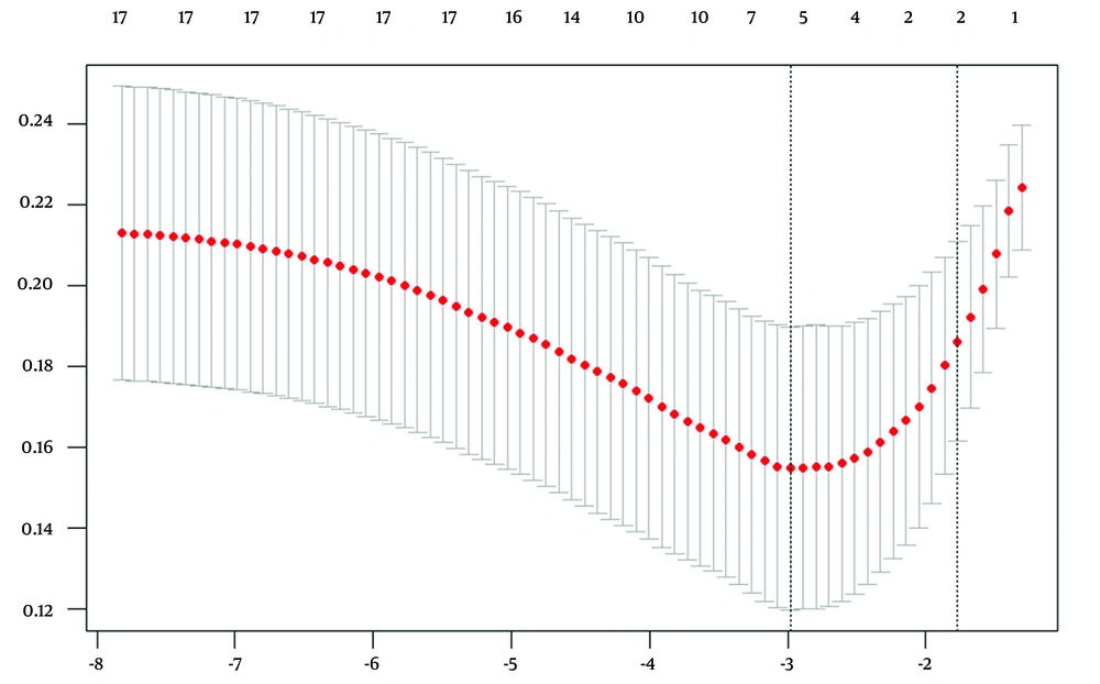
Due to the high mortality and disability rate associated with outcomes from BAO, patients with a 90-day mRS score of 3 are typically assigned to the good outcome group in most studies. However, to confirm the value of the available image of the basilar artery apex, we assigned them to the poor outcome group. In this study's LASSO regression analysis, cross-validation identified five key factors corresponding to Lambda min, including pc-CTA, the available image of the basilar artery apex, admission NIHSS score, smoking, and Glu. These factors are critically associated with the onset and prognosis of BAO. As the λ value increased, the available image of the basilar artery apex and admission NIHSS score emerged as the only two statistically significant variables in the model, which was subsequently validated in the ROC curve.
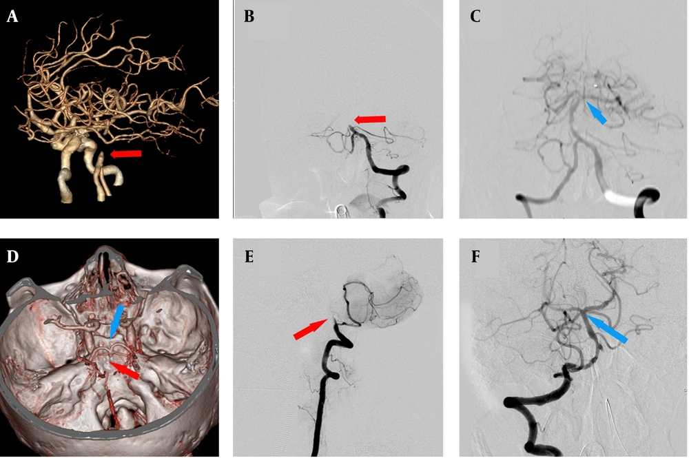
The basilar artery apex is the junction consisting of five branch vessels, located at the front of the junction between the pons and midbrain, with a diameter of approximately 2 mm. During our study, our neuroradiologists clarified the basilar artery apex by confirming the junction of the basilar artery, posterior cerebral artery, and superior cerebellar artery, thereby determining the available imaging for the basilar artery apex. In our study, the fetal-type posterior cerebral artery was quite common, mainly unilateral, and thus had a minimal impact on our assessment of the basilar artery apex. However, the presence of bilateral fetal-type posterior cerebral arteries often caused confusion in confirming the imaging of the basilar artery apex. Yet, such patients typically exhibited mild symptoms and rarely underwent EVT. Moreover, a clearer diagnosis through DSA imaging could reduce the confusion that such cases might introduce to our study.
The ability of basilar artery apex imaging and admission NIHSS score to predict prognosis post-thrombectomy in BAO analysis may be related to collateral circulation opening. In our study, the "available image of the basilar artery apex" was precisely defined as a clearly visualized anatomical junction of the basilar artery with the posterior cerebral arteries and superior cerebellar arteries, seen on either CTA or DSA. It is anatomically located at the anterior portion of the pons-midbrain junction, typically measuring approximately 2 mm in diameter. We considered the image as "available" only when this region was well visualized without motion artifacts, poor contrast, or thrombus obscuration. The visibility of this region may suggest lower clot burden, better perfusion, and preserved collateral pathways — factors that contribute to a more favorable prognosis. These characteristics, as shown in our multivariate and ROC analyses, indicate that the imaging clarity of the basilar artery apex is not merely a structural observation but a clinically meaningful marker linked to improved 90-day mRS scores.
The basilar artery apex configuration and its connection to the Circle of Willis could impact prognosis. However, our study does not clarify whether the "available image" has any anatomical or clinical correlation with these factors. The visibility of the basilar artery apex on imaging modalities such as CTA or DSA may serve as a surrogate marker for the integrity of collateral circulation. Anatomically, the basilar artery apex is a crucial junction where perforating arteries that supply vital regions, including the thalamus and midbrain, emerge. These regions are pivotal in maintaining cerebral function and may directly influence the outcome following endovascular therapy. Further investigation into the anatomical variations at this junction and their relationship with clinical outcomes could provide deeper insights into the mechanisms underlying the observed correlation. Addressing this relationship would strengthen the interpretability of our findings and provide a more robust scientific framework for our conclusions.
The ability of basilar artery apex imaging and admission NIHSS score to predict prognosis post-thrombectomy in BAO analysis may be related to collateral circulation opening. Following BAO, collateral circulation primarily involves the posterior communicating artery, anastomoses of the meningeal branches from the anterior circulation, and cerebellar arteries, predominantly through the posterior communicating artery. The basilar artery apex serves as a crucial transit point for these collateral circulations. Additionally, perforating arteries at the top of the basilar artery supply vital regions such as the thalamus and midbrain, providing valuable time for endovascular intervention and enhancing thrombectomy outcomes.
Cerebral stenting, performed acutely during endovascular therapy for persistent stenosis due to cerebral atherosclerotic disease and subacutely for severe, unresponsive stenosis, is becoming more accepted. Luo et al. (
15) observed 97 patients with severe symptomatic cerebral vertebrobasilar stenosis (70 - 99%), 30 had BA, 55 had V4, and 12 had V4-BA. Their group had 96 successful stent placements. Two fatalities, two brain hemorrhages, and seven ischemic events occurred. The Mori classification of intracranial artery stenosis was used to classify lesions as type A (less than 5 mm, concentric or moderately eccentric, not completely occlusive), type B (tubular lesions 5 - 10 mm, extremely eccentric or fully occluded), and type C. Mori C lesions were treated with self-expanding stents, while Mori A lesions using Apollo stents. Complications were more common in Mori C lesions compared to Mori A (P = 0.008) and Mori B (P = 0.047).
Another multicenter prospective registry by Gao et al. (
13) examined the safety and efficacy of stenting within 30 days for patients with 70 - 99% severe symptomatic stenosis. Of 159 balloon-mounted stent patients, 48 had basilar and intracranial vertebral artery stenosis. Of 141 patients who underwent balloon angioplasty and self-expanding stents, 46 had basilar artery stenosis and 25 had intracranial vertebral stenosis. The cumulative 30-day stroke, transient ischemic attack (TIA), and death rate was 4.3%. Revascularization was successful in 97.3%.
Acute stenting works for BAO thrombus retrieval failures. In 93 Chinese patients, rescue stenting following EVT failure was tested for safety and efficacy in BAO. Strokes were 76.2% atherosclerotic, 7.1% cardioembolic, and 16.7% other or unknown. TICI 2b/3 was achieved by 75 rescue stenting patients (92.6%). Additionally, rescue stenting improved 90-day mRS 0-3 outcomes (51.9% vs. 16.7%; P = 0.023) without increasing spontaneous intracerebral hemorrhage (sICH) incidence. Rescue stenting decreased mortality (18.5% vs. 58.3%; P = 0.006). The study has limitations such as observational technique, small sample size in the non-stented cohort, only including patients with NIHSS ≥ 10, and geographical variations in intracranial atherosclerotic disease (ICAD) prevalence in China (
15).
Futile recanalization in posterior circulation during EVT is prevalent in multicenter studies (
16). This study's ROC curve findings show a high probability for the available image of the basilar artery apex and admission NIHSS score in predicting prognosis, offering clinical assessment support to reduce futile recanalization before treatment initiation. The multivariate logistic regression analysis (
Table 2) indicated that the presence of a picture of the basilar artery apex (OR, 8.868; 95% CI, 1.683 - 36.727) could predict favorable outcomes following EVT for VBAO. This study reveals that the visibility of the basilar artery apex in CTA/DSA is directly linked to a favorable prognosis. Nevertheless, owing to the limited sample size and single institution, additional comparisons with other variables are imperative. Nonetheless, these streamlined image interpretations provide operational benefits in clinical environments.
This retrospective study may exhibit bias due to its limited sample size, highlighting the need for larger prospective investigations to validate its applicability. Recent randomized trials have had inconclusive outcomes, leaving lingering doubts. Future research employing standardized clinical evaluations and imaging methods is essential to enhance our comprehension of the efficacy, safety, selection criteria, and optimal technical methodologies for endovascular therapy in patients with BAO. Currently, a judicious strategy involves selecting patients exhibiting more severe stroke symptoms with reduced infarct loads. Clinical parameters related to posterior circulation stroke severity and outcomes may provide clarity, whereas enhanced imaging during the hyperacute period may offer numerous advantages for decision-making. Despite existing information gaps, the majority of professionals in our area concur that a reperfused basilar artery is preferable to an occluded one for patients with viable brain tissue to preserve (
17).
In conclusion, this study indicates that the available image of the basilar artery apex confirmed by CTA/DSA within 24 hours of onset in acute BAO patients predicts better clinical outcomes post- EVT, providing a convenient method for predicting clinical prognosis.