Coronary computed tomography angiography has become a widely used non-invasive imaging modality for evaluating coronary atherosclerosis. Current European Society of Cardiology (ESC) guidelines recommend CCTA as a primary diagnostic tool for CAD (
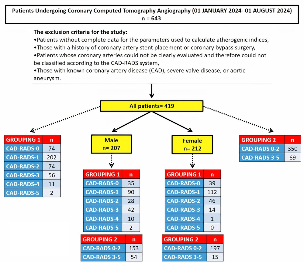
23). In this study, 643 patients who underwent CCTA between January 1 and August 1, 2024, were retrospectively analyzed. After applying exclusion criteria, 419 patients were included in the study. Patients were grouped based on their CAD-RADS scores using two different classification methods. In the first approach, they were divided into six groups to evaluate the relationship between CAD-RADS scores and parameters such as the MHR and other atherogenic indices. In the second approach, they were classified into two groups (CAD-RADS 0 - 2 and 3 - 5) to assess the predictive value of MHR and other atherogenic indices for the need for invasive angiography. Additionally, gender-based subgroup analyses were conducted, taking into account the known differences in HDL cholesterol levels.
Significant gender and age differences were observed between CAD-RADS score groups, with male and older patients having higher CAD-RADS scores. The relationship between advanced age, male gender, and increased risk of cardiac death or major cardiovascular events is well known. In our study, the association between higher CAD-RADS scores and male gender is consistent with previous studies. Popa et al. and Kadiyoran and Yilmaz reported that advanced age and male gender independently correlate with higher CAD-RADS scores (
24,
25).
Inflammation plays a key role in atherosclerosis, with monocytes contributing to plaque progression by transforming into macrophages (
3,
9). Gratchev et al. demonstrated that circulating monocytes are precursors to tissue macrophages and that these cells can become foam cells, which are markers for new atherosclerotic plaque development (
26). Similarly, Nozawa et al. revealed the critical role of monocytes in coronary plaque progression, particularly in acute myocardial infarction (
27). Cholesterol is also central to the atherosclerotic process. LDL contributes to plaque formation, while HDL has anti-atherosclerotic properties by preventing LDL oxidation and inhibiting monocyte activity (
28,
29). The MHR has been proposed as a marker of inflammation and atherosclerosis balance, with previous studies linking it to CAD severity (
30-
32). Moreover, MHR has been linked to other reliable atherosclerosis markers such as carotid intima-media thickness, flow-mediated dilation, slow coronary flow, and left ankle-brachial pulse wave velocity (
33-
36).
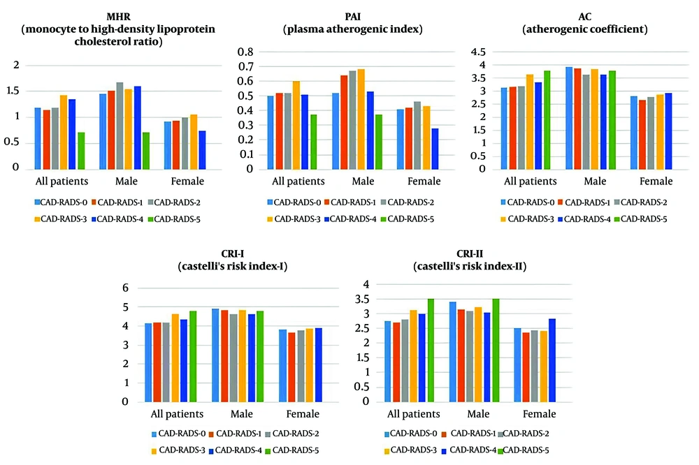
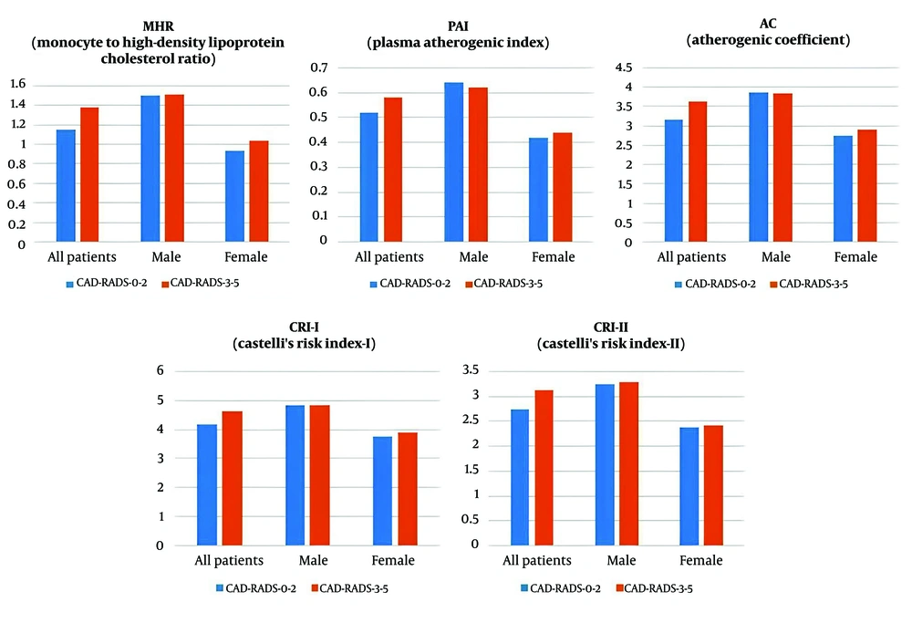
However, our results challenge the reliability of MHR as a predictive marker for CAD-RADS scoring, particularly when gender is considered. While no significant differences in MHR were found among the six CAD-RADS groups (P = 0.094), a significant difference emerged when patients were dichotomized into two groups (P = 0.029). The impact of gender on these results should not be ignored, as there were differences in gender distribution between groups. When gender-based analyses were conducted, MHR did not show significant differences within either male or female subgroups. These findings suggest that while MHR may initially appear predictive of the need for invasive angiography, their association with CAD-RADS scores is likely confounded by gender differences in HDL cholesterol reference levels. Our findings highlight the importance of controlling for gender differences in lipid levels when interpreting MHR-related results.
Kadiyoran and Yilmaz examined the relationship between MHR and CAD-RADS scores in CCTA patients and observed a significant association (
25). However, in this study, men were significantly more prevalent in higher CAD-RADS score groups, which actually makes interpreting MHR as an independent predictor inappropriate. In another study, patients were classified based on plaque types (no plaque, vulnerable plaque, stable plaque), and it was found that MHR was significantly higher in the vulnerable plaque group compared to the no plaque group (
22). The lack of a significant gender difference between groups in this study supports the reliability of the results. Similarly, in a study where patients were classified according to coronary calcium scores (CCS), MHR was significantly higher in patients with moderate and high CCS (
37). However, in this study, significant gender differences between the groups were reported, and the results should be interpreted considering the gender factor.
Considering the role of endothelial dysfunction and lipid profiles in the progression of atherosclerosis, accurate and non-invasive clinical assessment methods have long been investigated. Traditionally, markers such as total cholesterol, triglycerides, LDL-C, and HDL-C have been used to predict the development of atherosclerotic plaques in clinical and research settings (
38). However, these traditional markers may not fully reflect the severity of atherosclerosis (
38). This has led to the development of non-traditional lipid indices, such as CRI-I, CRI-II, AC, and PAI. Introduced in 1983, the Castelli indices were designed to assess cholesterol clearance efficiency via HDL-C (
39). The atherogenic coefficient further refines these measurements by considering the balance between atherogenic and anti-atherogenic lipids. PAI combines triglycerides and HDL-C into a single marker, providing a practical cardiovascular risk assessment method that can be calculated using routine lipid panel data (
40).
Non-traditional lipid indices have been proposed as superior predictors of atherosclerosis compared to traditional lipid markers. In our study, findings for atherogenic indices such as the Atherogenic Coefficient (AC), Castelli Risk Index-I (CRI-I), and Castelli Risk Index-II (CRI-II) exhibited a pattern similar to that of MHR. Therefore, our interpretation of MHR is also applicable to these parameters: While these results suggest that non-traditional lipid indices may initially predict the need for invasive angiography, their association with CAD-RADS scores is likely confounded by gender differences in HDL cholesterol reference levels. Previous studies have reported significant associations between these indices and the severity of CAD, whereas our results do not appear to be consistent with these studies. Our findings suggest that these indices should be interpreted with caution in studies with unequal gender distributions. Li et al. reported significantly higher CRI-I, CRI-II, and PAI values in the CAD group compared to the non-CAD group, but these higher values were likely due to lower HDL cholesterol levels in males, as 80% of the CAD group were male (
7). Similarly, in a study comparing control and atherosclerosis groups, we believe the higher PAI, CRI-I, and CRI-II values in the atherosclerosis group were influenced by the higher proportion of male participants (
41). Mahdavi-Roshan et al. found higher PAI, CRI-I, and CRI-II values in the CAD group compared to controls, with no gender differences, which strengthens the reliability of their results (
42).
One of the strengths of this study is its systematic approach to analyzing CAD-RADS scores in relation to MHR and atherogenic indices, incorporating both overall analysis and gender-based subgroup analyses. The six-group classification based on CAD-RADS scores allowed for a detailed assessment of trends across increasing CAD severity, while the two-group classification focused on evaluating the necessity of ICA, ensuring a comprehensive evaluation of these markers. However, several limitations should be acknowledged. First, the sample size calculation was not specifically powered for gender-based further analysis. This may have reduced the statistical power and affected the significance levels due to the smaller sample sizes within each gender subgroup. While we observed significant associations in the two-group analysis of the variables, the lack of significance in gender-based further analyses may potentially be attributed to the smaller sample sizes within each gender subgroup. However, rather than omitting gender-specific analyses entirely, we believe presenting these findings transparently allows for a more comprehensive interpretation of the data. Additionally, the explanation of this finding in light of general medical knowledge about known gender-based reference value differences in lipid levels, particularly HDL-C, suggests that the study provides meaningful contributions to the literature. Future studies should consider a priori sample size calculations that account for gender-based further analysis to enhance the power of such analyses and validate these observations.
A further limitation of this study is the small sample size in specific CAD-RADS categories, particularly in CAD-RADS-4 and CAD-RADS-5, which could impact the statistical power of subgroup analyses. Although we included all patients who underwent CCTA within the specified time period, the limited number of participants in these categories may reduce the ability to detect statistically significant differences. Nevertheless, we believe the findings, especially those highlighting gender-specific differences in lipid profiles, provide valuable insights. Future studies with larger sample sizes, particularly targeting these specific subgroups, are needed to provide more reliable and generalized insights into these categories.
Additionally, the retrospective design and missing or inconsistent data in patient records limited our ability to include important confounding variables such as comorbidities, medication use, lifestyle factors, and socioeconomic status. However, the key findings of our study highlight meaningful relationships based on the available data and offer valuable contributions to the literature. Future studies should prospectively collect these factors and control for them using multivariate analyses. Finally, as a single-center, retrospective observational study, it may be subject to selection bias. In particular, variations in clinical practices and patient populations across different centers may restrict the generalizability of our findings. Therefore, larger-scale, multi-center, and prospective studies are needed to strengthen these findings.
In conclusion, this study highlights the importance of considering gender differences when evaluating the relationship between MHR, atherogenic indices, CAD severity, and ICA necessity. While initial analyses suggested that these markers might predict the need for invasive angiography, gender-based subgroup analyses revealed no significant relationships, indicating that gender differences in HDL cholesterol levels may influence these findings. Future studies should incorporate gender-balanced populations and prospective methodologies to further validate these observations.