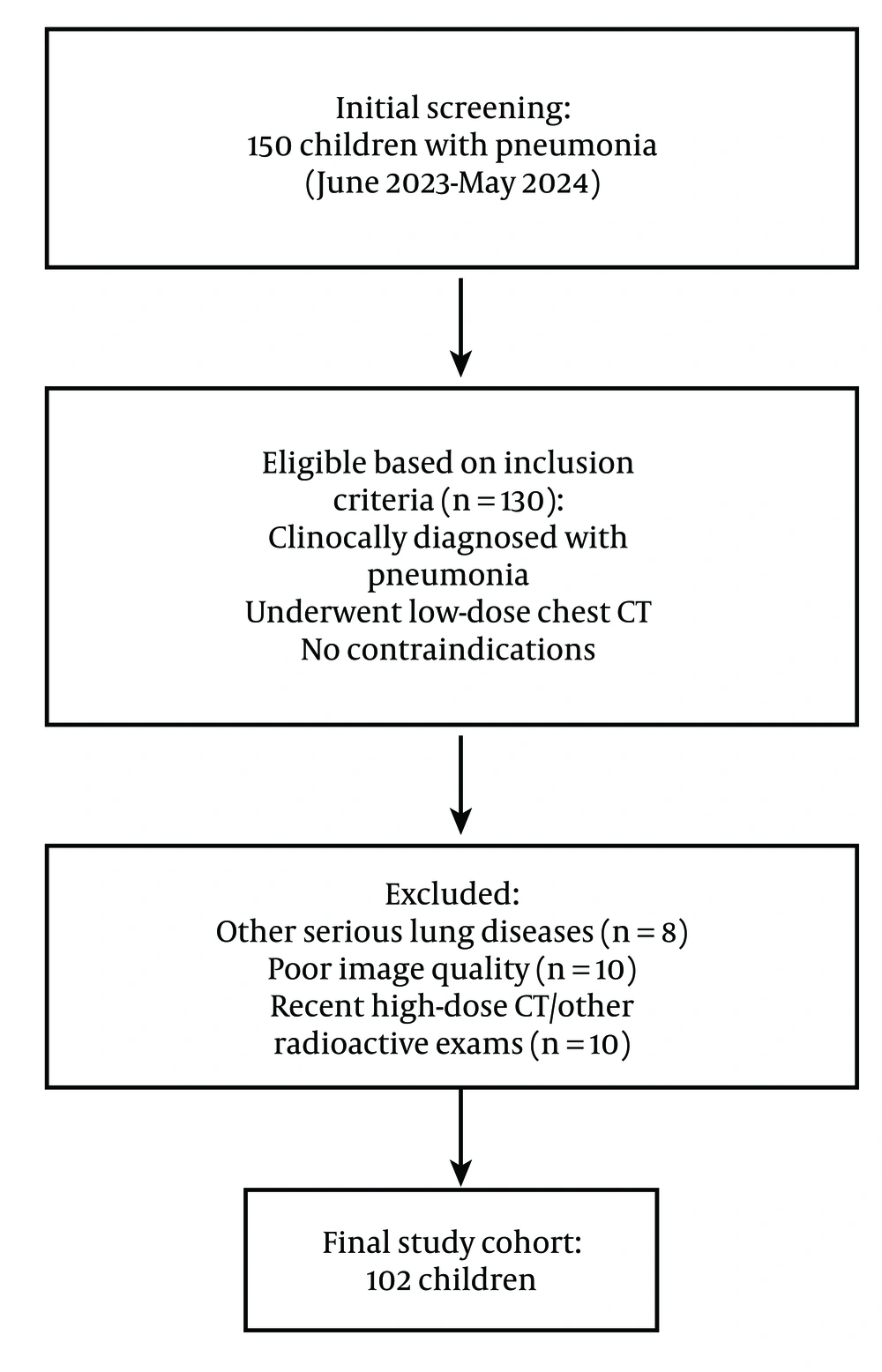
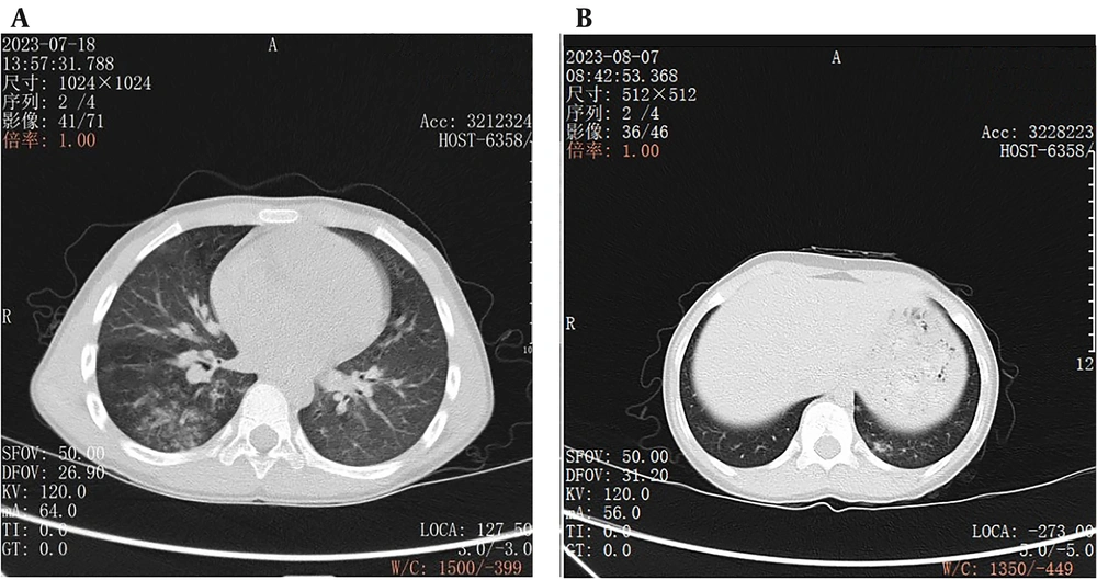
Chest CT scans play a key role in the diagnosis of childhood pneumonia. They provide detailed information about the anatomical structure and lesions in the lungs, assisting doctors in accurately identifying the type, scope, and severity of pneumonia. However, children are particularly sensitive to radiation, and long-term or high-dose radiation exposure may elevate the risk of cancer (
9). Therefore, low-dose chest CT scans are significant for diagnosing childhood pneumonia, as they reduce the radiation dose while maintaining image quality, thus minimizing potential health hazards (
10).
The MBIR algorithm is an advanced image reconstruction technique with several significant advantages. It establishes a precise physical model that considers complex factors such as X-ray transmission, detector response, and photon scattering (
11). During reconstruction, MBIR performs multiple iterative computations on raw data to continuously optimize image quality, effectively reducing image noise. Noise in low-dose CT images can affect image definition and readability (
12). The MBIR substantially attenuates image noise, increases SNR and CNR, and enhances image definition. It also improves contrast and resolution, particularly in low-dose chest CT images of children with pneumonia. The MBIR can display microstructure features of the lung parenchyma, which are valuable for accurately determining the scope and nature of pneumonia (
13). Additionally, MBIR enhances the definition of lesion edges, helping doctors accurately determine lesion scope and morphology, which is crucial for developing effective treatment protocols. Clear lesion edges assist in assessing lesion invasiveness and determining tissue infiltration, providing a reference for selecting therapeutic methods such as surgery, radiotherapy, and chemotherapy (
14). High-quality images from MBIR lower the risk of misdiagnosis and missed diagnosis, ensuring timely treatment (
15). Accurate determination of pneumonia type is essential for selecting appropriate therapies, and MBIR's high-quality images aid in identifying pneumonia types for individualized treatment protocols.
The ASIR algorithm is a statistical MBIR technique that uses prior knowledge and statistical information for iterative reconstruction, reducing noise and improving image quality (
16). However, ASIR is less capable than MBIR in enhancing image quality. In low-dose chest CT images of children with pneumonia, ASIR is inferior to MBIR in microstructure display, lesion edge definition, and overall image quality, though it reduces noise to some extent (
16). The ASIR may not display microstructures as clearly as MBIR, potentially affecting accurate judgment of lesion scope and nature. ASIR may also struggle to present lesion edges clearly, complicating the determination of lesion scope and morphology. Overall, ASIR-reconstructed images may be less clear and accurate than MBIR-reconstructed images, influencing diagnostic accuracy.
In this study, two experienced radiologists conducted subjective evaluations of the reconstructed images, showing high consistency and reliability. The evaluations covered image noise, microstructure display, lesion edge definition, and overall image quality. Results indicated that MBIR produced better images than ASIR in all aspects, aligning with objective measurements and confirming MBIR's advantages in optimizing low-dose chest CT image quality for children with pneumonia. Subjective evaluation is vital for clinical diagnosis (
17).
Radiologists primarily rely on image observation for diagnosis in their daily work, so image quality directly impacts diagnostic accuracy (
18). The MBIR algorithm can reconstruct higher-quality images, providing doctors with more distinct and accurate information. In diagnosing childhood pneumonia, accurate diagnosis is foundational for formulating effective treatment protocols. Inaccurate diagnosis may lead to improper treatment, disease exacerbation, or missed treatment opportunities, potentially resulting in serious consequences for children (
19). Therefore, improving image quality is vital for diagnosing and treating childhood pneumonia.
From the perspective of radiation dose, this study found that the MBIR group had lower DLP and ED than the ASIR group, suggesting that the MBIR algorithm also has advantages in decreasing radiation dose. Reducing radiation dose is especially important for child patients in clinical practice. Children in growth and development stages have immature organs and systems, making them more sensitive to radiation. Minimizing potential radiation hazards to children's health is crucial. Although low-dose CT scans reduce radiation dose, further reduction is possible with advanced image reconstruction algorithms like MBIR. This is of great clinical value for children requiring multiple CT scans, as it reduces cumulative radiation dose and lowers cancer risk. The reduction in radiation dose also aligns with medical ethics, reflecting care and protection for patients.
Signal-to-noise ratio and CNR are vital objective indexes of image quality. This study revealed increases in SNR and CNR in the MBIR group compared to the ASIR group, indicating that MBIR-reconstructed images excel in SNR and contrast between lung parenchyma and mediastinum. High SNR and CNR facilitate visualizing lung lesions and enhance diagnostic accuracy. In low-dose chest CT images of children with pneumonia, the MBIR algorithm enhances SNR and CNR, allowing doctors to observe differences between pneumonia lesions and surrounding normal tissues more clearly, aiding in accurate identification of lesion scope, nature, and characteristics (
20). Clear contrast between lesions and surrounding tissues helps define lesion boundaries and determine invasiveness. High SNR and CNR assist in identifying lesions with unclear boundaries, avoiding missed diagnoses.
However, this study has limitations. The relatively small sample size might impact the generalizability of the results. Future studies could expand the sample size to further validate MBIR's strengths in optimizing low-dose chest CT image quality for children with pneumonia. An enlarged sample size could better reflect conditions across different age groups and severities, enhancing generalizability and reliability (
21). Additionally, confounding factors such as operator experience, differences in CT equipment or settings, and clinical context were not controlled, which may influence result generalizability. Standardization efforts were made, but these variables could introduce variability. Future multicenter studies with diverse clinical contexts and equipment will help further validate the results.
In conclusion, the MBIR algorithm shows significant advantages in optimizing low-dose chest CT image quality for children with pneumonia, reducing radiation dose while ensuring diagnostic accuracy. These improvements in image quality can lead to more accurate lesion detection, better visualization of subtle abnormalities, and more confident decision-making by clinicians, ultimately contributing to more effective management and treatment of pediatric pneumonia.