I J Radiol

Image Credit:I J Radiol

When a Psoas Abscess Is No Longer an Abscess: Computed Tomography Angiography in Detection of Type I Endoleak with Aorto-Psoas Fistula After Endovascular Aortic Repair

Author(s):
Shang-Chun HungShang-Chun HungShang-Chun Hung ORCID1, Meng-Hsuan ChiangMeng-Hsuan ChiangMeng-Hsuan Chiang ORCID2,*
1Department of Medical Imaging, Kaohsiung Medical University Hospital, KMU, Kaohsiung, Taiwan
2Department of Neuroradiology, E-Da Hospital, I-Shou University, Kaohsiung, Taiwan

IJ Radiology:Vol. 22, issue 4; e168741
Published online:Apr 17, 2026
Article type:Case Report
Received:Dec 09, 2025
Accepted:Mar 24, 2026
How to Cite:Hung S, Chiang M. When a Psoas Abscess Is No Longer an Abscess: Computed Tomography Angiography in Detection of Type I Endoleak with Aorto-Psoas Fistula After Endovascular Aortic Repair. I J Radiol. 2025;22(4):e168741. doi: https://doi.org/10.5812/iranjradiol-168741

Abstract

Introduction:

Aorto-psoas fistulas resulting from infected stent grafts are rare but carry high mortality rates. This case report highlights the critical necessity of distinguishing between a simple abscess and a vascular fistula in patients with prior aortic interventions to prevent catastrophic procedural complications.

Case Presentation:

An 84-year-old man with a history of lung cancer and prior endovascular aneurysm repair (EVAR) performed 5 years ago presented to the emergency department with severe low back pain. He had a known history of persistent spondylodiscitis and psoas abscess diagnosed 4 months ago, for which he had undergone multiple drainages. The initial admission assessment suggested a recurrent abscess, and percutaneous drainage was arranged. However, the patient's condition rapidly deteriorated with sudden shock and massive gastrointestinal bleeding. An emergency arterial-phase computed tomography angiography (CTA) was performed during resuscitation. The CTA demonstrated contrast medium leakage from the proximal edge of the stent graft, establishing a Type Ia endoleak with fistulous communication to the psoas abscess and the duodenum. Consequently, the contraindicated drainage was withheld. Due to the catastrophic hemorrhage and hemodynamic collapse, the patient died shortly after the diagnosis.

Conclusion:

This case underscores the necessity of routine contrast-enhanced CTA prior to any image-guided drainage in patients with prior aortic stent grafts. Early identification via CTA is essential to modify management strategies and avoid iatrogenic hemorrhage

1. Introduction

Endovascular aneurysm repair (EVAR) is a standard treatment for abdominal aortic aneurysms; however, late complications such as endoleaks and graft infections remain significant clinical challenges (1). Aorto-psoas fistulas resulting from infected stent grafts are extremely rare, with limited cases reported in the literature, yet they carry high mortality rates (2). These complex pathologies can deceptively mimic recurrent psoas abscesses, particularly in patients with chronic infections or malignancies. This case report highlights the critical necessity of distinguishing between a simple abscess and a vascular fistula in patients with prior aortic interventions to prevent catastrophic procedural complications.

2. Case Presentation

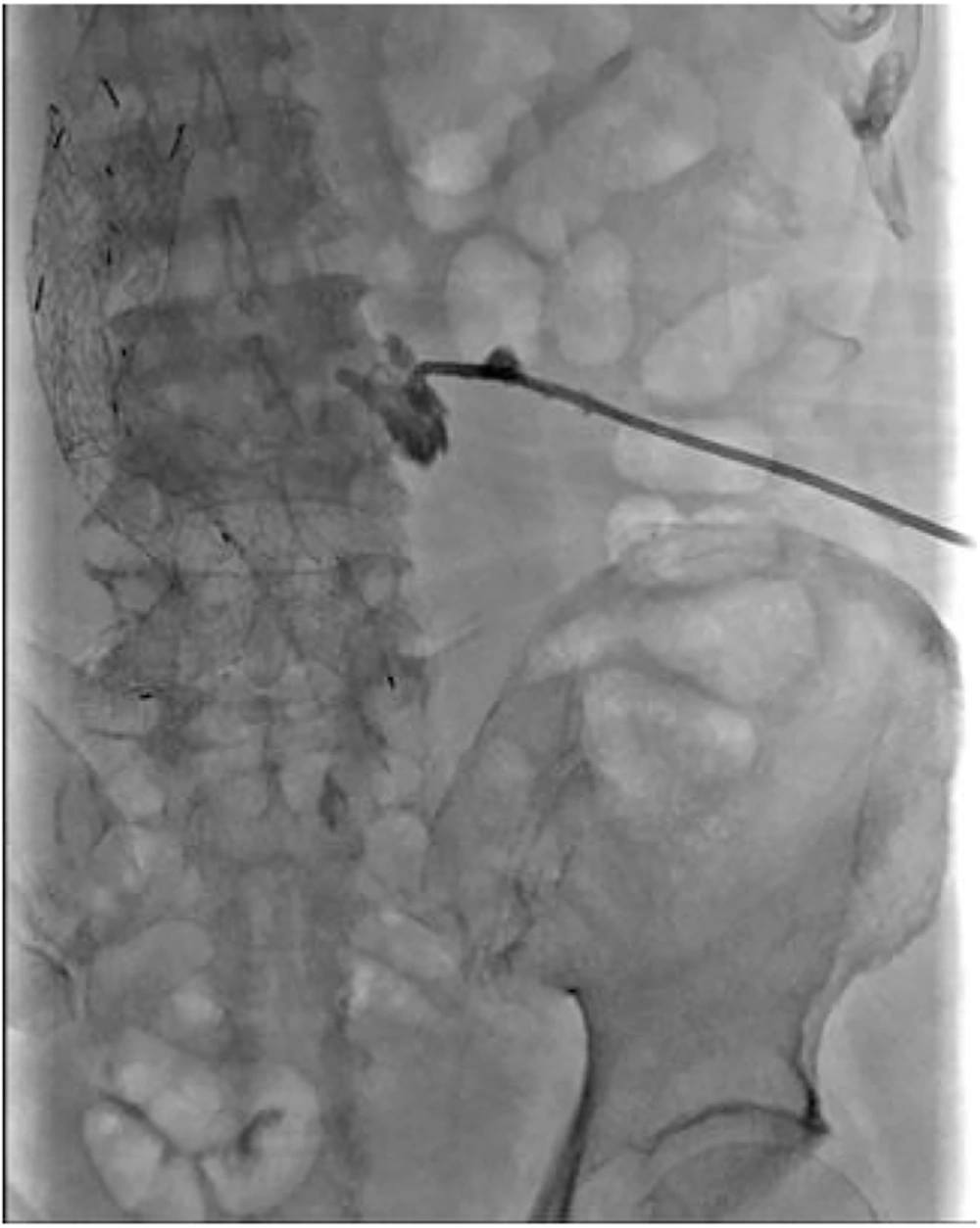
An 84-year-old man with a history of lung cancer and EVAR performed 5 years earlier for an abdominal aortic aneurysm presented to the emergency department (ED) with severe low back pain. He had been followed in the infectious disease clinic for persistent L3–L4 spondylodiscitis and chronic vertebral osteomyelitis due to Streptococcus intermedius for the past 4 months. He had undergone multiple percutaneous drainages (Figure 1) of the diagnosed bilateral psoas abscess 4 months ago. Previous percutaneous drainages had consistently yielded purulent fluid without any bloody return, and the drainage tube had been removed 3 months ago prior to this presentation.
Fistulography of left psoas abscess drainage revealed opacification of a simple abscess cavity without fistula.
Figure 1.

Fistulography of left psoas abscess drainage revealed opacification of a simple abscess cavity without fistula.

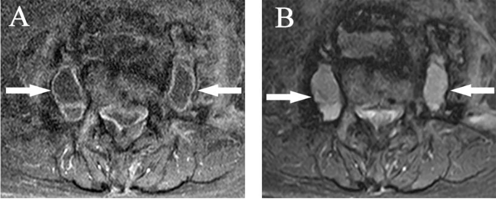
Upon arrival at the ED (day 1), the patient’s vital signs were stable: blood pressure 138/70 mmHg, heart rate 71 bpm, and body temperature 37.1°C. Physical examination revealed tenderness over the left flank and lumbar region. Laboratory investigations showed leukocytosis (white blood cell [WBC] count: 17.71 × 10³/µL), elevated C-reactive protein (CRP: 63.73 mg/L), and mild anemia (hemoglobin: 9.1 g/dL). Notably, there was no clinical evidence of gastrointestinal bleeding such as hematemesis or melena at the initial presentation. Given the patient's history and signs of infection, recurrent psoas abscesses were first suspected. Besides, the previous magnetic resonance imaging (MRI) had disclosed the bilateral psoas abscesses 3 weeks earlier (Figure 2). Therefore, on the afternoon of day 2, an interventional radiologist was consulted to arrange percutaneous drainage for infection control. However, the clinical course changed drastically later that night.
Previous magnetic resonance imaging (MRI) (3 months before). T1-weighted images with contrast medium injection (2A) showed rim-enhanced lesions in the bilateral psoas muscles, suggestive of abscesses (white arrows). T2-weighted images (B) showed hyperintense signal in the abscesses.
Figure 2.

Previous magnetic resonance imaging (MRI) (3 months before). T1-weighted images with contrast medium injection (2A) showed rim-enhanced lesions in the bilateral psoas muscles, suggestive of abscesses (white arrows). T2-weighted images (B) showed hyperintense signal in the abscesses.

At approximately 22:00 on day 2, the patient suffered a sudden alteration in consciousness (Glasgow Coma Scale [GCS] E1V1M1) and profound hypotension (BP 52/40 mmHg). He was immediately transferred to the resuscitation room. Endotracheal intubation and nasogastric (NG) tube insertion were performed; notably, massive fresh blood was drained from the NG tube, indicating active upper gastrointestinal bleeding. Aggressive resuscitation was initiated with a fluid challenge, blood transfusion (packed red blood cells [PRBC), broad-spectrum antibiotics (Doripenem), and vasopressors (Norepinephrine). Despite these measures, the patient progressed to pulseless electrical activity (PEA), and cardiopulmonary resuscitation (CPR) was started early in the morning on day 3.
Return of spontaneous circulation (ROSC) was achieved after resuscitation. Follow-up laboratory results showed worsening anemia (hemoglobin dropped to 7.0 g/dL), severe lactic acidosis (lactate 21 mmol/L), and coagulopathy. Crucially, abdominal distension was noted during CPR. Suspecting internal hemorrhage or hollow organ perforation, an emergency abdominal computed tomography angiography (CTA) was arranged at 04:00 am.
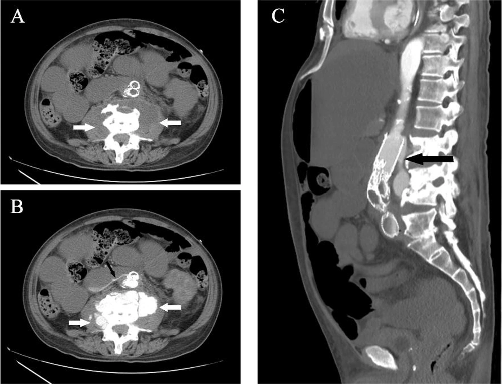
The non-enhanced CT phase demonstrated a left psoas fluid collection abutting the aortic stent graft (Figure 3A). The arterial phase (Figure 3B and C) revealed the catastrophic cause: contrast medium leakage from the proximal upper edge of the stent graft (Type Ia endoleak) communicating with the psoas abscess, and a simultaneous aorto-enteric fistula involving the duodenum.
A, Non-contrast axial CT. Low-attenuation fluid-density collections are in the bilateral psoas/paravertebral compartment (white arrows). Note the "Red Flag" sign: The loss of the fat plane between the abscess and the aortic stent graft, which necessitates further vascular evaluation. B, Arterial-phase axial CTA. IV-iodinated contrast opacifies the bilateral psoas abscess cavities (white arrows), confirming the "Red Flag" of active fistula communication. Besides, an aorto-enteric fistula is revealed (black arrow). C, Sagittal view of arterial-phase CTA. Type Ia endoleak at the edge of the aortic graft with fistula connection to the left psoas abscess cavity (black arrow).
Figure 3.

A, Non-contrast axial CT. Low-attenuation fluid-density collections are in the bilateral psoas/paravertebral compartment (white arrows). Note the "Red Flag" sign: The loss of the fat plane between the abscess and the aortic stent graft, which necessitates further vascular evaluation. B, Arterial-phase axial CTA. IV-iodinated contrast opacifies the bilateral psoas abscess cavities (white arrows), confirming the "Red Flag" of active fistula communication. Besides, an aorto-enteric fistula is revealed (black arrow). C, Sagittal view of arterial-phase CTA. Type Ia endoleak at the edge of the aortic graft with fistula connection to the left psoas abscess cavity (black arrow).

The CTA confirmed that the "abscess" was now a complex vascular fistula actively hemorrhaging. Unfortunately, shortly after the scan, the patient went into PEA again. Despite extensive efforts and vascular surgery consultation, the patient could not be stabilized for surgical intervention. He expired at 04:52 am, less than an hour after the definitive diagnosis.

5. Discussion

Endovascular aneurysm repair-related complications, such as endoleaks and graft infections, remain significant clinical challenges (1). While aorto-psoas fistulas resulting from these infections are extremely rare, they carry high mortality rates exceeding 25 - 50% (2). Secondary aortoenteric fistula (SAEF) is another devastating complication often linked to graft infection. Current literature indicates that mortality for SAEF remains extremely high, largely attributable to diagnostic delays and complex surgical requirements (3, 4). Although our patient initially presented with a psoas abscess, the subsequent massive nasogastric bleeding aligns with the "herald bleed" phenomenon — a sentinel hemorrhage that often precedes fatal rupture and necessitates urgent intervention (3-5). Our case is unique due to its "double-hit" pathology driven by a Type Ia endoleak. The mechanism is consistent with reports indicating that persistent endoleaks can cause progressive sac expansion and erosion into adjacent structures over years (6, 7). Similar to our case, only one case report in the literature described an endoleak with endograft infection, combined with aorto-psoas abscess and concomitant aortoenteric fistula (1). Linn YL et al. presented that the patient survived this critical condition with an extra-anatomical bypass, graft explant, and bowel repair. Lifelong surveillance is required for complications of the aortic stump and bypass patency.
Consequently, in patients with prior aortic interventions, a psoas abscess should not be viewed merely as a spinal infection but as a potential sentinel sign of underlying graft infection or fistula formation (1, 2, 8). While psoas abscess drainage is required, pre-procedural imaging evaluation cannot rely solely on non-enhanced CT or prior imaging. Accurate diagnosis requires identifying specific "red flags" on CTA, which are pivotal indicators for distinguishing vascular fistulas from simple abscesses (9):
- Rapid fluid re-accumulation despite drainage.
- Loss of the distinct fat plane between the aorta and psoas muscle.
- Contrast extravasation into the abscess cavity.
In conclusion, as guidelines recommend, immediate CTA is mandatory to rule out an aorto-psoas abscess fistula before any drainage procedure is attempted in patients with prior aortic stent grafts (10).

Footnotes

References

  • 1.
    Linn YL, Ng NZP, Tang TY, Chong TT. Endoleak Complicated by communicating psoas abscess and aorto-enteric fistula in an immunocompromised patient. Ann Vasc Surg. 2022;78:378 e23-9. [PubMed ID: 34487807]. https://doi.org/10.1016/j.avsg.2021.06.033.
  • 2.
    Ma JW, Hu SY, Lin TC, Tsai CA. Psoas abscess associated with aortic endograft infection caused by bacteremia of Listeria monocytogenes: A case report and literature review (CARE Complaint). Medicine (Baltimore). 2019;98(45). e17885. [PubMed ID: 31702663]. [PubMed Central ID: PMC6855657]. https://doi.org/10.1097/MD.0000000000017885.
  • 3.
    Rey J, Bornak A, Montoya C, Polania C, Kenel-Pierre S, Kang N, et al. Aortoenteric Fistulas Following Endovascular Aortic Aneurysm Repair: A Review. Vasc Endovascular Surg. 2025;59(7):695-705. [PubMed ID: 40340624]. https://doi.org/10.1177/15385744251339966.
  • 4.
    Wiangphoem N. Secondary Aorto-Colonic Fistula: A Case Report and Literature Review of a Rare Complication after EVAR. Case Rep Surg. 2022;2022:8412460. [PubMed ID: 36530176]. [PubMed Central ID: PMC9754831]. https://doi.org/10.1155/2022/8412460.
  • 5.
    Hassan A, Khan A, Huasen B, Banihani M. Aortoenteric fistula after endovascular mycotic aortic aneurysm exclusion: lessons learned during the COVID-19 era. BMJ Case Rep. 2021;14(2). [PubMed ID: 33547124]. [PubMed Central ID: PMC10577721]. https://doi.org/10.1136/bcr-2020-238875.
  • 6.
    Koda Y, Murakami H, Yoshida M, Matsuda H, Mukohara N. Secondary Aorto-enteric Fistula and Type II Endoleak Five Years after Endovascular Abdominal Aortic Aneurysm Repair. EJVES Short Rep. 2019;43:12-7. [PubMed ID: 31193640]. [PubMed Central ID: PMC6536776]. https://doi.org/10.1016/j.ejvssr.2019.04.003.
  • 7.
    Smeds MR, Duncan AA, Harlander-Locke MP, Lawrence PF, Lyden S, Fatima J, et al. Treatment and outcomes of aortic endograft infection. J Vasc Surg. 2016;63(2):332-40. [PubMed ID: 26804214]. https://doi.org/10.1016/j.jvs.2015.08.113.
  • 8.
    Walter C, Taher F, Rieger H, Assadian A, Falkensammer J. Endograft Infection Due to Secondary Aortoenteric Fistula Treated With Custom-Made Bovine In Situ Aortic Bifurcation Graft. Vasc Endovascular Surg. 2019;53(2):165-9. [PubMed ID: 30336737]. https://doi.org/10.1177/1538574418805868.
  • 9.
    Bruggink JL, Slart RH, Pol JA, Reijnen MM, Zeebregts CJ. Current role of imaging in diagnosing aortic graft infections. Semin Vasc Surg. 2011;24(4):182-90. [PubMed ID: 22230672]. https://doi.org/10.1053/j.semvascsurg.2011.10.007.
  • 10.
    Chakfe N, Diener H, Lejay A, Assadian O, Berard X, Caillon J, et al. Editor's Choice - European Society for Vascular Surgery (ESVS) 2020 Clinical Practice Guidelines on the Management of Vascular Graft and Endograft Infections. Eur J Vasc Endovasc Surg. 2020;59(3):339-84. [PubMed ID: 32035742]. https://doi.org/10.1016/j.ejvs.2019.10.016.

Crossmark
Crossmark
Checking
Share on
Cited by
Metrics

Ordering Reprints

Articles are published under the Creative Commons license stated on each article. No permission or royalty fee is required for uses permitted by that license. CCC handles optional bulk and customized reprint orders. Any quotation covers production and delivery services only, not copyright permission. > Request Reprints from CCC 

Search Relations

Author(s):

Related Articles