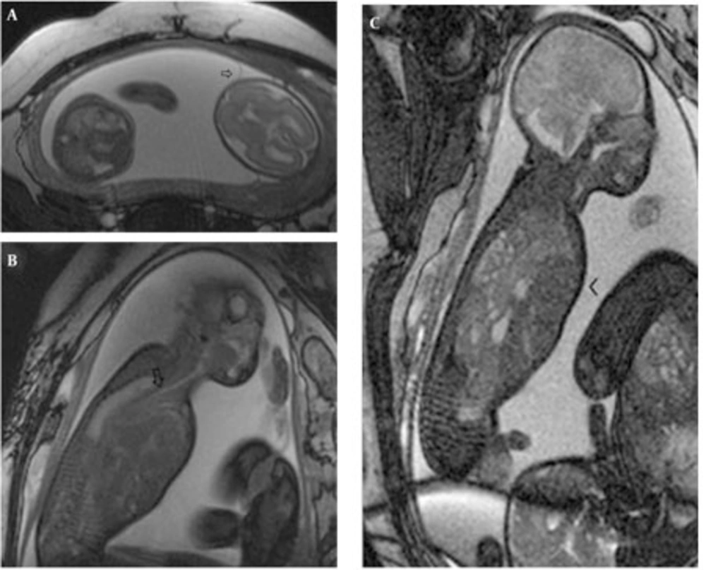
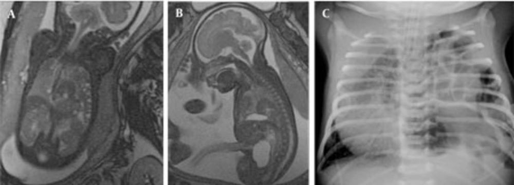
In cases of intrathoracic space occupying lesions, survival after birth is greatly dependent on adequate assessment of the lesion and development of pulmonary hypoplasia in the antenatal period. Our study demonstrated that the differential diagnosis of the most common three fetal chest lesions; namely, CCAM, BPS and CDH may reliably be made by fetal MRI and follow-up MRI has utmost importance since the lesions may completely involute or progress to exhibit additional complications such as hydrops and mediastinal shift. Accurate diagnosis supports parental decisions about the course of pregnancy by differentiating the lesions with a possibility of involution such as CCAM and BPS from CDH that requires postnatal surgery. Besides, fetal MRI can accurately guide the postnatal surgery by depicting all herniated organs including the thoracic kidney in CDH and identifying the extent of disease in CCAM, obviating the necessity for extensive postnatal imaging.Accurate prenatal diagnosis of chest lesions is important because the natural history of these lesions and their treatment vary substantially (
1). On US, the classic differential diagnosis for an echogenic lung mass is CCAM, sequestration, or CDH. Accompanying anechoic structure may be the stomach, intestines or cyst, and sometimes the systemic blood supply is visualized (
5). However, sonographic differential diagnosis is not always possible and fetal MR imaging can be helpful when the diagnosis is unclear by easily differentiating CDH from CCAM and BPS (
1,
3).
CCAM is a rare lesion with a prevalence of 9:100,000 total births (
6,
7). It is characterized by a multicystic mass of pulmonary tissue with abnormal proliferation of bronchiolar structures that connect to the normal bronchial tree and also called as congenital pulmonary airway malformation (
1,
5,
8). The vascular supply of CCAM is from the pulmonary artery with drainage into the pulmonary veins. Hence, the vascular supply of classical CCAM is not observed in any of our cases with MRI. BPS is generally thought to result from an abnormal accessory tracheobronchial bud arising from the foregut accounting for up to 6% of congenital lung malformations (
9,
10,
11). Sequestration may be extralobar (more diagnosed in the prenatal and neonatal period) and intralobar (more diagnosed in the childhood period) (
1). It has its own vascular supply from the systemic circulation and if a vessel arises from the aorta, the lesion is presumed to be a sequestration. The vasculature supplying BPSs were visualized on MRI in all our cases including the hybrid cases. However, there is a wide spectrum of these anomalies with much overlap. Stocker et al. (
4) categorized CCAM into three histologic types; type 1 consists of large cysts (3-10 cm), type 2 numerous cysts smaller than 2cm and type 3 is microcystic lesions (0.2cm) that appears as a solid mass (
12). Although MR characteristics are known for the initial three types, type 0 and type 4 were later added to the classification (
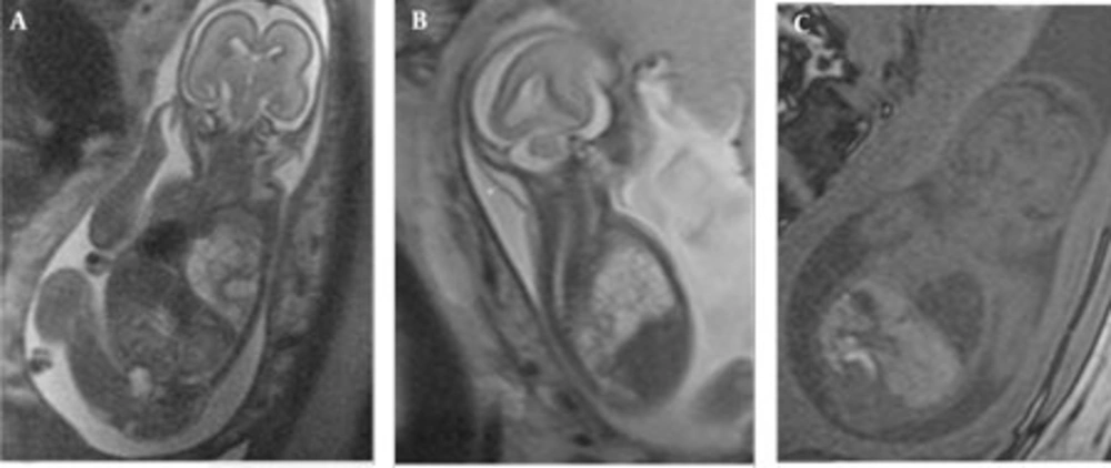
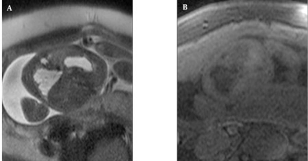
13). On MR imaging, CCAM and BPS exhibited higher signal intensity than normal adjacent lung tissue on T2-weighted imaging and lower signal intensity than normal lung on T1-weighted imaging. Type 1 and type 2 CCAM are very high signal on T2-weighted images almost equal to amniotic fluid; however, the signal intensity of BPS was not that high, yet still higher than the lung tissue. Type 3 CCAM is relatively homogeneous and has moderately high signal intensity that is higher than that of normal lung, but not as high as that of the amniotic fluid. On MRI, pure fetal BPS appears as a well-defined, triangular, homogeneous and hyperintense mass (
5). In our cases, isolated BPSs had well-defined contours, but hybrid lesions contained peripheral small cysts causing irregular contour appearance. The hybrid cases had feeding vessels from the aorta. Sequestrations are frequently located in the lower lobes so were our cases. One of the CCAMs in this series developed hydrops because of the mediastinal shift. The prognosis of CCAM lesions is variable, depending on the size rather than the histological type (
1). If large, they can cause mediastinal shift, pulmonary hypoplasia, vascular compromise and hydrops. Hydrops is a harbinger of impending fetal death and this fetus was terminated. When adjacent normal lung is compressed by a pulmonary mass, such as a CCAM or sequestration, it can be visualized on MR as of slightly lower signal intensity than adjacent normal lung. In addition, polyhydramniosis may develop due to impaired swallowing, caused by esophagus compression by the mass. Although rare, CCAM and BPS may involute partially or completely (
1,
14,
15). Even rarer, complete in utero regression may be observed. In one of our cases with two small pulmonary hyperintense lesions that may indicate CCAM type 3, the lesions completely disappeared in utero in the follow-up period. The lesions may disappear on sonography, but still be visible on MR imaging. Liu et al. state that other than large CCAMs, all CCAMs showed some degree of regression in their series (
2). As CCAM and BPS lesions regress, their signal intensity decreases. Partially regressed BPS tends to have a lobulated margin with a decrease in signal intensity and signal inhomogeneity. Levine (
5) et al. found that larger lesions did not regress as much as smaller ones. A pleural effusion may be visualized transiently as the lesion decreases in size, but this was not evident in our case.
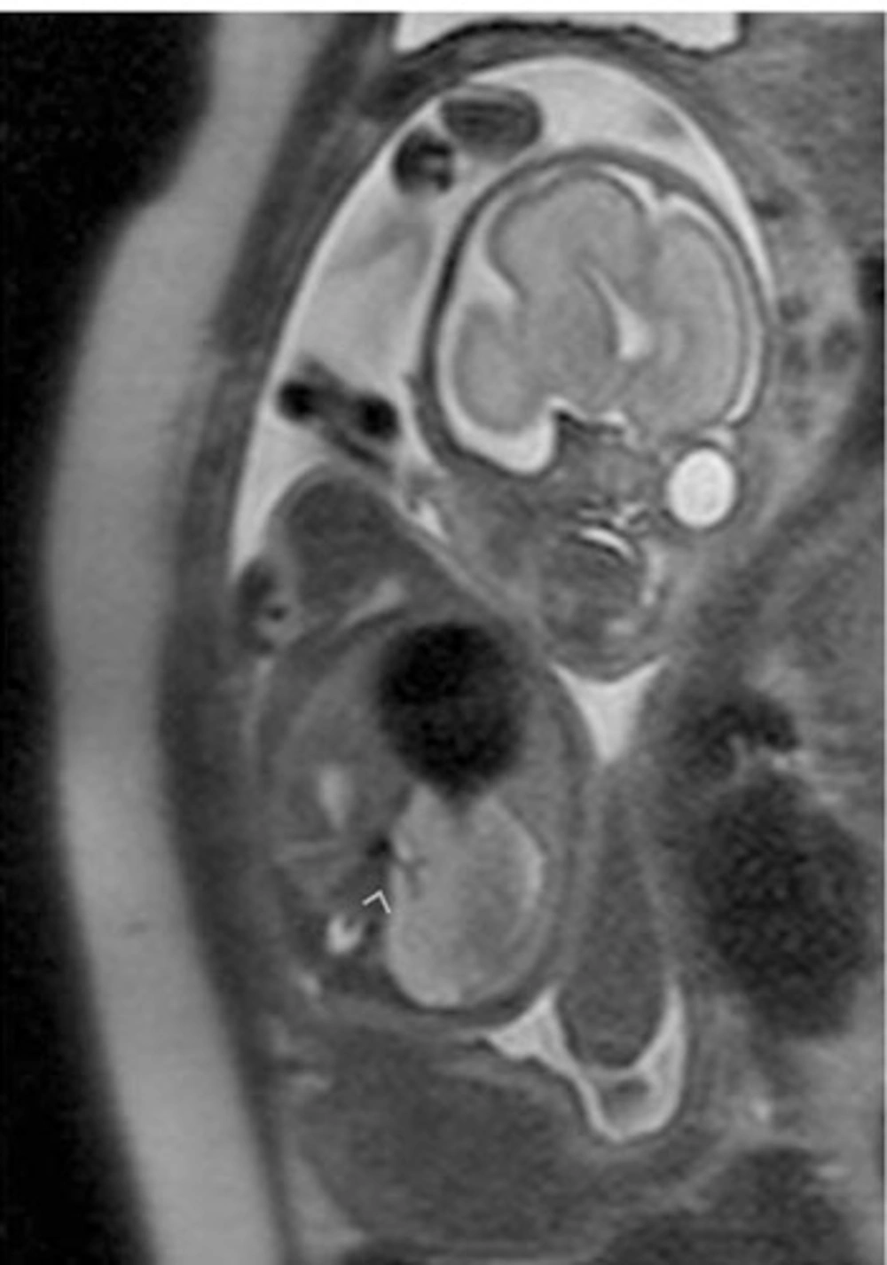
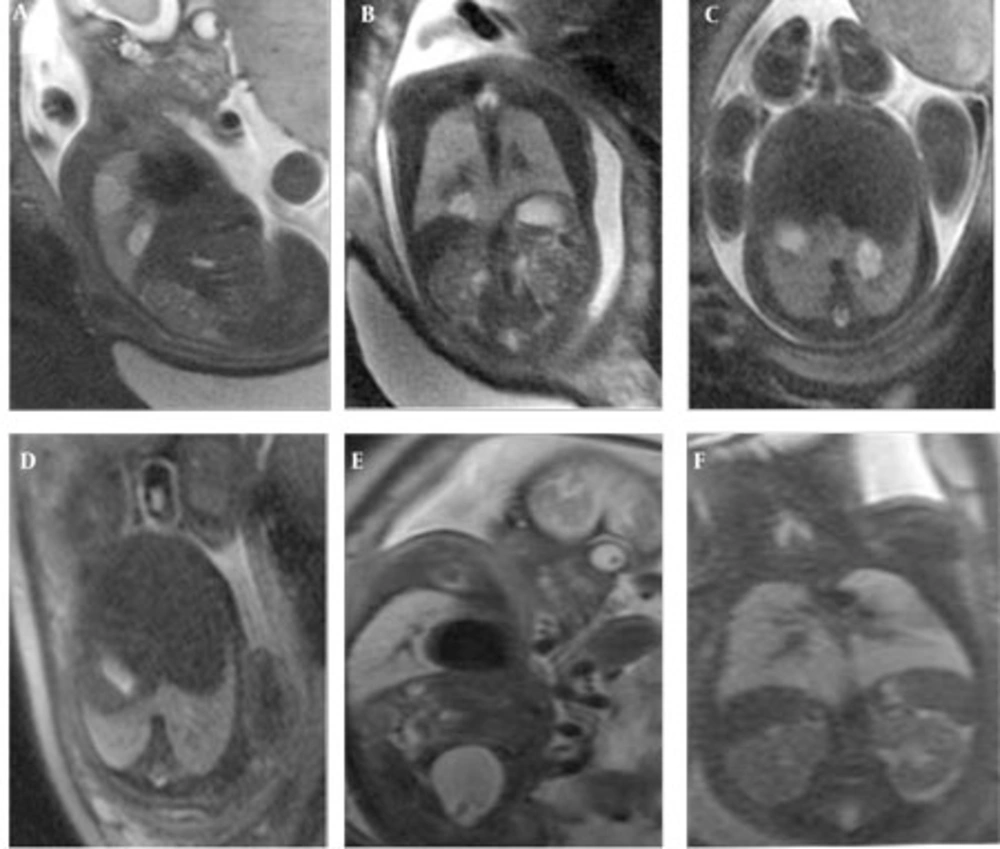
Congenital diaphragmatic hernias occur in approximately 1 in 2,200–12,500 live births and typically occur in the posterolateral left hemidiaphragm (
16). The reported cases in this series were also located on the left side. Right-sided, bilateral, paraesophageal and pericardial hernias may also occur (
5). The classic US signs are cystic thoracic lesion with a mediastinal shift and occasionally an absence of a normally positioned fluid- filled stomach (
17). However, these sonographic findings do not always warrant a sufficient differential diagnosis and MR may be used for clarification of the findings. The contours of the diaphragm may easily be traced in the sagittal and coronal MR images. Besides, herniated organs like stomach, intestines, spleen and the liver can be identified by their characteristic MR appearances (
18,
19). In this series, herniated liver and spleen were seen in 50% of the CDH cases. The liver is observed on MR imaging as a slightly low signal intensity structure on T2-weighted imaging that is of higher signal intensity on T1-weighted imaging. The high morbidity associated with CDH is due to pulmonary hypoplasia resulting from the compression of the developing lungs by the herniated viscera (
5,
20). For diaphragmatic hernias in general, the position of the liver (above or below the diaphragm) is one of the best positive predictive values for survival, especially liver in the chest is associated with a worse prognosis than when the liver is completely intra-abdominal (
5,
21). Colon, with a high signal intensity on T1-weighted imaging and low signal intensity on T2-weighted imaging, the small bowel with fluid-filled loops, the stomach and the spleen, all can be well-visualized in hernias (
5). In this series, one case (case no. 17) was reported to have an intra-thoracic kidney. The incidence of intra-thoracic kidney as a result of congenital diaphragmatic hernia is extremely rare with a prevalence of less than 0.01% (
20,
22,
23). Incorrect prenatal diagnosis may have poor clinical results as many infants with space-occupying lesions suffer from respiratory problems. Fetal MRI may obviate postnatal CT or MRI necessity, which requires sedation to some extent especially when performed in the late gestational period. Our CDH cases no. 12, 13, and 16 were operated with prenatal planning. Termination of fetuses with US diagnosis of space-occupying lesions of the chest limits the number of fetuses referred for prenatal MRI. The assessment of space-occupying lesions of the chest with larger series would provide additional information of involuting lesions as well as providing guidance for postnatal surgery.
In conclusion, fetal MRI is helpful for reliable differential diagnosis of most common space occupying lesions of the chest. Follow-up MRI is important since the lesions may completely involute in utero or may be complicated by hydrops and mediastinal shift. Accurate differential diagnosis enabled by the multiplanar capability of MRI may help parental counseling; moreover, this may guide the immediate postnatal surgery obviating the necessity for extensive postnatal imaging.