The incidence of IPA has been reported as between 0.05% and 7.7% according to several reports (
2-
6). Typical access site for diagnostic and/or therapeutic catheterization is the common femoral artery (
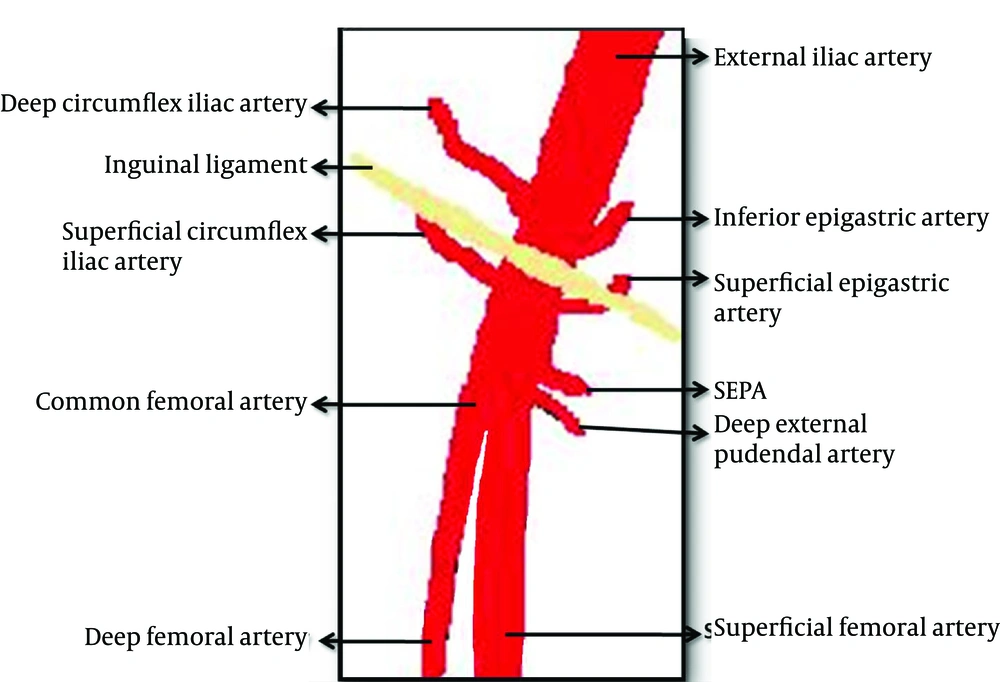
Figure 4) (
5). Therefore, IPA occurrence following catheterization is most commonly seen in the common femoral artery neighborhood. Since gray scale and Doppler US are the most important and non-invasive tools in the diagnosis of IPA, Doppler US should be the first line choice in patients suspected of IPA (
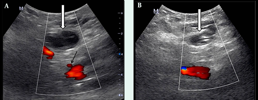
4). In Doppler US examination of IPA, the following signs might be observed: hypoechoic aneurysm sac around the femoral artery and to and fro type high velocity waveform in the aneurysm sac or near the neck of the aneurysm (
6).
Vascular diagram of the inguinal region (7)
There are different treatment modalities for the iatrogenic pseudoaeurysm of the femoral artery (
3). US-guided compression treatment is an expensive, safe, and non-invasive method among these modalities (
5,
6). In case this treatment fails, or when IPA exceeds a size of 4 cm, minimally invasive percutaneous treatment techniques including thrombin, saline or collagen injection, coil embolization or insertion of covered stent should be considered (
4-
6,
8). All of these techniques have their own disadvantages. Although minimally invasive techniques are more effective than US-guided compression; they are more expensive and could cause various complications (including thrombotic complication, bleeding, anaphylaxis and infection) (
3). Moreover, surgical treatment might result in serious complications, and should be performed only when minimal invasive techniques have failed (
6). For these reasons, US-guided compression was our first choice in the presented case and complete healing was achieved in two sessions.
There are various factors that have an impact on IPA development. Among these, arterial hypertension, improper puncture site, thrombolytic-anticoagulant agent, and the larger size of the introducer used are the most important ones (
2). Moreover, Popovic et al. demonstrated that the access side is an important predictive factor for IPA development (
2). A right-handed interventionist could have problem in directing the needle in the right position and puncturing the artery in the proper position when working on the left side of the patient, as in the presented case.
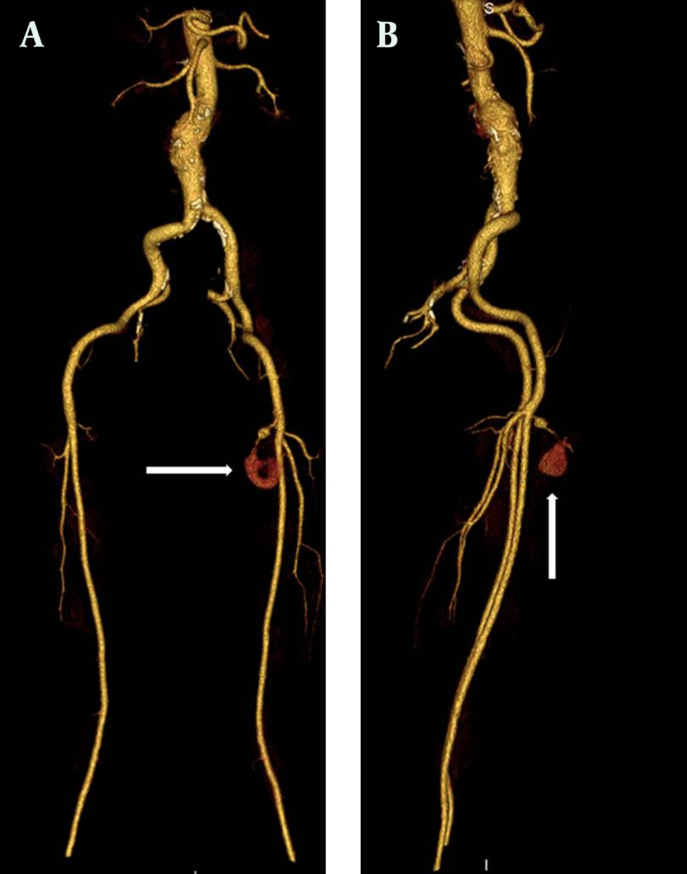
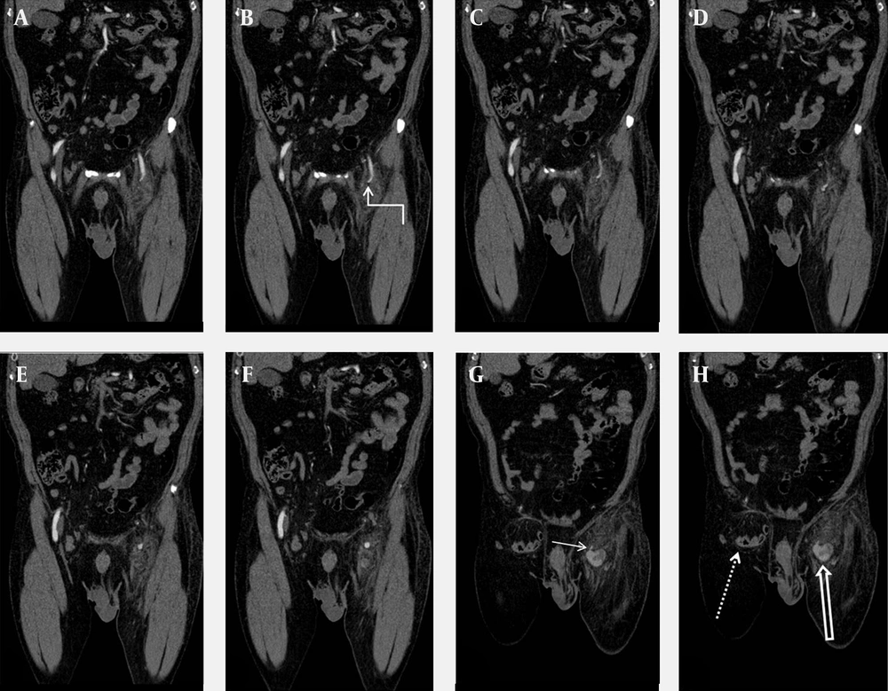
The superficial external pudendal artery is located at the level of the femoral artery bifurcation, originates from the medial side of the common femoral artery, and proceeds to the perineal region (
7). In MDCTA examination, we observed that the neck of IPA originated from SEPA. Therefore, we thought that the intervention at the bifurcation level of the femoral artery leading to this situation was performed by a right-handed cardiologist from the right side with an improper angle. Detection of the puncture area of the introducer at the medial side of the neck of the femur in US examination supported our thought. In the presented case, due to inguinal hernia on the right side, the left groin had been accessed. In patients in whom therapeutic angiography will be performed from the left groin, to prevent these types of complications, arterial puncture should be done under Doppler US guidance before intervention. It was reported that this maneuver could reduce the incidence of IPA development significantly (
9). Furthermore, the exact position of the needle should be determined with fluoroscopy before arterial puncture. The needle should enter the skin with a 45° angle from the conjunction of the femoral head and neck, and the femoral artery should be punctured exactly in the mid-femoral head level. In this level, the femoral artery is supported by the femoral head posteriorly; therefore, post procedure hemostasis could be easily provided with compression (
5).
It has been reported that pseudoaneurysm of the pudendal artery is associated with endorectal prostate biopsy, penetrating gluteal trauma, ischial pressure wound with secondary infection, and blunt pelvic trauma (
10). To the best of our knowledge, IPA of the pudendal artery due to angiographic catheterization has not been reported in the literature. In our case, the diagnosis of pudental artery IPA was made by MDCTA with 3D reconstructions. Doppler US examinations may have limitations in detecting small pseudoaneurysms of the proximal extremities and localizing the neck of the pseudoaneurysm correctly (
10). MDCTA has many benefits, including being accurate, rapid, and relatively operator independent (
10). Moreover, MDCTA is cheaper, less invasive with a lower complication rate when compared with conventional angiography, which is accepted as the gold standard (
10). MDCTA enabled detailed diagnostic evaluation sufficient for pretreatment planning of IPA (
10).
In conclusion, the incidence of iatrogenic pseudoaneurysm has increased recently due to the increased number of therapeutic catheterization and/or the improper technique. In the current patient, the pseudoaneurysm arose from a different vessel than usually seen. In patients in whom therapeutic angiography will be performed from the left groin, arterial puncture should be done under Doppler US guidance to reduce the risk. Doppler US examination can be an effective, non-invasive, and sufficient tool for IPA diagnosis and may also be part of the treatment when using the compression technique (
5). In case we cannot evaluate IPA morphology, MDCTA can be used as an effective and safe technique. US-guided compression technique for IPA therapy is a noninvasive, inexpensive, easy, and well tolerable method, with low mortality and a high success rate in selected patients (
5). Minimally invasive percutaneous techniques should be used when US-guided compression is unsuccessful.