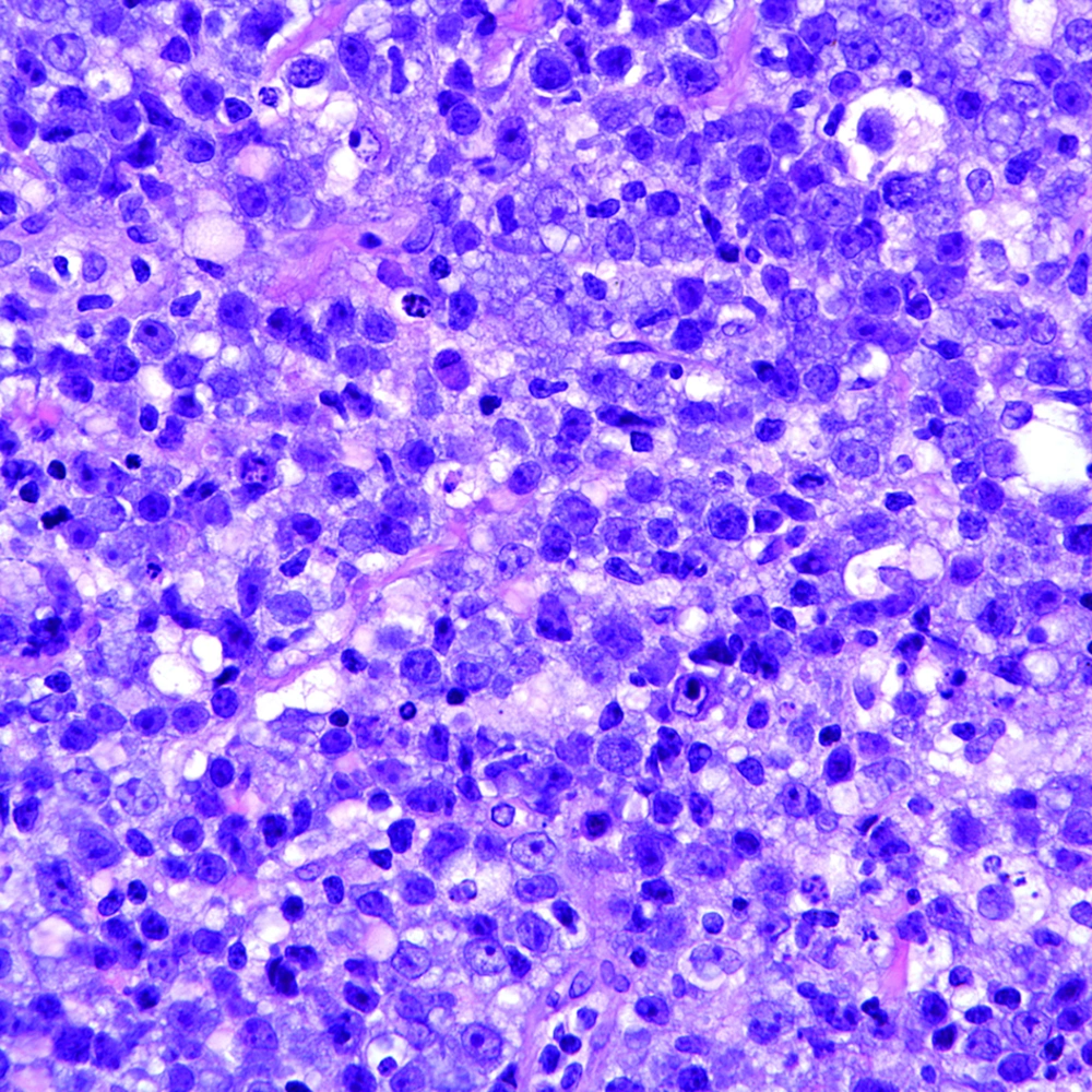
Musculoskeletal involvement affects about 7% of NHL patients, typically as metastatic disease and infrequently as a primary lymphoma of either the bone or soft tissues (
2-
4). Presentation with joint involvement is very rare (
2). To the best of our knowledge, there are only 16 cases of joint involvement with synovial infiltration reported in English literatures (
1-
4), most of which had known pre-existing NHL (
1,
2). Of the reported cases, males and females were equally affected with a wide age range of 13 to 76 years old (
1). The knee joint (11 cases) is the most commonly involved joint followed by the elbow (two cases), sternoclavicular joint (one case), shoulder (one case) and wrist (one case) (
1). Joint involvement in NHL usually presents with symptoms suggestive of an inflammatory arthropathy or low grade infection such as joint pain, swelling and limitation of movement affecting a single joint rather than multiple joints (
2,
5,
6). A minority of patients also has systemic symptoms such as weight loss, night sweats and fever (
2,
7). About one-third of all cases are immunocompromised (
8-
10). Serological tests are usually non-contributory. Similarly, synovial fluid analysis may not always help in distinguishing joint lymphoma from a low-grade chronic septic arthritis or inflammatory arthropathy since all these conditions can lead to mild synovial leukocytosis with lymphocytic predominance (
1). Synovial involvement of lymphoma usually occurs through direct extension from the bone (
1,
2). In this case, the lymphoma seemed to be primarily located in the medial femoral condyle with secondary infiltration of the synovium on the medial aspect of the knee. In a few of the previous cases of intra-articular lymphoma, the lymphoma seemed to be primarily synovial in origin as there was no bone involvement as judged by MRI (
11,
12). In the absence of serial histological sectioning, MRI is the only reliable means to determine whether there is co-existing bone marrow involvement (
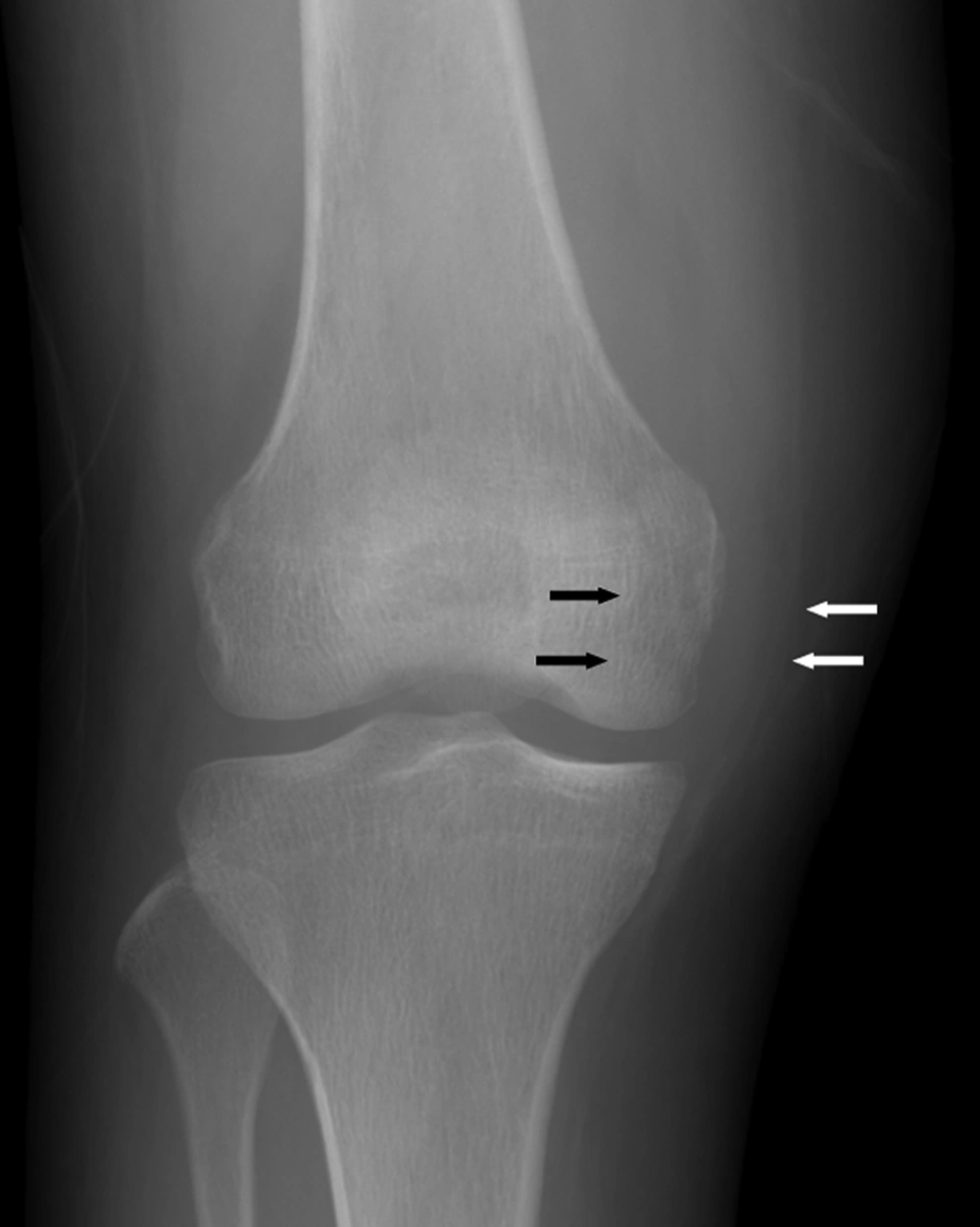
11,
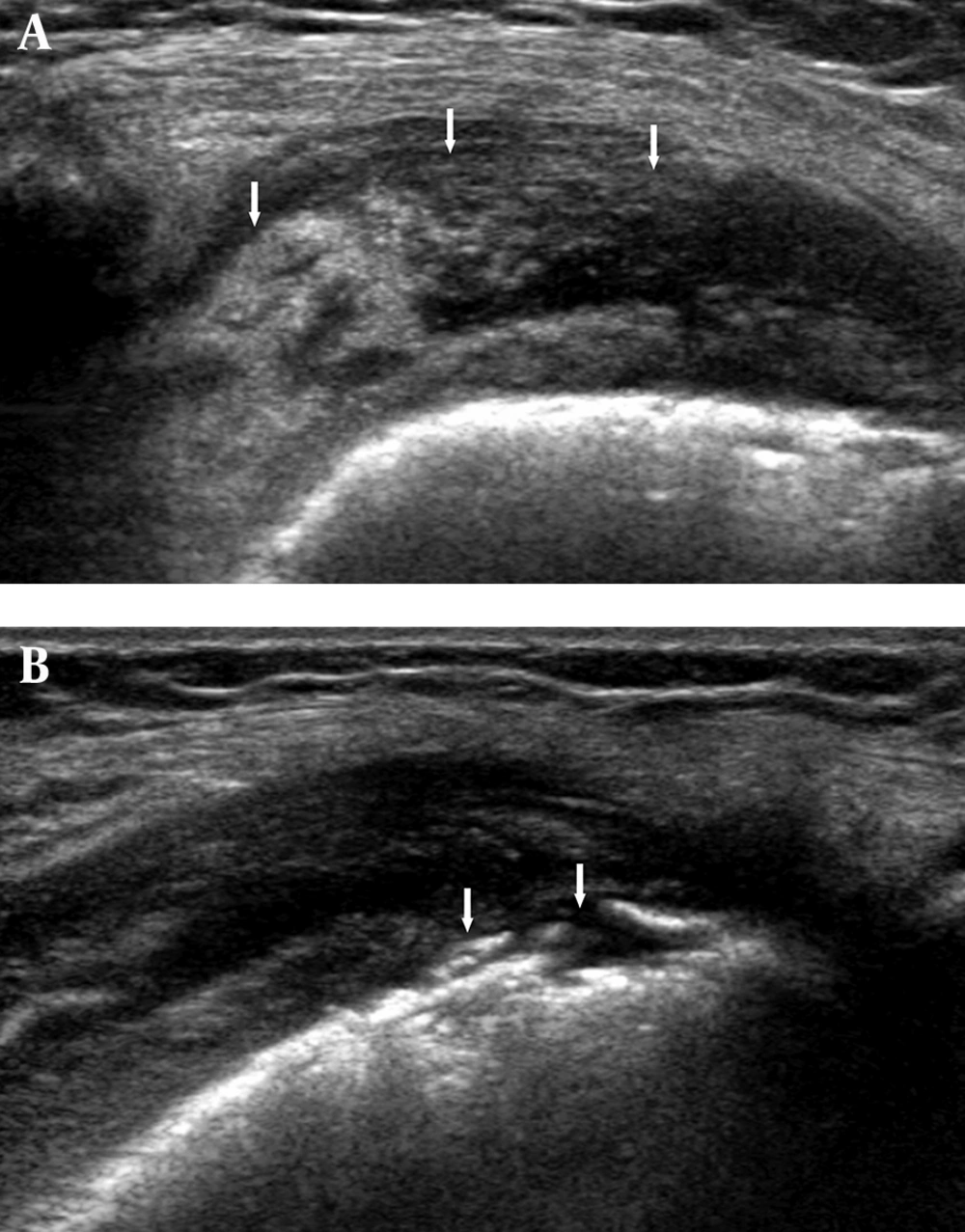
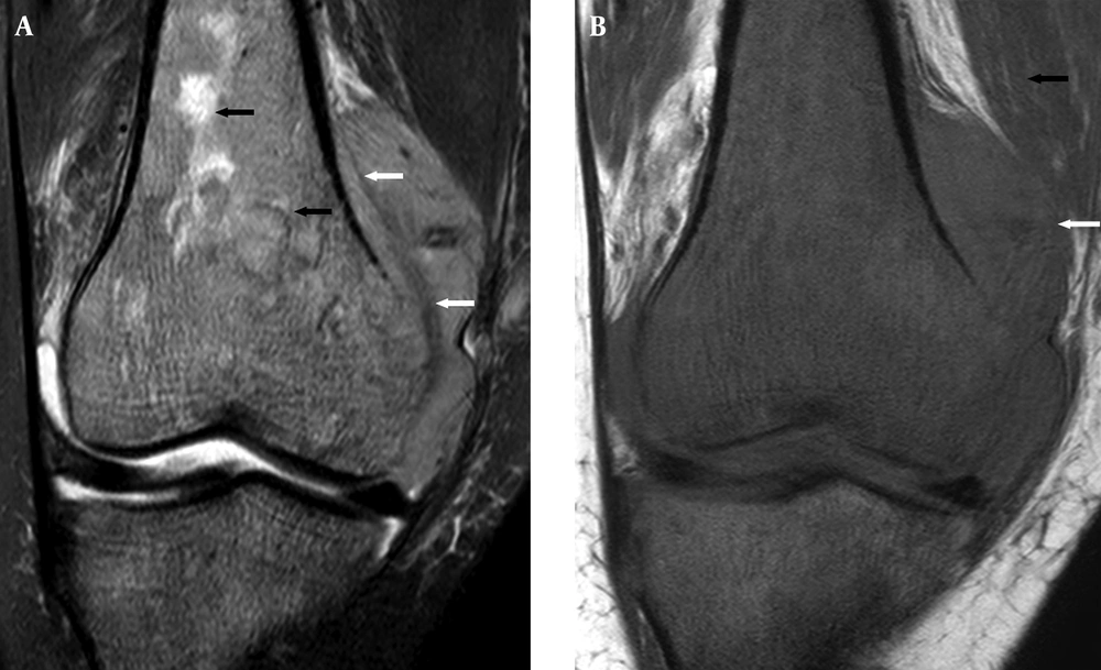
12). Radiography may reveal para-articular bony erosion or osteolysis though in half (8 out of 16 cases) of the reported cases, radiography will be unremarkable except for soft tissue swelling (
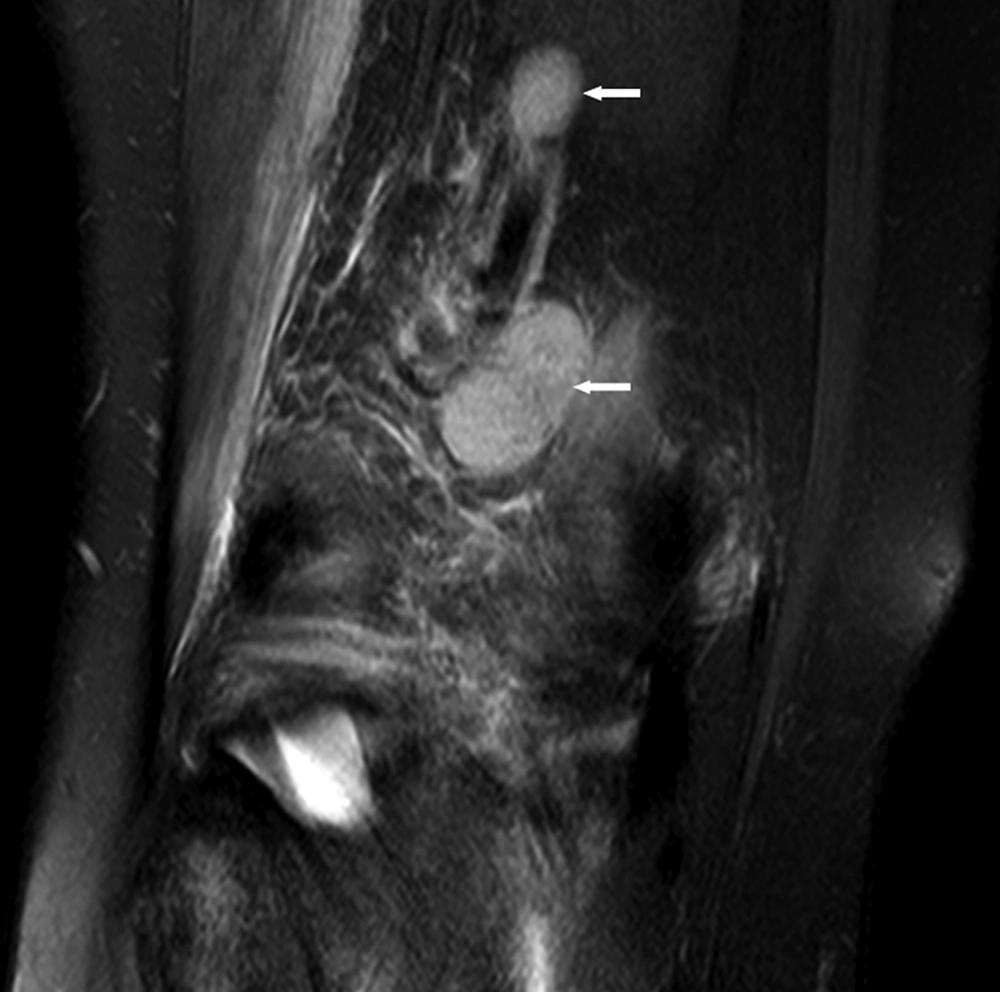
1). Ultrasound is useful in distinguishing effusion from synovial thickening. Focal synovial thickening as in this case is a useful marker of an infiltrative disorder such as lymphoma as TB and inflammatory arthropathy would usually give rise to more diffuse synovial thickening. Ultrasound also facilitates imaging-guided synovial biopsy. Ultrasound findings of joint lymphoma have not previously been reported. MRI allows a complete assessment of the bone marrow, bone cortex, joint, and lymph node involvement. Similar to all the previous reported cases, the signal intensity of lymphoma in this case is also non-specific (T1 Whypointense and T2W hyperintense) though the signal characteristics are helpful in distinguishing lymphoma from other synovial disorders such as pigmented villonodular synovitis or synovial osteochondromatosis (
13). The presence of enlarged regional lymph nodes is only seen with tuberculous infection, lymphoma, and malignancy, which is helpful in further narrowing the differential diagnosis. Only 2-4% of musculoskeletal sarcomas have nodal involvement at presentation (
14,
15). On the other hand, nodal enlargement with musculoskeletal lymphoma is very common (
16,
17).The differential diagnosis of focal synovial thickening includes synovial chondromatosis, pigmented villonodular synovitis (PVNS), focal nodular synovitis, chronic infection such as tuberculosis, crystal or amyloid depositional disease and rarely synovial sarcoma (
18). The MR appearances also distinguish most of these entities from one another. Synovial chondromatosis is associated with T1-weighted and T2-weighted hypointense nodules or nodules with a center showing fat signal (osteochondral metaplasia). Pigmented villonodular synovitis (PVNS) is associated with hypointense foci due to hemosiderin deposition on T2*-weighted sequences. Nodular synovitis will not cause periosteal elevation, medullary infiltration or nodal enlargement as seen in this case. Chronic infection with bone marrow involvement is the main consideration though it is usually associated with more marked peri-articular inflammation and a synovium of higher T2-weighted signal intensity than lymphoma as seen in this case. Gout and amyloid are often hypointense on T2-weighted sequences. Intra-articular synovial sarcoma is rare and may have similar imaging features and clinical presentations. However, it usually presents as a peri-articular rather than articular mass and would infiltrate the medullary canal as a late feature (
12). As the imaging findings are not pathognomonic, ultrasound-guided synovial biopsy was helpful in confirming the diagnosis of lymphoma. The absence of systemic work-up (such as abdominopelvic CT scan) is the main limitation of this case report as the patient refused further investigation, though clinically there was no systemic adenopathy, hepatosplenomegaly or constitutional symptoms to indicate involvement beyond the knee region. Intra-articular lymphoma with synovial involvement is very rare although it can be broadly distinguished from other synovial diseases by MRI. However, the imaging findings are not entirely specific and image -guided synovial biopsy is required for definitive diagnosis.