


1. Introduction
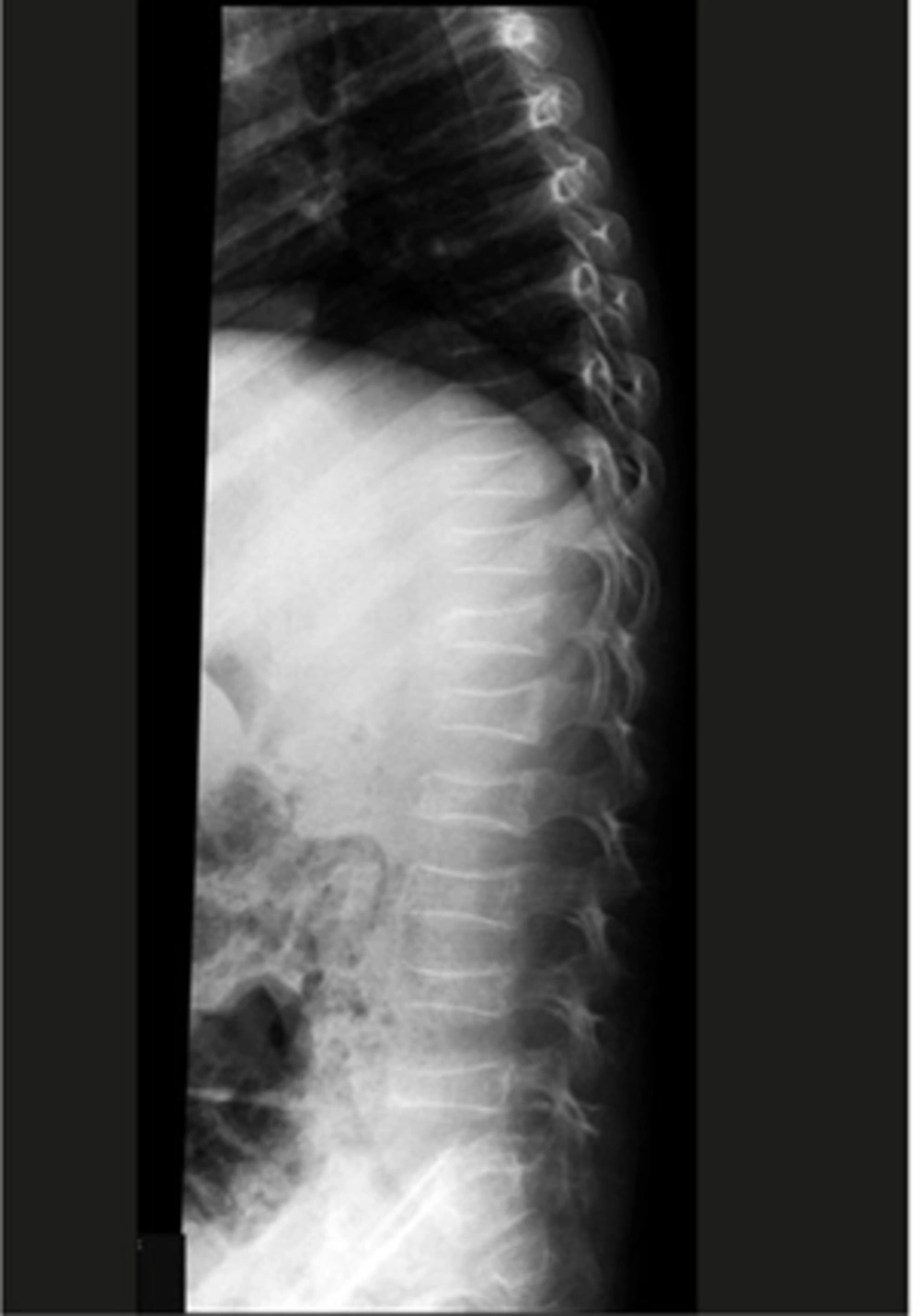
Lateral thoracolumbar X-ray reveals multiple collapsed vertebrae.
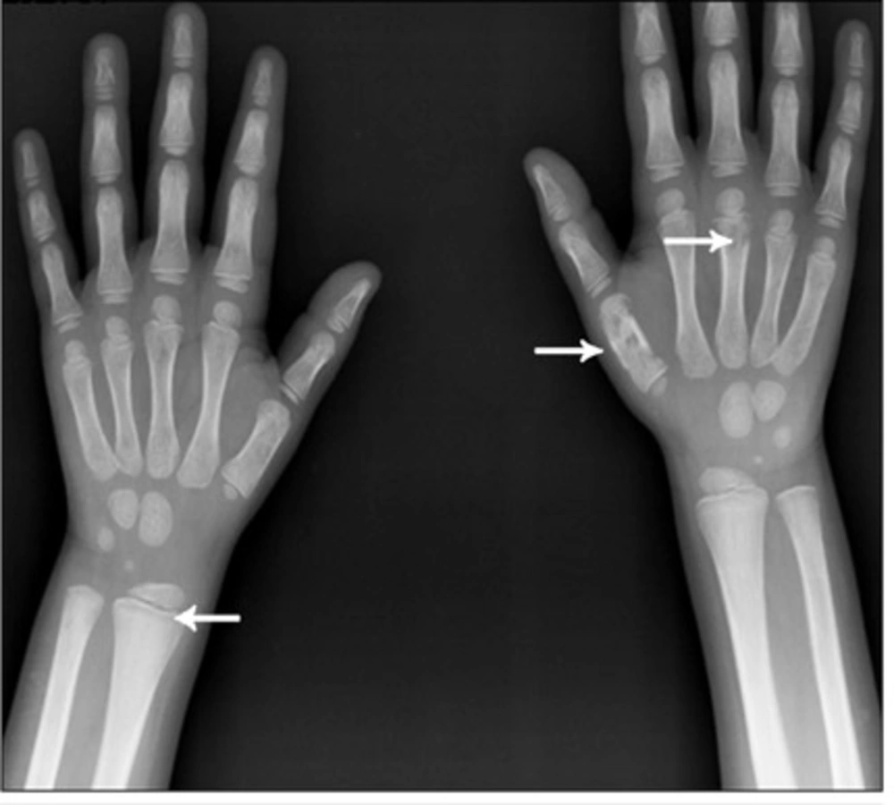
Wrist X-ray reveals metaphyseal lucent band (arrow head) in radial and ulnar metaphyses. Note the lucent bony lesions of the first and third metacarpal bone on the right side (arrows).
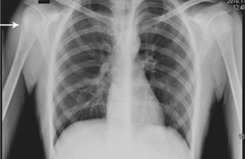
CXR reveals permeative bone lesion in the right humerus (arrow).
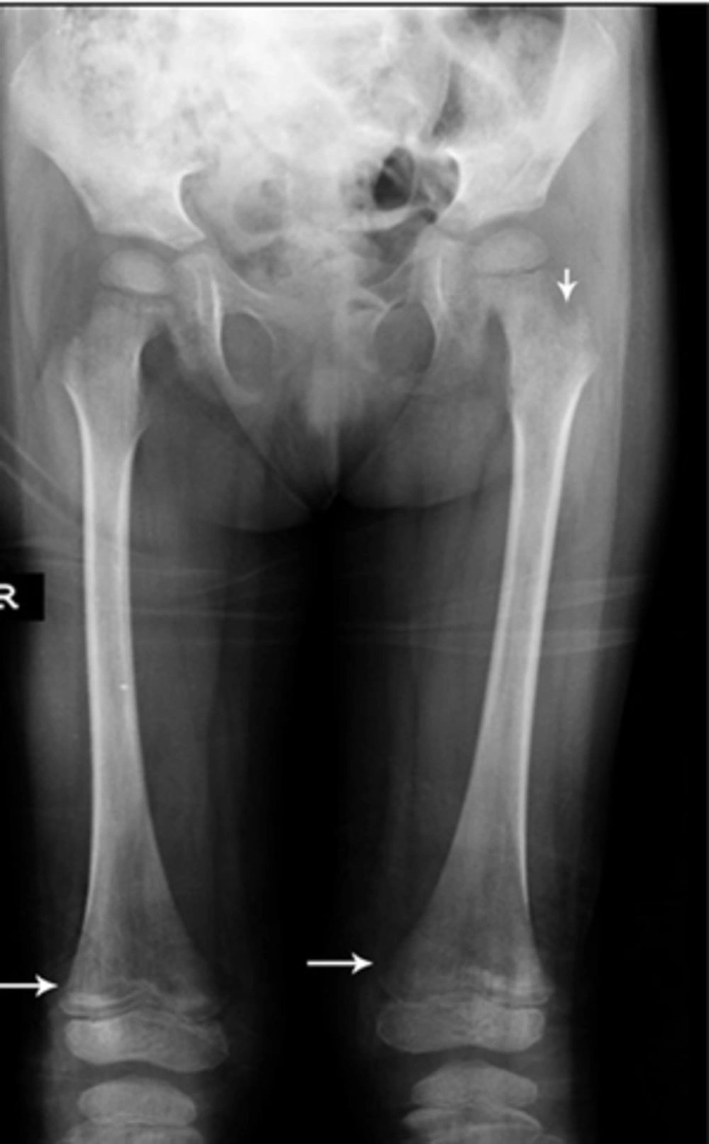
Radiography reveals metaphyseal hypodensity on both femurs with sclerotic lesions (arrows). There are also lytic lesions in the left femoral neck (arrowhead).
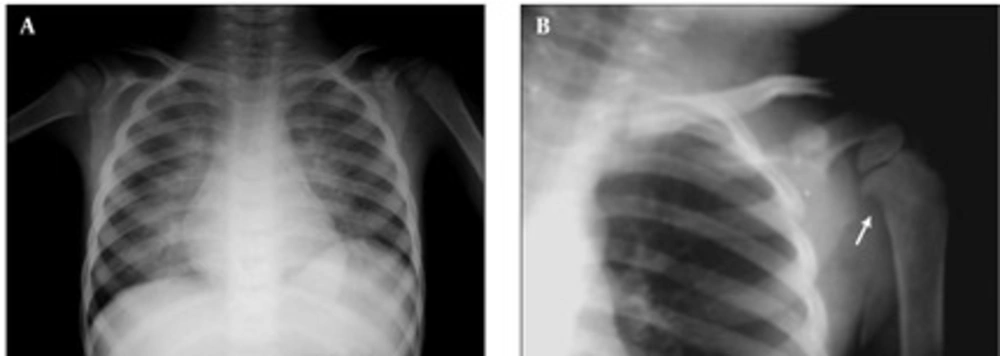
A, CXR reveals bilateral parahilar alveolar infiltration in both lung fields. There is suspicious right humeral metaphyseal erosion; B, Magnification view reveals erosion more clearly with reduced adjacent bone density (arrow).
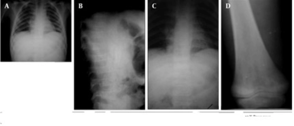
A, CXR reveals perihilar and retrocardiac infiltration. Abnormal shaped vertebra is suspicious.; B and C, Thoracolumbar AP and Lat X-Rays reveal multiple collapsed vertebrae; D, Knee X-xay reveals hypodensity around the knee joint with metaphyseal translucency.
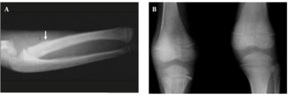
A, Forearm X-Ray reveals periosteal reaction of radial proximal metaphysis with distal metaphyseal translucency(arrow);B, AP X-ray of knee joints reveals metaphyseal translucency in both distal femurs and proximal tibiae more prominent in the latter.
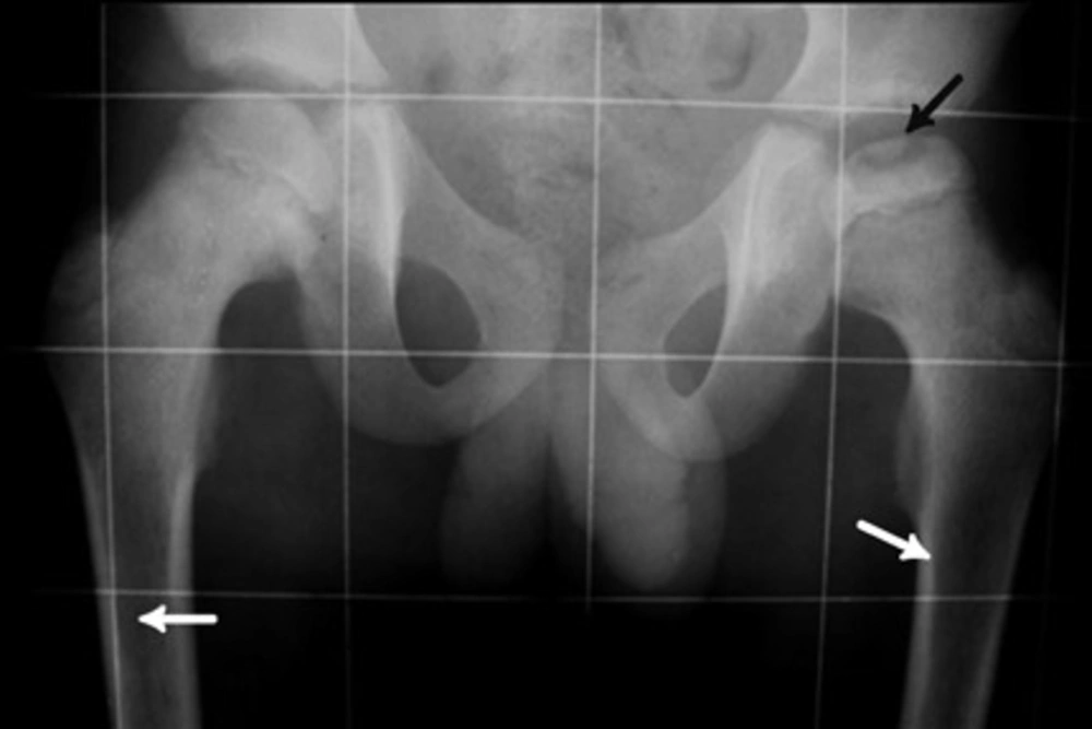
Pelvic X-Ray of this case reveals reduced height of femoral epiphysis with osteochondral fracture on the left side due to avascular necrosis (black arrow). Permeative lesion in both femoral proximal metaphyses are noted (white arrows).
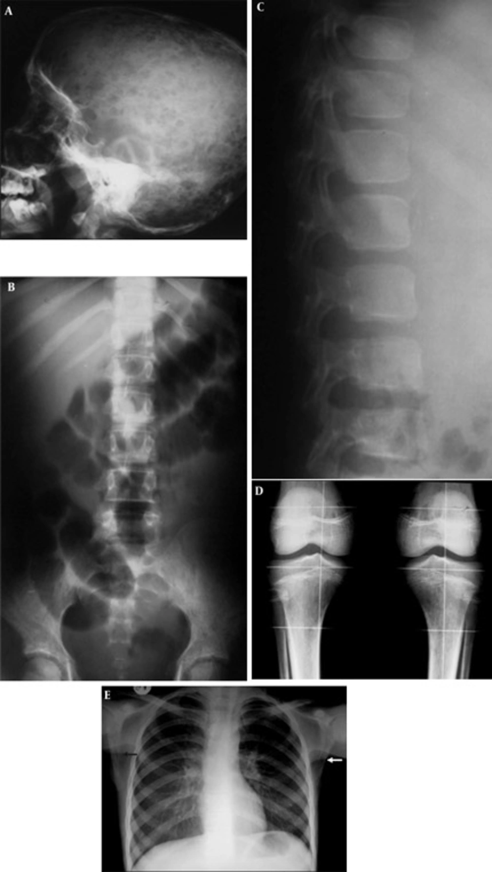
A, Skull X-ray reveals multiple lytic lesions; B, Lumbosacral AP X-ray reveals permeative bony lesions in iliac bones with reduced vertebral height of L5; C, Lumbosacral lateral X-ray reveals reduced vertebral height of L5 with reduced bone density and lucent zone beneath the vertebral endplate; D, Knee X-ray reveals reduced bone density with permeative appearance; E, CXR reveals permeative bony lesions in bilateral scapular bones (arrows).

A, CXR reveals anterior mediastinal widening with double contour thymus (arrow) and right pleural effusion; B, Magnification view reveals metaphyseal erosion in proximal medial humeral metaphysis (arrow).
