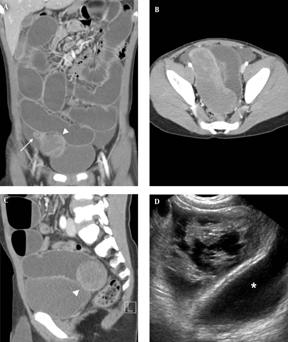
Small bowel hematoma in children is generally caused by trauma, anticoagulation therapy, bleeding disorders, vasculitis, and tumors (
1-
5). Because of the rich submucosal vascular supply, hemorrhage usually originates in the submucosal layer of the bowel (
1). As the bleeding continues slowly, intramural, intraluminal, mesenteric, and retroperitoneal hemorrhage may occur (
1). It may also be associated with hemorrhagic ascites (
1).
The imaging features of small bowel hematoma vary depending on its location and age. For acute intramural small bowel hematoma, US may show circumferential thickening of the bowel wall with hyperechogenicity (
1). An acute intraluminal hematoma may appear as an avascular mass with diffuse or inhomogeneous echogenicity (
6). As liquefaction occurs, the hematoma shows mixed echogenicity with internal septation, thickened hyperechoic walls, or mural nodules (
6), and eventually becomes anechoic. The US or magnetic resonance imaging can reveal the multilocular nature of the mass with fine septation. On CT images, an acute small bowel hematoma appears as a thickened bowel wall or mass with hyperdensity (
7). The hyperdense area can be observed during the first ten days and subsequently evolves into a hypodense area (
7).
The differential diagnosis of a bowel-related cystic mass in children should include duplication cyst, Meckel’s diverticulum, lymphangioma, and small bowel hematoma, which may present with intussusception or volvulus. Lymphangiomas are usually multilocular and infrequently located in the bowel (
8). Occasionally, a duplication cyst might be multilocular.
Traumatic small-bowel hematomas most commonly involve the duodenum, whereas nontraumatic ones most commonly affect the jejunum, followed by the ileum and duodenum (
1,
3,
5). Abbas et al. reported that nontraumatic spontaneous small-bowel hematomas appear longer than traumatic hematomas (
4). Gaines et al. reported that traumatic injury of duodenum is unusual in children and child abuse should be suspected in a young child with duodenal injury (
5). Although traumatic ileal hematoma is uncommon, a tentative cause in our case was blunt trauma. Several imaging features are helpful in distinguishing spontaneous small-bowel hematomas from other types. However, the differential diagnosis is challenging and blunt trauma in children can be unwitnessed.
In conclusion, although intraluminal small bowel hematoma is rare in children, it can present as an intraluminal cystic mass and should be considered as a rare cause of small bowel obstruction. The US and CT findings of submucosal ileal hematoma could be useful for the diagnosis of such cases in the future.