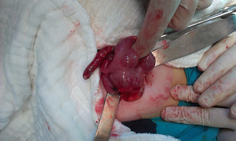
The appendix is located on the posteromedial side of the caecum, at approximately 3 cm below the ileocecal valve (
5). There is an invariable connection between the bottom of the appendix and the caecum, but the free end of the appendix is mobile, which accounts for its variable location in the abdominopelvic cavity (
6). A mobile ascending colon and caecum are uncommon reasons of acute abdomen (
7,
8). In embryogenesis, the failure of the right colonic mesentery to fuse with the lateral peritoneum occurs because of this abnormal feature. Therefore, the ascending colon and caecum are unassociated and can revolve freely. In spite of this, the ascending colon and caecum commonly have a normal anatomic condition (
9). This may be described as the likely cause of normal US and barium studies in this patient.
The US exhibits high specificity and sensitivity in the diagnosis of acute appendicitis. Nevertheless, in US examination, a mobile caecum may be misdiagnosed as Meckel’s diverticulitis, duplication or mesenteric cyst, infected urachal cyst, or sigmoid diverticulitis (
10). The most common congenital anomaly of the gastrointestinal system is Meckel’s diverticulitis (
11). In the general population, duplications of gastrointestinal tract are uncommon (
12). The mesenteric cysts that are found within the abdomen are benign lesions, with an incidence of less than 1 : 100.000 cases (
13). Diverticulitis, which is the most common complication of diverticulosis, is defined as the inflammation or infection of diverticula. On average, 20% of cases are symptomatic (
14).
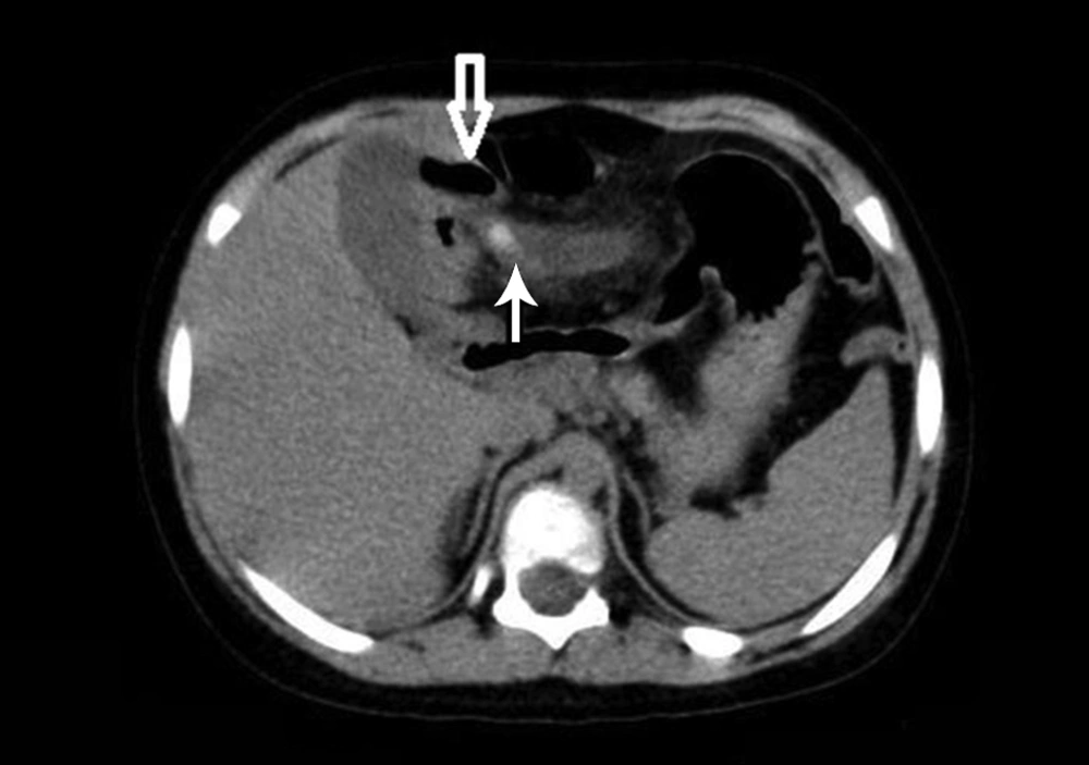
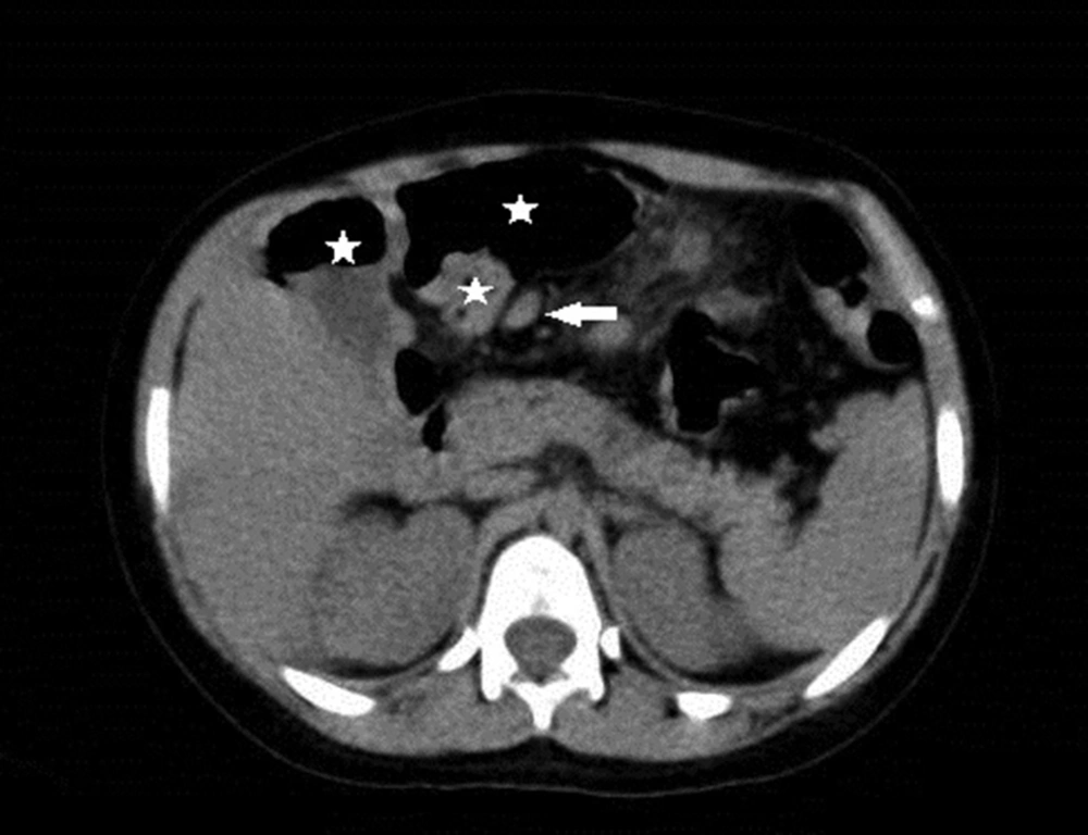
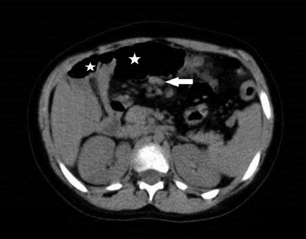
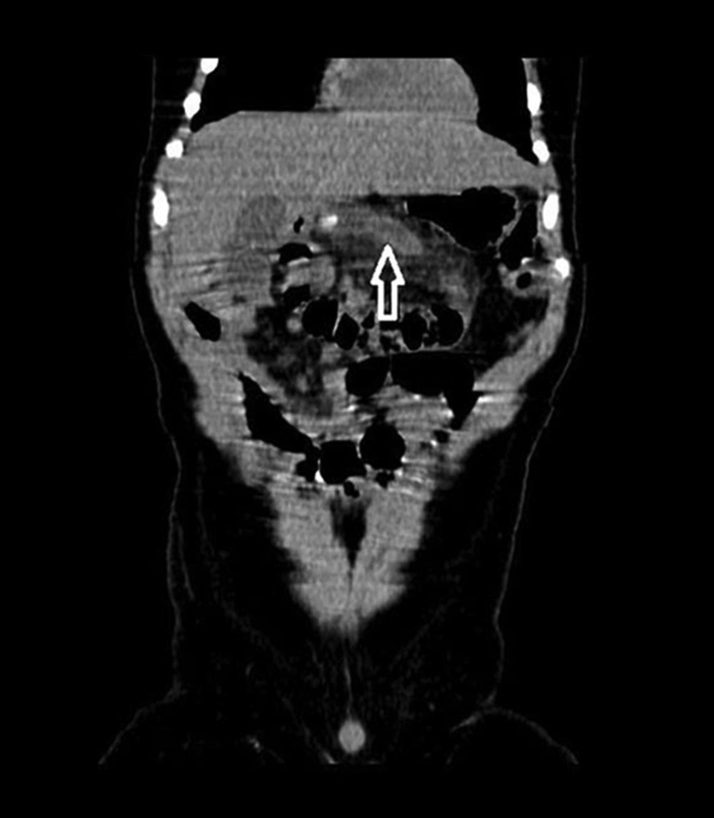
In our case, US examination did not show the appendix. On CT examination, the position of the caecum and ascending colon were shown to follow the enteric sections sequentially from proximal to distal. In the literature, Toprak et al. reported two cases of mobile caecum with acute appendicitis in adults. They found that the right hemicolon and caecum were located in the left upper quadrant, adjacent to the splenic flexure and descending colon (
15). We found that the caecum was located in the epigastric area, adjacent to the left lobe of liver and gallbladder. Printen et al. and Makama et al. reported several mobile caecum cases in adults (
3,
4). Makama et al. found an abnormal position of the caecum and ascending colon in barium study. They did not observe abnormal findings on abdominopelvic US (
4). In contrast to their case, we found acute gangrenous appendicitis in a child, extending laterally to the left upper quadrant.
When there are atypical clinical findings and no US findings in patients with abdominal pain, the CT is beneficial in helping to recognizing the situation of the appendix and caecum, and to prevent misdiagnosis in children.