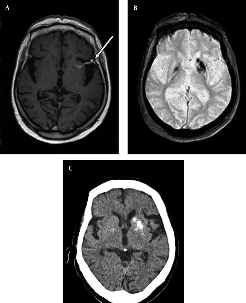
DVA is the most common vascular malformation of the brain occurring in up to 4% of general population and is usually discovered incidentally by contrast-enhanced MRI (
1,
4,
7). Some authors consider DVAs as embryological variants of venous drainage system (
4,
5,
7). They are characterized by radially oriented dilated veins draining normal parenchyma, which are typically likened to “caput medusa” and a larger collector vein, which drains into superficial or deep venous system. Although DVAs are usually asymptomatic, headache, seizures and focal neurological deficits have been claimed to be associated with DVAs (
4).
Some recent studies have focused on the parenchymal changes surrounding DVAs. In one study, locoregional atrophy, white matter changes, cavernous malformations (CM) and dystrophic calcifications were found surrounding two thirds of DVAs (
3). Another study showed that 12.5% of the lesions had subjacent fluid-attenuated inversion recovery (FLAIR) abnormalities (
6).
Dehkharghani et al. (
1), presented 6 cases with unilateral caudate and putamen calcifications in DVA drainage territories. All these authors believed that venous hypertension was responsible for these abnormalities. Decrease in outflow, usually caused by the stenosis of the collector vein causes chronic increase in the venous pressure within the DVA (
5). Venous hypertension can also occur even without frank stenosis of the collector veins. Progressive thickening and hyalinization of the walls of DVA vessels results in increased resistance, decreased compliance and venous hypertension (
1,
3). Chronic venous hypertension brings about chronic ischemia, edema, microhemorrhages, gliosis, dystrophic calcifications, and atrophy. Indeed, CMs have been shown to be caused by repeated microhemorrhages surrounding DVAs (
4). Only 7 cases of unilateral caudate and putamen calcifications associated with DVAs that have deep venous drainage have been reported (
1,
3). The occurrence of dystrophic calcifications in the drainage territory of DVAs drained into deep venous system rather than those drained into superficial venous system can be explained by the higher venous pressure in the former. Indeed, deep venous drainage is an increased risk factor for hemorrhage in an arteriovenous malformation related to increased intranidal pressure (
8). On the contrary, in our first case, drainage was to superficial venous system. However, in our patient, a narrowing of the collector vein at the site of drainage into superficial venous system was noted, although it was not very reliably shown due to the lack of angiography or 3D T1-weigted post contrast images. We can nevertheless postulate that narrowing of the collector vein might have caused chronic venous hypertension, subsequent venous congestion, parenchymal injury and dystrophic calcification. Our first case represents the only example of unilateral putamen and caudate calcification in the vascular territory of a DVA drained into superficial venous system and shows that this pattern of calcification can be seen regardless of the drainage site of the DVA. It also provides further evidence to the concept that chronic venous hypertension might be the mechanism causing parenchymal changes surrounding DVAs.
Bilateral punctate basal ganglia calcifications, usually involving globus pallidi symmetrically, is considered physiological. It has an incidence of 0.3 - 1.5% and is seen more frequent in elderly (
2,
9). Metabolic diseases with abnormal calcium/phosphate balance, infection or inflammation, ischemic or toxic injury are the main reasons for bilateral calcifications in corpus striatum. Differential diagnosis of unilateral corpus striatum calcification is more limited and includes unilateral involvement by infection or neoplasm, in which case calcification is expected to be irregular (
1). However, in both of our cases, basal ganglia calcifications were coarse but well- circumscribed and regular and there were no imaging or laboratory findings to suggest metabolic, infectious or neoplastic processes.
Although CT is the method of choice to diagnose calcification, it is being more frequently noticed on MRI scans especially after the routine use of GRE or SWI sequences has become common in daily practice. Most DVAs are diagnosed incidentally by routine contrast enhanced MRI scans or by SWI, even without the use of contrast agent (
10). We may expect that increasingly higher number of cases with unilateral or asymmetrically dominant corpus striatum calcifications will be noticed through the routine use of GRE or SWI images. Therefore, it becomes important to know that these calcifications may be related to DVAs.
In conclusion, unilateral caudate and putamen calcifications in the drainage territory of DVAs have rarely been reported. It is postulated that chronic venous hypertension, usually caused by the outflow restriction of the collector vein, leads to parenchymal injury and dystrophic calcification. It is expected that this pattern of calcification will be more frequently recognized by MRI after the routine use of GRE or SWI sequences. Therefore, it is of great importance to be aware of this entity to avoid unnecessary diagnostic work-up and to relieve patient anxiety.