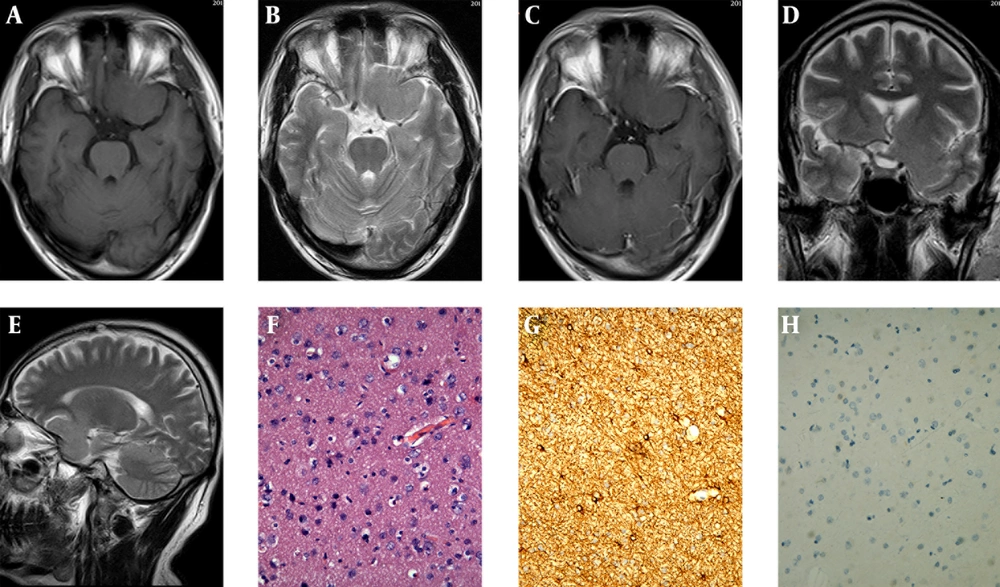
Intracranial gliosis as an uncommon non-neoplastic disease is the brain’s way of reacting to injury and insult. It predominantly results from proliferation of astrocytes (
1-
4). Intracranial gliosis typically manifests in children or young adults, and may involve both gray and white matter, especially in the frontal and temporal lobe. The lesion in this report that originated from the gray matter of the anterior perforated substance of brain may be related to the history of the craniofacial trauma that happened 25 years ago.
Intracranial gliosis is a non-specific change that can clinically and radiologically mimic a neoplastic or non-neoplastic mass lesion. It is even common to observe at least some degree of reactive astrocytosis adjacent to or associated with these conditions. Therefore, the distinction between gliosis and other mass lesions is often the most difficult challenge of diagnostic radiology, even neuropathology, especially for the routine evaluation of small biopsy specimens (
5-
7).
MRI is still of critical importance for gliosis. On MRI, intracranial gliosis, similar to inflammation or low-grade astrocytoma, is usually depicted as an ill-defined margin mass with slight hypo- or isointense signal on T1-weighted images, hyperintense signal on T2-weighted images, nonenhancing or patchy, linear enhancement, and no obvious or minimal mass effect and perilesional brain edema (
8). The lesion has no calcification or cystic change. The MRI appearances of intracranial gliosis and inflammation are alike somewhat depending on infiltration of inflammatory cells in the area of gliosis. The lesion enhancement results from variable breakdown of the blood-brain barrier and abnormality of vascular permeability. However, the lesion in this report showed the same isointense signal as the gray matter on T2-weighted image, no hyperintense signal as inflammation, low-grade astrocytoma or other intra-axial lesions, and no enhancement, which may be related to the absence of inflammatory cell infiltration and integrity of vascular permeability. Moreover, low-grade astrocytoma often occurs in or near the corticomedullary junction, and is seldom located in the surface of the brain parenchyma and becomes mass like an extra-axial lesion. Thirdly, the gliosis we reported is also easily misdiagnosed as meningioma due to its isointense signal on T1 and T2-weighted images, and cerebrospinal fluid interface around the lesion; whereas, the mass attachment to the brain base with a pedunculation on coronal and sagittal T2-weighted image points towards an intra-axial lesion. A typical meningioma is a markedly enhanced lesion with a broad base of dural attachment, and a dural tail is often seen. However, the lesion in this report shows no apparent contrast enhancement or meningeal enhancement on gadolinium-enhanced T1-weighted image. Magnetic resonance spectroscopy may be helpful for the differential diagnosis of gliosis.
The diagnosis of intracranial gliosis depends on its histopathology analysis and the differential diagnosis should be made from low-grade astrocytoma (
6,
7). Microscopically, similar to low-grade astrocytoma, gliosis may result in increased cellularity. The increased cellularity associated with gliosis is generally evenly distributed, but astrocytoma is not. The differences between gliosis and astrocytoma may not be readily apparent on routine hematoxylin eosin staining and may require immunohistochemical stains to be clearly visualized. The most commonly used markers to differentiate them are GFAP, proliferation markers (e.g. Ki-67) and p53. Gliosis has a feature of evenly spaced astrocytes with multiple, thin long radiating glial processes. In contrast, astrocytic cells of astrocytoma cluster and have shorter and thicker processes. Gliosis usually has a lower proliferation index than astrocytoma for Ki-67. Astrocytoma usually has strong and diffuse p53 staining; however, gliosis may show negative. Furthermore, the distinction between gliosis and low-grade astrocytoma is often the most difficult challenge, even neuropathologically. Several studies demonstrated IDH1-specific immunohistochemistry may be useful for further differential diagnosis, but a negative IDH1 immunostain does not exclude an infiltrating glioma.
Intracranial gliosis may not necessarily require surgery. For lesion of uncertain diagnosis or difficult to distinguish from glioma, stereotactic biopsy is feasible. Most of the patients with gliosis have good prognosis, and long-term survival.
In summary, intracranial gliosis is easily misdiagnosed for other neoplastic or non-neoplastic lesions. The age of the patient, a previous history of disease relating to the brain especially radiation therapy or trauma, location and the radiologic presentation of the lesion, are all important and very useful information. The case we reported has a unique feature of origin, growing pattern and MR signals. Bearing in mind that gliosis can present this way may be useful in differentiating it from other diseases.