A 60-year-old male presented with gradually progressive history of lower abdominal pain associated with an intermittent fever for the past 6 months. Past medical history included presence of skeletal tuberculosis involving the ribs. Physical examination of the abdomen revealed no focal signs. Blood investigations revealed an elevated ESR and total leukocyte count with an increased lymphocyte count. Three sputum samples were positive for acid fast bacilli. On this basis, further treatment plan included category-two protocol of antituberculous antibiotic therapy including a three-month intensive phase of isoniazid (H), rifampicin (R), pyrazinamide (Z), ethambutol (E); two months of streptomycin (S); and five months continuation phase of HRE.
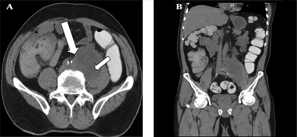
Gray scale ultrasound (US) of the abdomen revealed a large well-defined multilocular lesion with internal anechoic areas along the left psoas muscle. The US did not delineate the exact extent of the lesion. On Doppler study, the lesion showed no evidence of internal flow. The patient was further evaluated using cross-sectional imaging. A baseline nonenhanced computed tomography (NECT) scan followed by a contrast enhanced computed tomography (CECT) scan was acquired using a 16 slice scanner (General Electric Brightspeed, Milwaukee, Wisconsin, USA) by injecting 120 mL of intravenous contrast iohexol (Omnipaque, General Electric Healthcare) through an eighteen gauge needle in the right antecubital vein at the rate of 3 mL/sec. The scanning parameters used were a tube current of 105 milliampere seconds (mAs), and a tube voltage of 130 peak kilovoltage (kVp). Acquisition was performed at the slice thickness of 5 mm. Image acquisition was done during the venous phase (70 - 90 sec). The 5-mm-thick axial images were reformatted into thinner sections in three orthogonal planes (0.6 mm thick). On NECT, a large well-defined smoothly marginated multi loculated lesion of fluid attenuation (average HU 15 - 20) was noted in the left retroperitoneum involving the left psoas muscle (
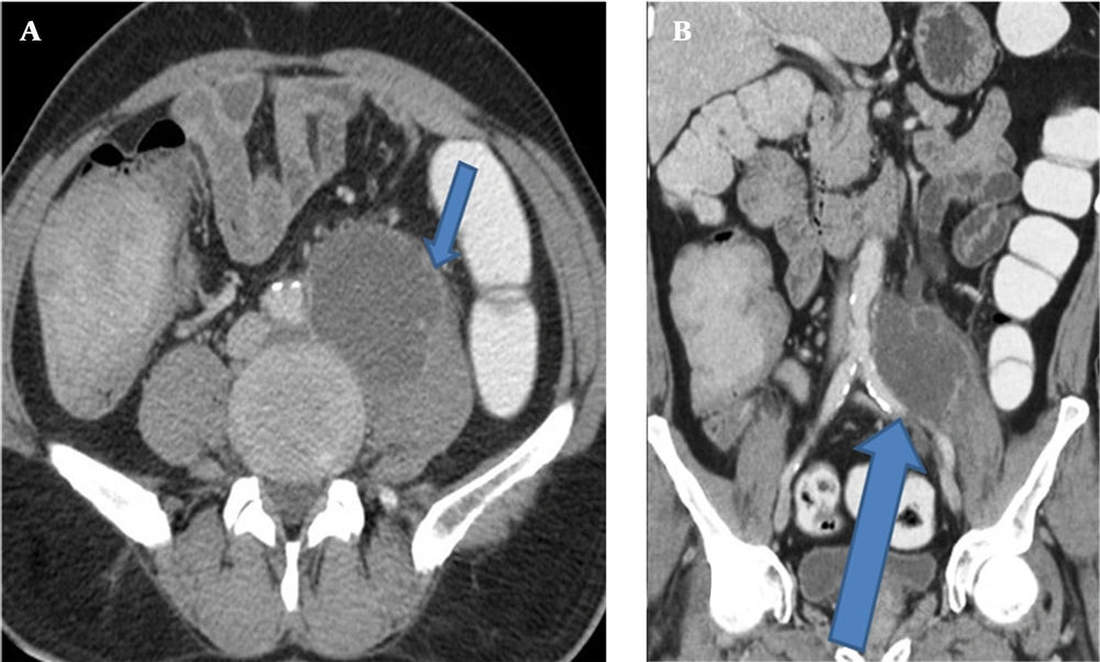
Figure 1). The lesion was 5.8 × 6.5 × 8.4 cm in size (AP × ML × CC). On CECT images, the lesion showed a peripherally thin, continuous rim enhancement in continuity with the left psoas muscle margin. The lesion caused inferomedial displacement of the left common iliac vessels; however, no evidence of invasion of adjacent retroperitoneal structures (including vessels) was noted (
Figure 2). No evidence of vertebral erosion was noted.
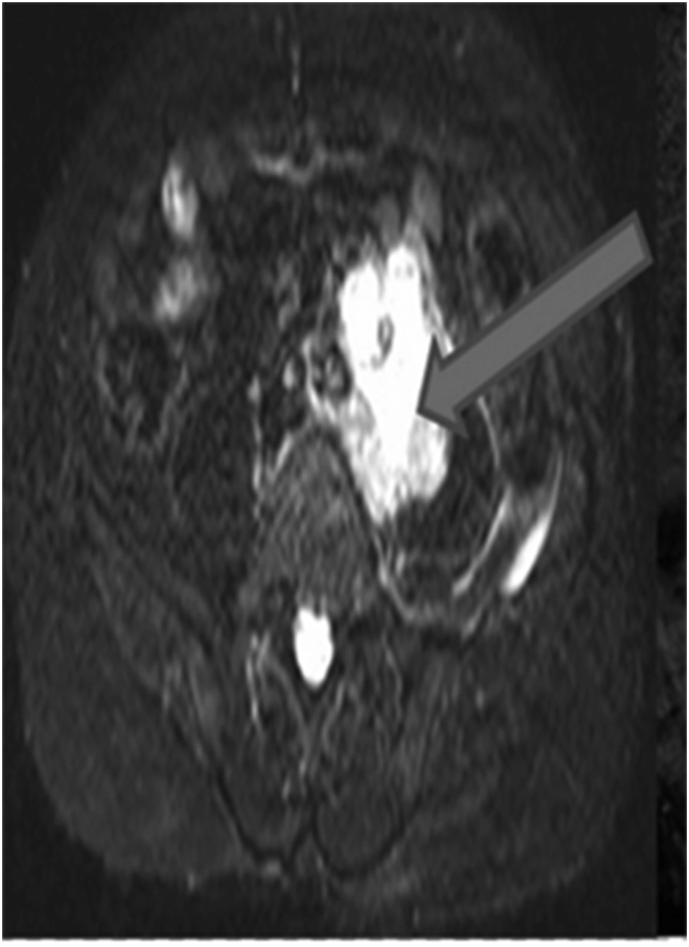
Magnetic resonance imaging (MRI) was performed with a 1.5 Tesla 8 channel scanner (Siemens Magnetom Essenza, Erlangen, Germany). A dedicated phased-array body coil was used. Non-enhanced MRI demonstrated a well-defined lesion with fluid signal intensity on short tau inversion recovery (STIR) images [TR 3750 ms, TE 30 ms, slice thickness 3 - 5 mm] (
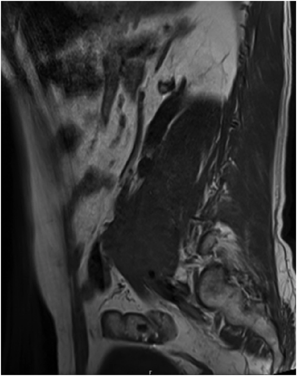
Figure 3) and hypointense signal lesion on T1 weighted images [TR 119 ms, TE 4.8 ms, slice thickness 5 mm] in the psoas muscle (
Figure 4). No evidence of spinal canal extension was noted. No evidence of focal vertebral marrow lesions was noted.
Based on combined clinical history, laboratory investigations and imaging findings, a benign infective lesion such as psoas cold abscess was considered as the diagnosis.
Ultrasound guided needle aspiration of the lesion revealed blood stained turbid fluid with debris. Laparoscopic debridement of the lesion was done and the specimen was sent for histopathological examination.
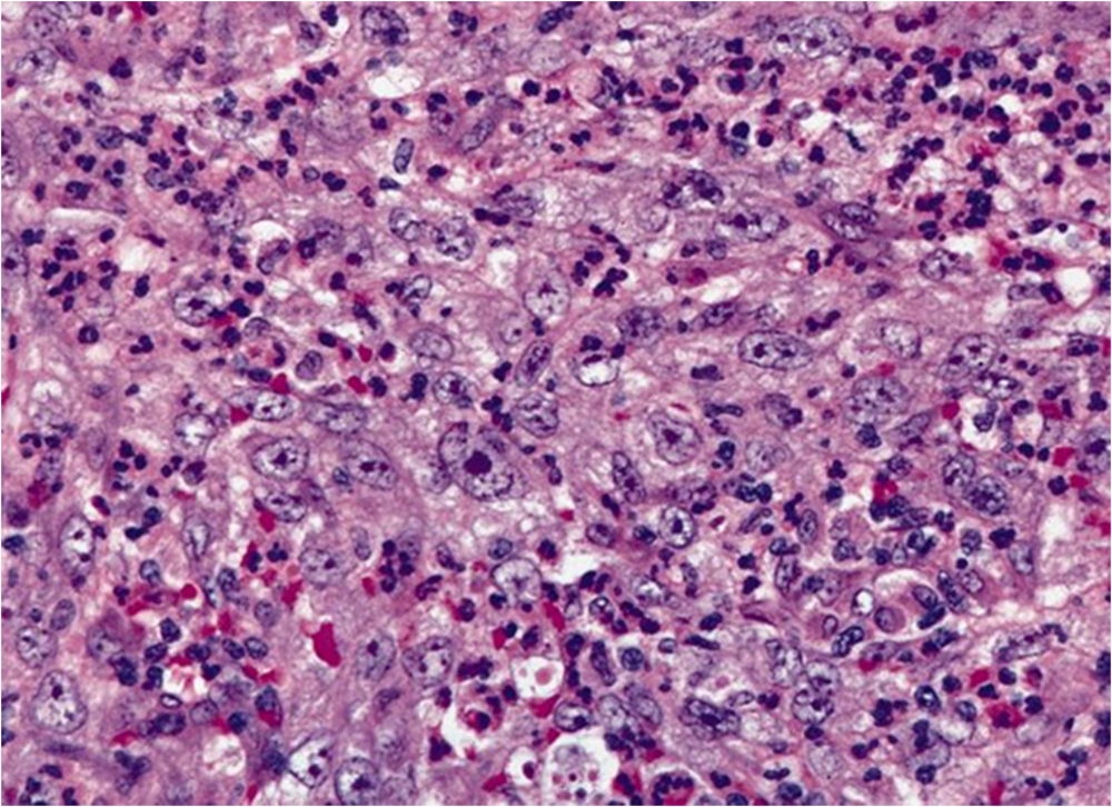
Histopathological examination showed spindle cells and fibroblast-like cells arranged in short fascicles and loosely arranged whorls in storiform pattern (
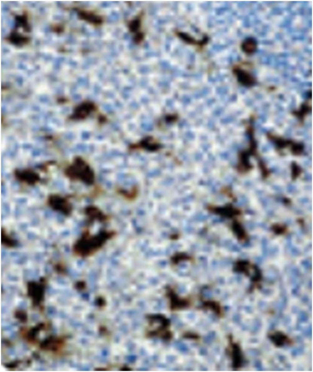
Figure 5). Histiocytes with an increased nucleus-cytoplasm ratio and prominent nucleoli were noted. On immunohistochemical examination, the tumor cells showed positivity for CD68, lysozyme and negative staining for S-100, and smooth muscle antigen (SMA) (
Figure 6). The patient was treated with adjuvant chemo-radiotherapy and is currently doing well. No cross sectional imaging follow-up has been performed for the patient yet.