


1. Introduction
2. Case Presentation
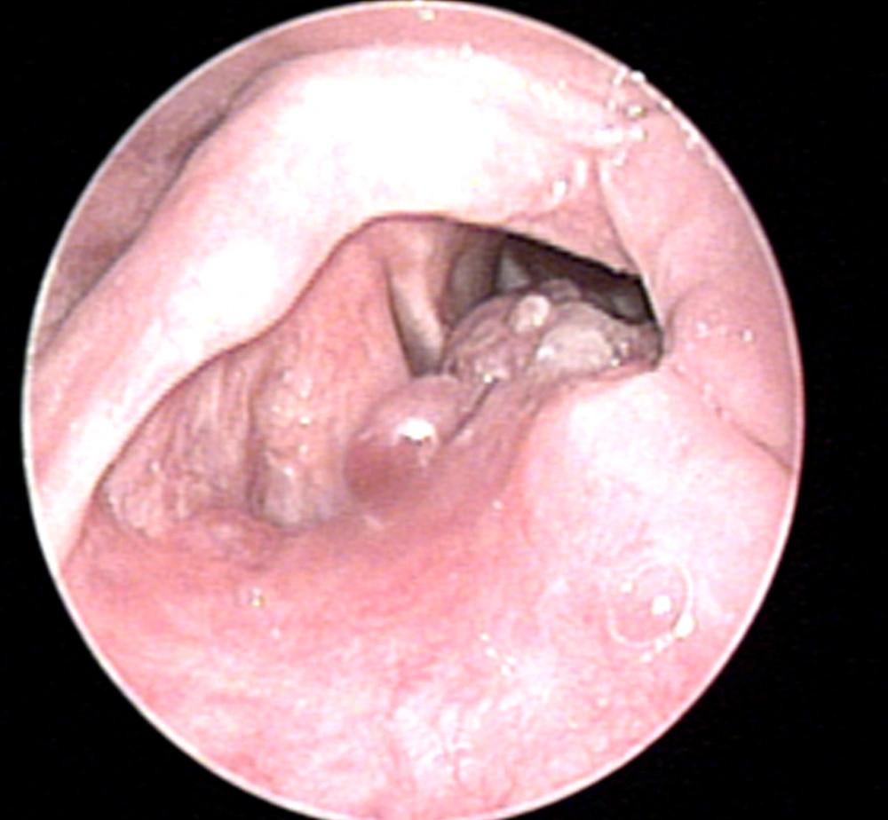
. Flexible fiberoptic laryngoscopy. A submucosal mass on the left aryepiglottic fold obstructing about 30% of the airway
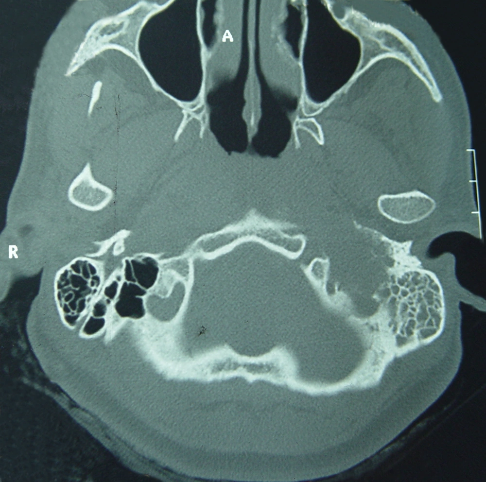
Temporal bone CT-scan without contrast (bone window, axial view). A lytic-expansile mass isodense with the brain is seen in the left jugular foramen that has eroded the petrous apex and the osseous septum between the carotid canal and this foramen. Opacity of the left tympanic cavity and mastoid air cells is also seen.
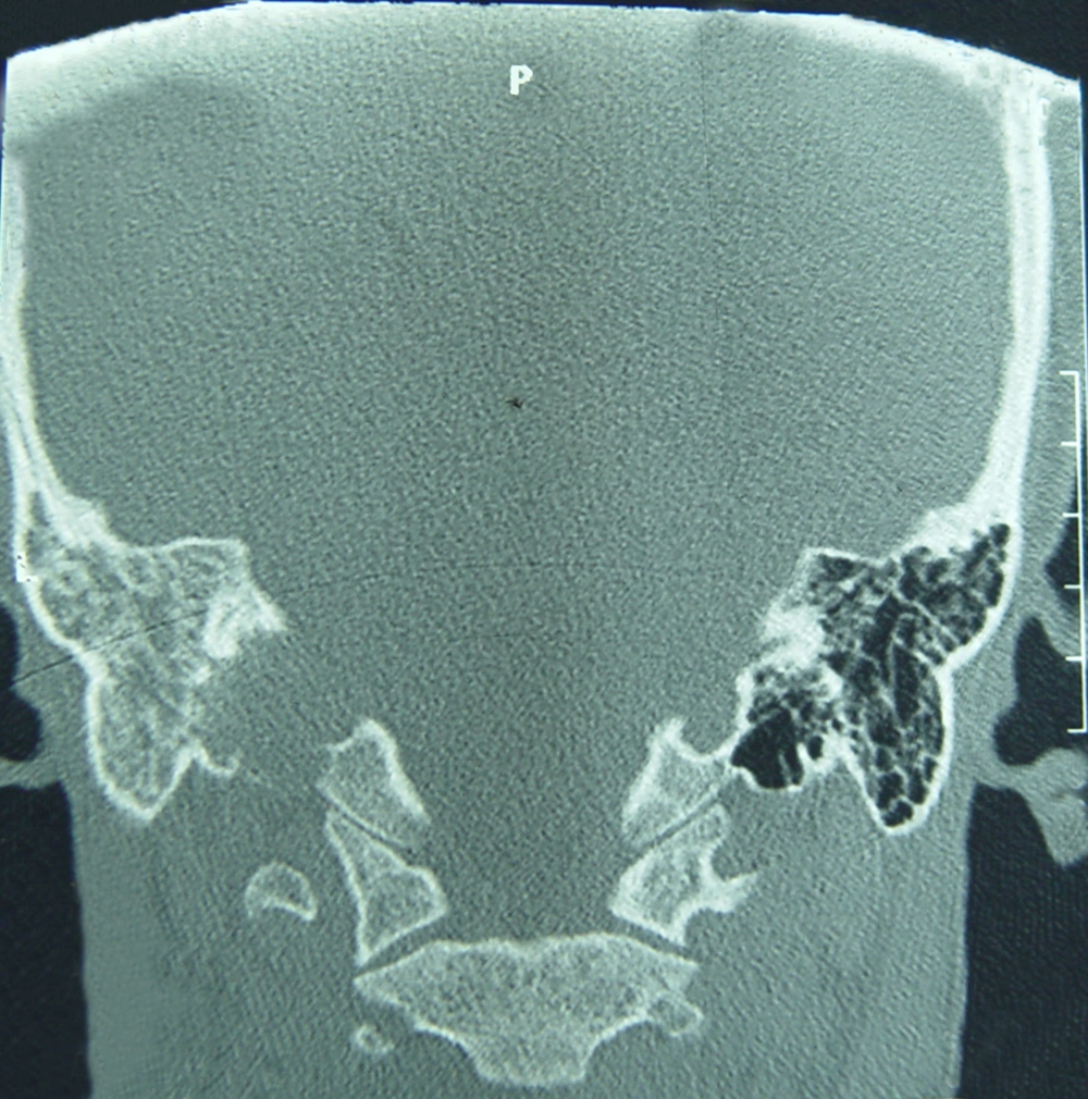
Temporal bone CT-scan without contrast (bone window, coronal view). A lytic-expansile mass isodense with the brain is seen in the left jugular foramen that has eroded the floor of the hypotympanic cavity and osseous labyrinthine nearby and shows extension within the tympanic cavity. Erosion of the inferior cortex of the petrous apex is noted. Opacity of the tympanic cavity and mastoid air cells is also seen.
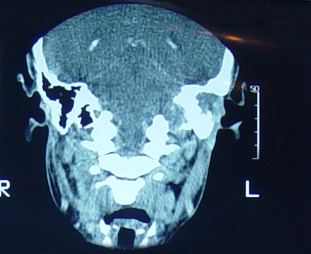
Contrast-enhanced temporal bone CT-scan (coronal view). A lytic expansile relatively enhanced mass in the left jugular fossa without any significant intracranial extension.
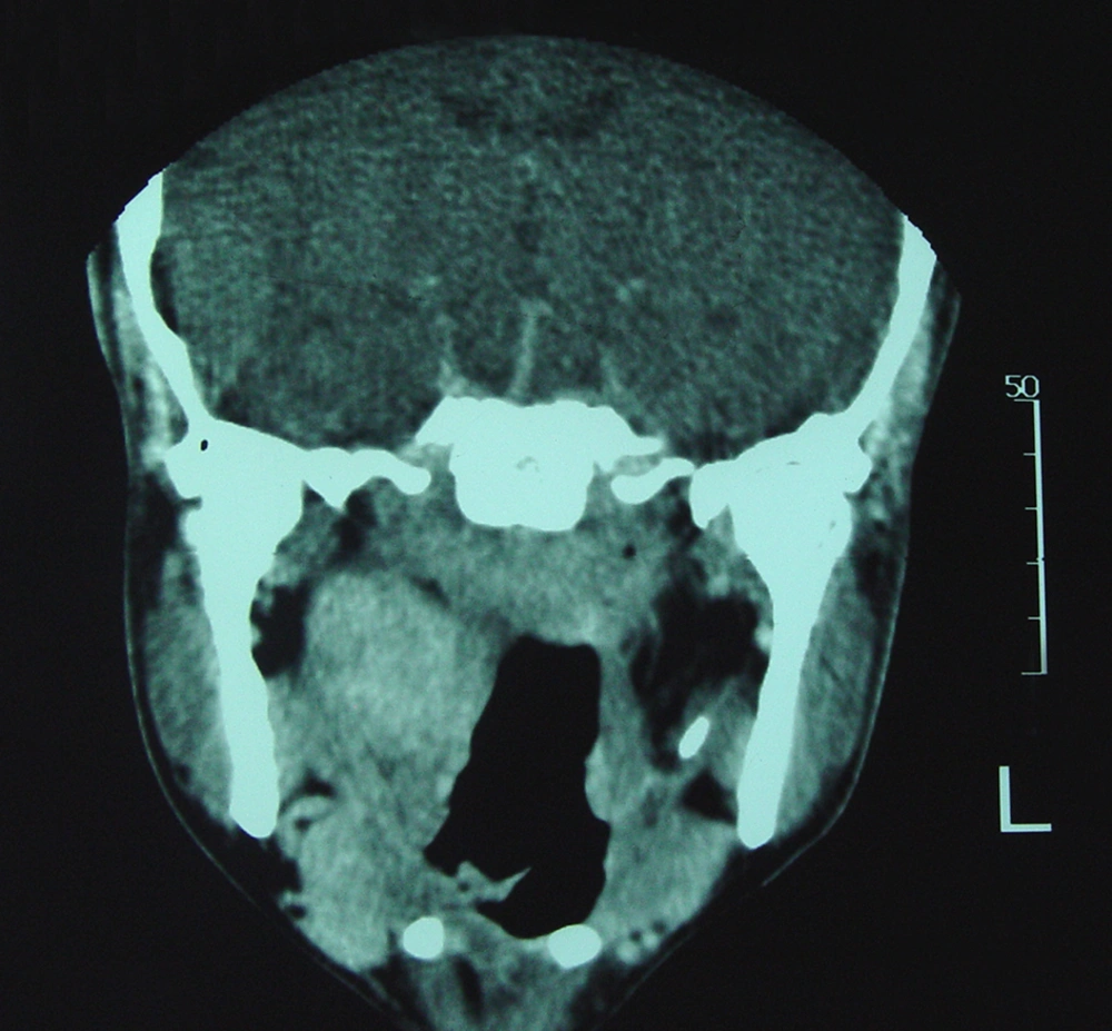
Contrast-enhanced PNS CT-scan (coronal view). Well-defined homogeneously enhanced soft tissue mass (35 × 25 mm) in the right parapharyngeal space is seen that has not shown any clear boundary with the right pharyngeal mucosal space of the nasopharynx.
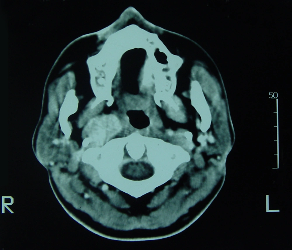
Contrast-enhanced neck CT-scan (axial view). A soft tissue mass (20 × 15 mm) with homogeneous marked enhancement and well-defined borders in the right vascular space.
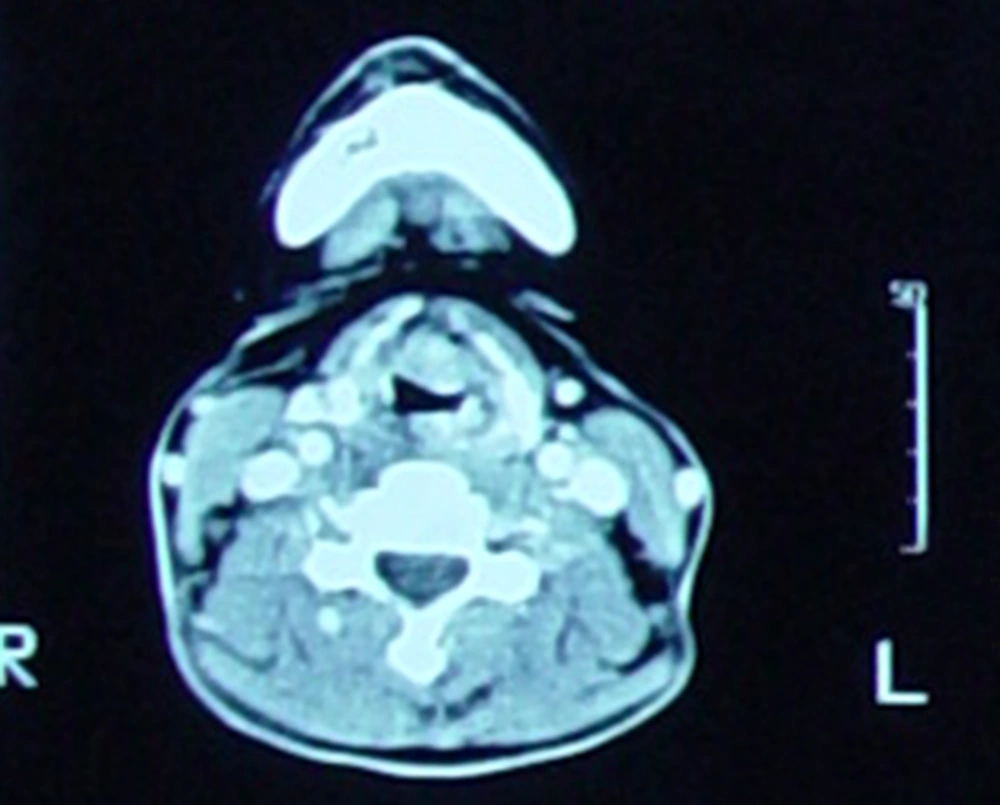
A soft tissue mass (17 × 10 mm) with homogeneous marked enhancement and well-defined borders on the midline and left side of the preepiglottic space is seen that has a mass effect on the epiglottis.
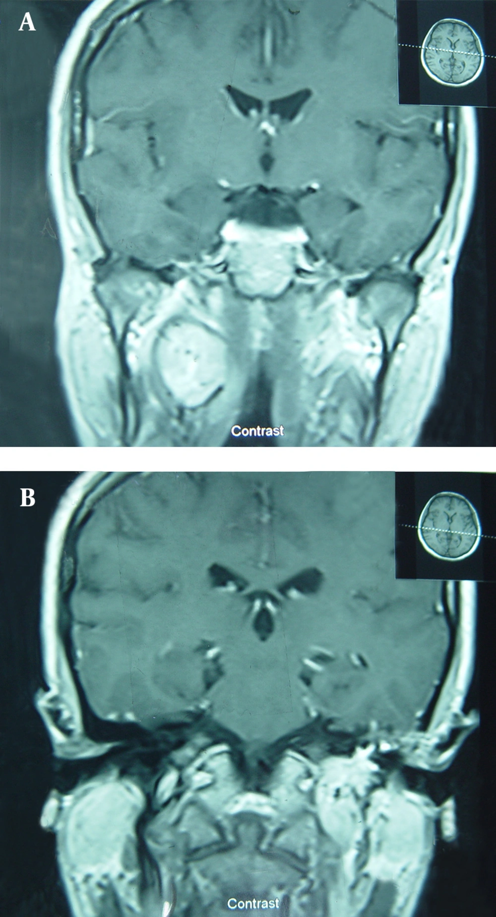
Contrast-enhanced brain MRI (coronal view). A) A well-defined relatively homogeneous mass (35 × 25 mm) with moderate enhancement is seen in the right parapharyngeal space. B) A markedly enhancing mass (30 × 25 mm) with lobulated borders is seen in the left jugular foramen.
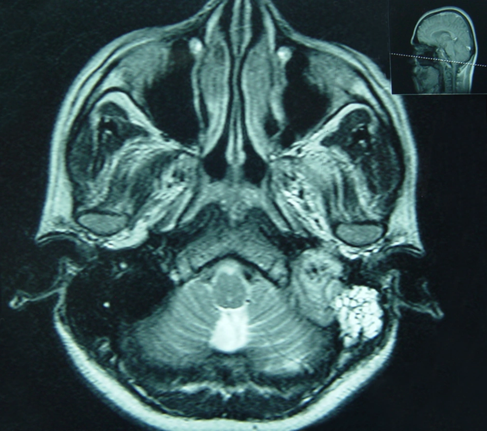
T2-weighted brain MRI (axial view). A relatively heterogeneous signal mass with well-defined lobulated borders (30 × 25 mm) in the left jugular foramen is seen that shows isointense signal to the brain cortex. Abnormal signal isointense to CSF in the left mastoid air cells and the tympanic cavity is also seen.
