


1. Background
2. Objectives
3. Patients and Methods
| Patients’ Characteristics | Height, cm | Weight, kg | Age, y | BMI, kg/m2 |
|---|---|---|---|---|
| Mean | 166.7 | 64.1 | 55.5 | 22.8 |
| Standard deviation | 7.4 | 11.4 | 11.8 | 2.8 |
| Minimum value | 151.5 | 43.6 | 26 | 17.4 |
| Maximum value | 180.2 | 88.7 | 74 | 30.3 |
aAbbreviation: BMI = body mass index.
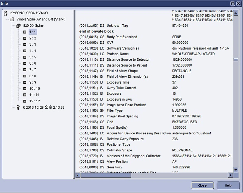
DICOM tag information by Centricity workstation. Information from each image DICOM tag indicated the image area dose product by tag number 0018.115E.
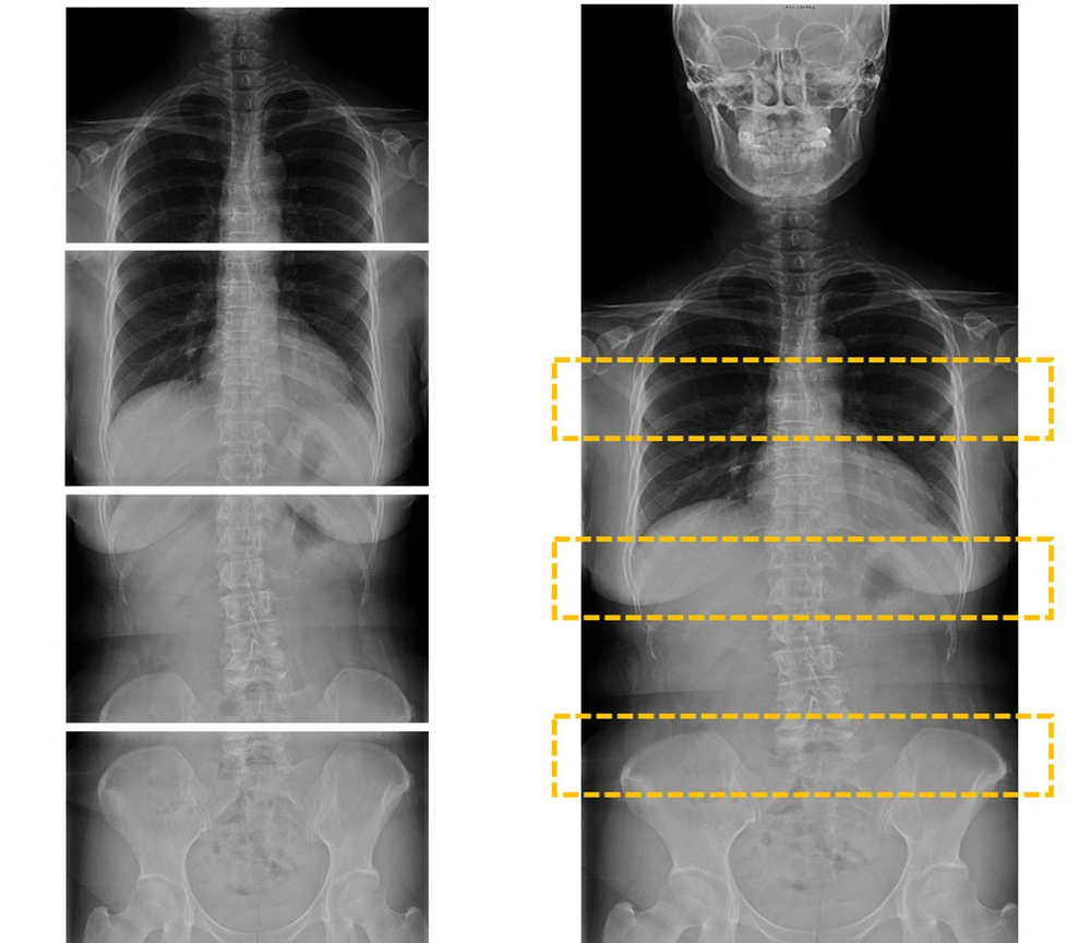
Automated acquisition and automated pasting of DR images
4. Results
| Gender | AP DAP (dGy cm2) | LAT DAP (dGy cm2) | AP ED (mSv) | LAT ED (mSv) |
|---|---|---|---|---|
| Female (n = 22) | ||||
| Mean (SD) | 2559.00 (711.81) | 9507.09 (2794.20) | 0.43 (0.10) | 0.53 (0.14) |
| Range (Min - Max) | 2699.75 (1542.64 - 4242.39) | 10865.84 (5197.34 - 16063.18) | 0.38 (0.28 - 0.66) | 0.5 (0.36 - 0.86) |
| Var | 506683.21 | 7807564.61 | 0.01 | 0.02 |
| Male (n = 28) | ||||
| Mean (SD) | 5591.95 (2200.78) | 12975.88 (2493.97) | 0.78 (0.27) | 0.78 (0.21) |
| Range (Min - Max) | 10370.93 (2589.83 - 12960.76) | 11420.1 (7740.83 - 19160.93) | 1.23 (0.38 - 1.61) | 0.8 (0.46 - 1.26) |
| Var | 4843460.94 | 6219896.31 | 0.07 | 0.04 |
| Total (n = 50) | ||||
| Mean (SD) | 4257.45 (2280.09) | 11449.61 (3130.28) | 0.62 (0.28) | 0.67 (0.22) |
| Range (Min - Max) | 11418.12 (1542.64 - 12960.76) | 13963.59 (5197.34 - 19160.93) | 1.33 (0.28 - 1.61) | 0.9 (0.36 - 1.26) |
| Var | 5198833.27 | 9798695.17 | 0.07 | 0.05 |
aAbbreviations: AP, antero-posterior; DAP, dose area product; ED, effective dose; LAT, lateral; Max, maximum; Min, minimum; SD, standard deviation; Var, variance.
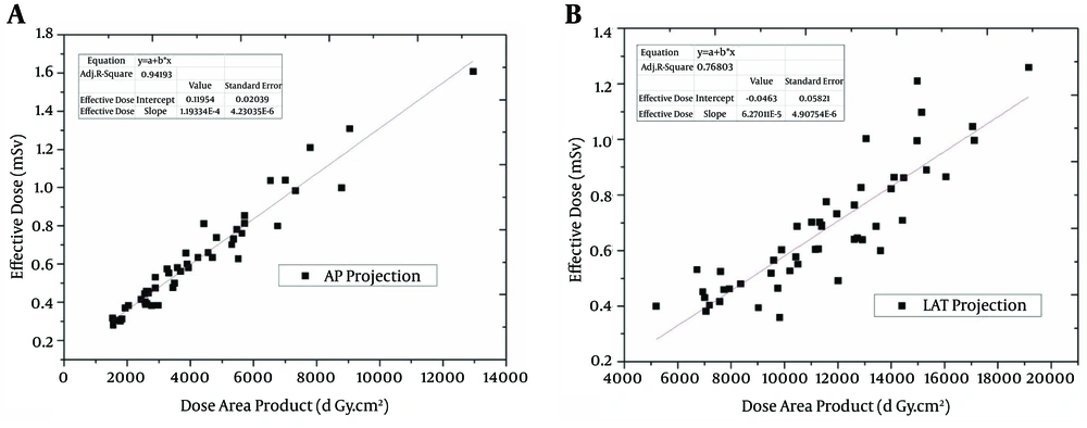
A, Antero-posterior and B, lateral projection proportional correlations.

| Group | LAT DAP, dGy cm2 | LAT ED, mSv |
|---|---|---|
| AEC (n = 30) | ||
| Mean (SD) | 10439.99 (3412.91) | 0.60 (0.23) |
| Range (Min - Max) | 13963.59 (5197.34 - 19160.93) | 0.9 (0.36 - 1.26) |
| Var | 11647999.32 | 0.05 |
| Manual (n = 20) | ||
| Mean (SD) | 12964.05 (1862.27) | 0.77 (0.17) |
| Range (Min - Max) | 6848.27 (10212.44 - 17060.71) | 0.61 (0.49 - 1.1) |
| Var | 3468072.37 | 0.03 |
| Total (n = 50) | ||
| Mean (SD) | 11449.61 (3130.28) | 0.67 (0.22) |
| Range (Min - Max) | 13963.59 (5197.34 - 19160.93) | 0.9 (0.36 - 1.26) |
| Var | 9798695.17 | 0.05 |
aAbbreviations: AEC, auto exposure control; DAP, dose area product; ED, effective dose; LAT, lateral; Max, maximum; Min, minimum; SD, standard deviation; Var, variance.
| Group | Correlation of DAP with ED | Significance Level |
|---|---|---|
| Total | ||
| AP projection | 0.971 | 0.01 |
| LAT Projection | 0.879 | 0.01 |
| Female | ||
| AP projection | 0.951 | 0.01 |
| LAT Projection | 0.841 | 0.01 |
| Male | ||
| AP projection | 0.954 | 0.01 |
| LAT Projection | 0.852 | 0.01 |
| Obese BMI | ||
| AP projection | 0.953 | 0.01 |
| LAT Projection | 0.764 | 0.01 |
| Normal BMI | ||
| AP projection | 0.957 | 0.01 |
| LAT Projection | 0.924 | 0.01 |
| AEC Mode | ||
| AP projection | 0.96 | 0.01 |
| LAT Projection | 0.887 | 0.01 |
| Manual Mode | ||
| AP projection | 0.947 | 0.01 |
| LAT Projection | 0.79 | 0.01 |
aAbbreviations: AEC, auto exposure control; AP, anterior-Posterior; BMI, body mass index; DAP, dose area product; ED, effective dose , BMI, body mass index, AEC, auto exposure contro.
5. Discussion
| Study | Number of Patients | Age, y | Effective Dose (Min-Max) | Method |
|---|---|---|---|---|
| Hallen et al. (13) | 7 | 9 - 25 | PA and lateral: 0.12 mSv | Single exposure |
| Chamberlain et al. (3) | 61 | 9 - 55 | AP: (0.14 - 0.18) mSv | Single exposure |
| Lee et al. (2) | 54 | 4 - 17 | PA: (0.081 - 0.123) mSv; lateral: (0.124 - 0.207) mSv | Single exposure |
| Geijer et al. (14) | Alderson phantom | PA: 0.16 mSv | Digital fluoro-graphic image reconstruction | |
| Mogaadi et al. (11) | 13 | 16 - 22 | AP: 0.798 (0.545 - 1.305) mSv; lateral: 0.597 (0.344 - 1.187) mSv | Single exposure |
| Hansen et al. (12) | 22 | 13 - 18 | PA female: 0.97 mSv; PA male: 0.65 mSv; lateral female: 1.2 mSv; lateral male: 1.2 mSv | Single exposure |
| This Study | 50 | 26 - 74 | AP female: 0.43(0.28 - 0.66) mSv; AP male: 0.78 (0.46 - 1.26) mSv; lateral female: 0.53 (0.36 - 0.86) mSv; lateral male: 0.78 (0.46 - 1.26) mSv; AEC used lateral: 0.6 (0.36 - 1.26) mSv; AEC not used lateral: 0.77(0.49 - 1.1) mSv | Auto Image Paste |
