


1. Background
2. Objectives
3. Subjects and Methods
3.1. Study Subjects
3.2. MR Imaging Protocol
3.3. VR and MIP Post-Processing
| Direction No. | X a | Y b | Z c |
|---|---|---|---|
| 0 | 0.000000 | 0.000000 | 0.000000 |
| 1 d | 1.000000 | 0.000000 | 0.000000 |
| 2 | 0.000000 | 1.000000 | 0.000000 |
| 3 | -0.031984 | 0.799591 | 0.599693 |
| 4 | 0.856706 | 0.493831 | -0.148949 |
| 5 | 0.834429 | 0.309159 | 0.456234 |
| 6 | 0.834429 | -0.309159 | 0.456234 |
| 7 | 0.856706 | -0.493831 | -0.148949 |
| 8 | 0.822228 | 0.000000 | -0.569158 |
| 9 | 0.550834 | 0.425872 | -0.717784 |
| 10 | 0.468173 | 0.834308 | 0.281963 |
| 11 | 0.515933 | 0.808894 | 0.281963 |
| 12 | 0.391890 | 0.515855 | 0.761785 |
| 13 | 0.478151 | 0.000000 | 0.878278 |
| 14 | 0.391890 | -0.515855 | 0.761785 |
| 15 | 0.515933 | -0.808894 | 0.281963 |
| 16 | 0.468173 | -0.834308 | -0.291108 |
| 17 | 0.550834 | -0.425872 | -0.717784 |
| 18 | 0.111012 | -0.264029 | -0.958105 |
| 19 | 0.111012 | 0.264029 | -0.958105 |
| 20 | 0.031984 | 0.799591 | -0.599693 |
a X-axis represents the anterior-posterior direction.
b Y-axis represents the left-right direction.
c Z-axis represents the superior-inferior direction.
d Direction 1 with the coordinates (1, 0, 0) represents the anterior-posterior direction.
3.4. Fiber Tractography
3.5. Imaging Quality Evaluation
3.6. Statistical Analysis
4. Results
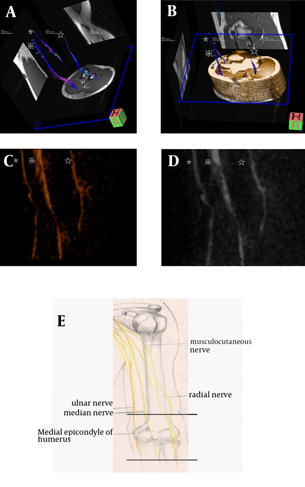
Fiber tractography and VR/MIP reconstruction of nerves from DT. The median nerve (※) radial nerve (☆), and ulnar nerve (*) can be seen clearly. A, Fiber tractography. B, Fiber tractography of DTI with reference of T1-weighted images. C, VR reconstruction. D, MIP reconstruction. E, Reference picture of the three main nerves at the elbow region. The scanned section is located between the two parallel lines.
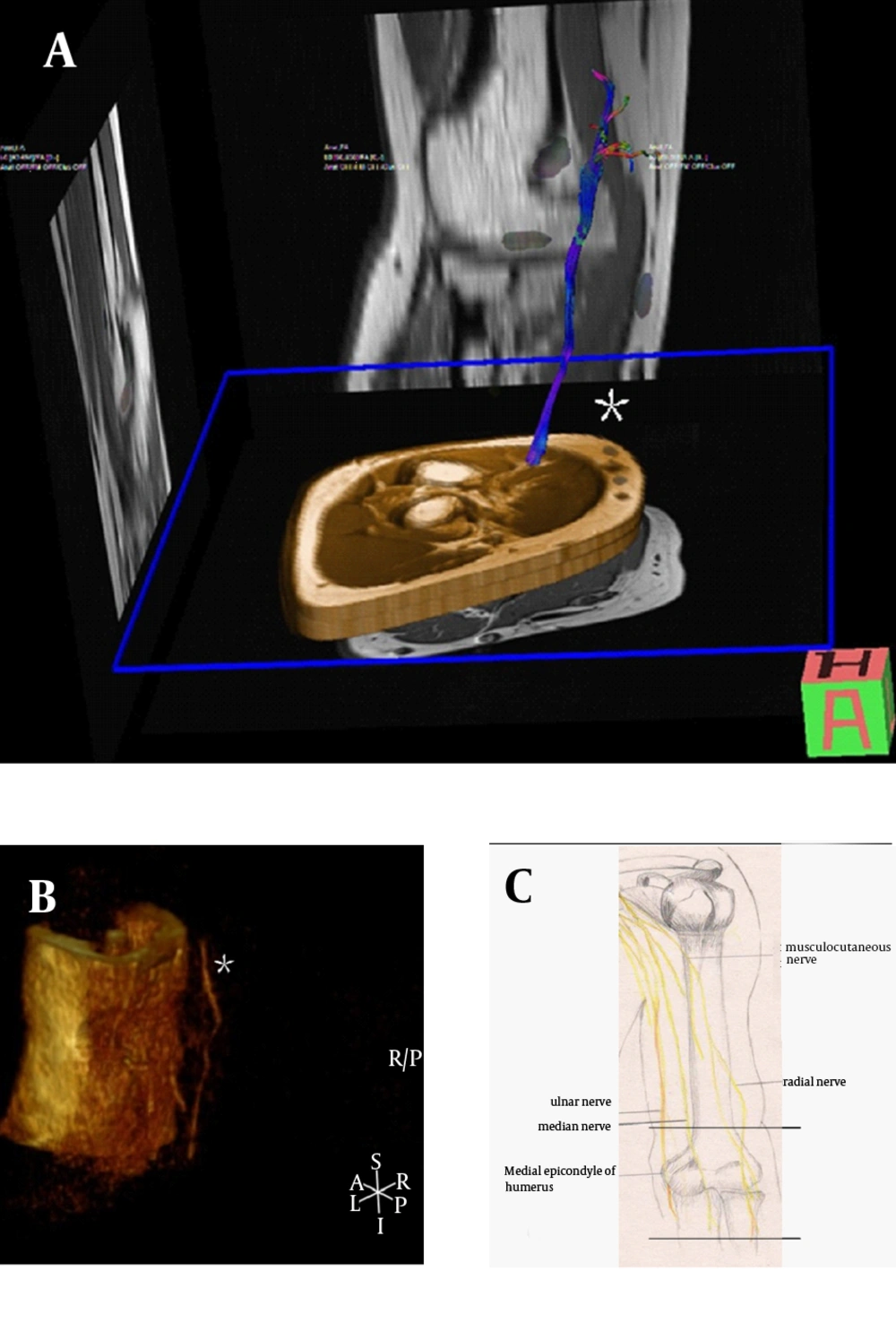
Fiber tractography and VR/MIP reconstruction of nerves from DTI. A, Fiber tractography. Only the ulnar nerve (*) can be seen clearly, whereas the median nerve and radial nerve cannot be tracked. B, VR reconstruction (without noise removal). Only the ulnar nerve (*) can be seen, which is located outside of the outline of the elbow. The median and radial nerve are covered by the outline of the elbow. C, Reference picture
| Median Nerve | VR | MIP | FT | P Value b | ICC | κ1 | κ2 |
|---|---|---|---|---|---|---|---|
| Ob1 | |||||||
| Mean. (SD) | 1.75 (0.532) | 1.79 (0.509) | 1.83 (0.482) | 0.674 | |||
| Median. (range) | 2 (0 - 2) | 2 (0 - 2) | 2 (0 - 2) | 0.368 | 0.838 | ||
| Ob2 | |||||||
| Mean. (SD) | 1.75 (0.532) | 1.79 (0.509) | 1.79 (0.509) | 0.83 | |||
| Median. (range) | 2 | 2 (0 - 2) | 2 (0 - 2) | 0.779 | 0.79 | ||
| Ob2 Sec. | |||||||
| Mean.(SD) | 1.71 (0.550) | 1.75 (0.532) | 1.79 (0.509) | ||||
| Median.(range) | 2 (0 - 2) | 2 (0 - 2) | 2 (0 - 2) | 0.223 | 0.901 | ||
| Ulnar Nerve | |||||||
| Ob1 | |||||||
| Mean. (SD) | 1.38 | 1.42 (0.584) | 1.46 (0.588) | 0.711 | |||
| Median. (range) | 1 | 1 (0 - 2) | 1.50 (0 - 2) | 0.368 | 0.877 | ||
| Ob2 | |||||||
| Mean. (SD) | 1.54 (0.509) | 1.54 (0.509) | 1.46 (0.588) | 0.733 | |||
| Median. (range) | 2 (1 - 2) | 2 (1 - 2) | 1.5 (0 - 2) | 0.513 | 0.709 | ||
| Ob2 Sec | |||||||
| Mean. (SD) | 1.38 (0.576) | 1.54 (0.509) | 1.46 (0.588) | ||||
| Median. (range) | 1 (0 - 2) | 2 (1 - 2) | 1.5 (0 - 2) | 0.135 | 0.736 | ||
| Radial Nerve | |||||||
| Ob1 | |||||||
| Mean. (SD) | 1.04 (0.690) | 1.04 (0.690) | 0.92 (0.654) | 0.909 | |||
| Median. (range) | 1 (0 - 2) | 1 (0 - 2) | 1 (0 - 2) | 0.105 | 0.88 | ||
| Ob2 | |||||||
| Mean. (SD) | 1.04 (0.690) | 1.17 (0.761) | 0.96 (0.690) | 0.845 | |||
| Median. (range) | 1 (0 - 2) | 1 (0 - 2) | 1 (0 - 2) | 0.066 | 0.812 | ||
| Ob2 Sec | |||||||
| Mean. (SD) | 1.08 (0.717) | 1.21 (0.721) | 1.00 (0.722) | ||||
| Median. (range) | 1 (0 - 2) | 1 (0 - 2) | 1 (0 - 2) | 0.066 | 0.815 |
a Abbreviations: FT, fiber tractography; ICC, intraclass correlation coefficient; κ1, interobserver κ value; κ2, intraobserver κ value; Ob1, observer 1; Ob2, observer 2; Ob2 Sec., observer 2 second time.
b P Values were calculated by Friedman test.


