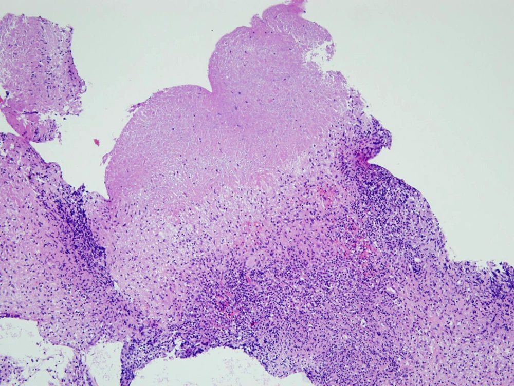
Upper airway TB is an uncommon clinical condition and is usually combined with pulmonary involvement (
1). Moreover, primary TB of the upper respiratory tract, without lung involvement, is rare (
1,
2). The nasopharynx is the least common site for TB involving the upper respiratory tract and comprises <1% of upper respiratory tract TB (
4). However, it is interesting to note that recent large scale studies, analyzing nasopharyngeal TB, have reported that primary nasopharyngeal TB is more common than secondary involvement (
7). Such discrepancy is probably derived from a limited ability to assess the nasopharynx by physical examination and a low clinical suspicion for nasopharyngeal TB.
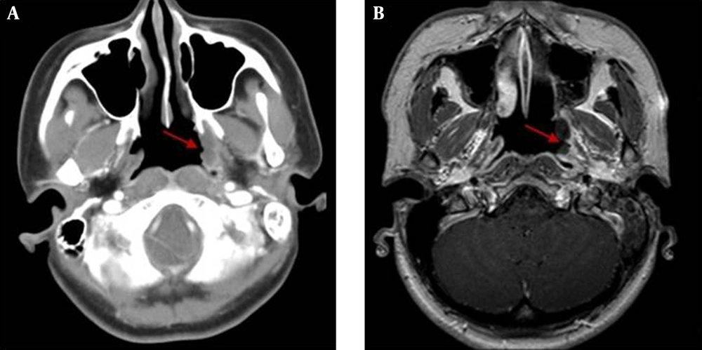
Radiographically, nasopharyngeal TB presents with two main patterns: 1) polypoid masses and 2) diffuse mucosal thickening (
3,
7,
8). Most cases of nasopharyngeal TB have been identified by a polypoid mass, which has been shown to indicate the proliferative phase (
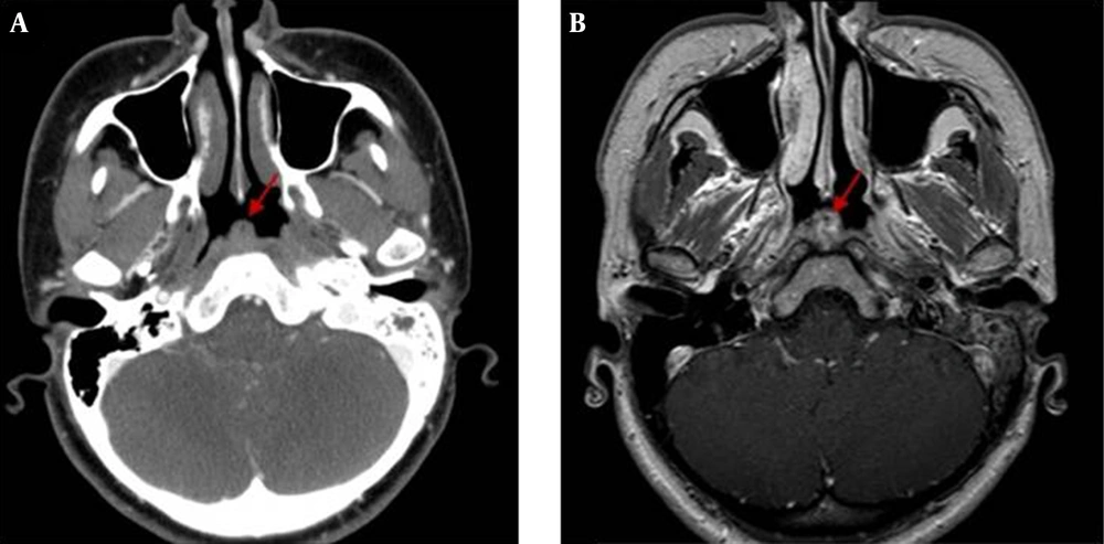
7). The CT and MR imaging show the characteristic findings of nasopharyngeal TB, including presence of necrosis and striped pattern in nasopharyngeal lesions, a lack of invasion of regional structures and peripheral enhancing cervical lymphadenopathy (
7). The nasopharyngeal mass or diffuse mucosal thickening may reveal intermediate signal intensity on T1 weighted and T2 weighted images, with moderate contrast enhancement on MR images (
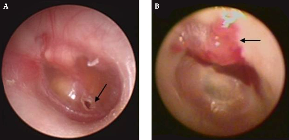
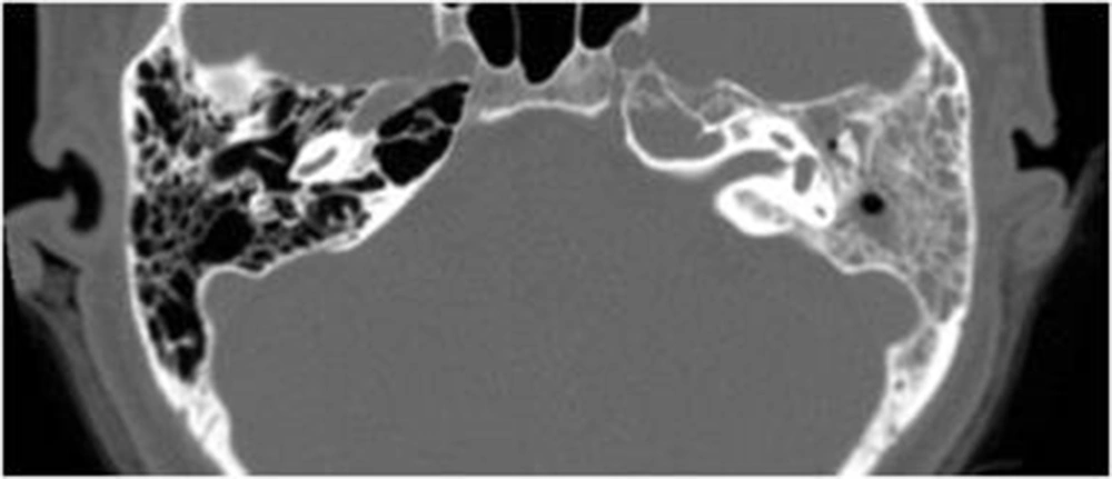
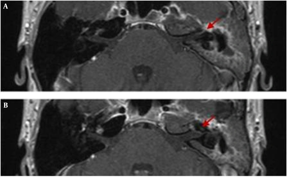
8). In the present case, there were also soft tissue densities, in the entire middle ear and mastoid antrum. It has been reported that radiologic characteristics of TOM are soft tissue in the entire middle ear cavity, preservation of mastoid air cells, without any sclerotic change, mucosal thickening of the external auditory canal, with intact scutum (
9), which are consistent with our case.
Differential diagnosis can vary, according to radiologic patterns. Lymphoid hyperplasia, nasopharyngeal carcinoma, lymphoma and Castleman’s disease should be considered in the differential diagnosis for the polypoid mass. Previous studies have reported that an isolated polypoid mass, with central necrosis centered on nasopharyngeal roof, suggests high probability of nasopharyngeal TB (
8). For the second pattern of nasopharyngeal TB, diffuse nasopharyngeal wall thickening, various benign (Wegener’s granuloma, syphilis, fungal infection) and malignant lesions (early local stage of nasopharyngeal carcinoma, lymphoma, minor salivary gland tumor) should be considered for the differential diagnosis (
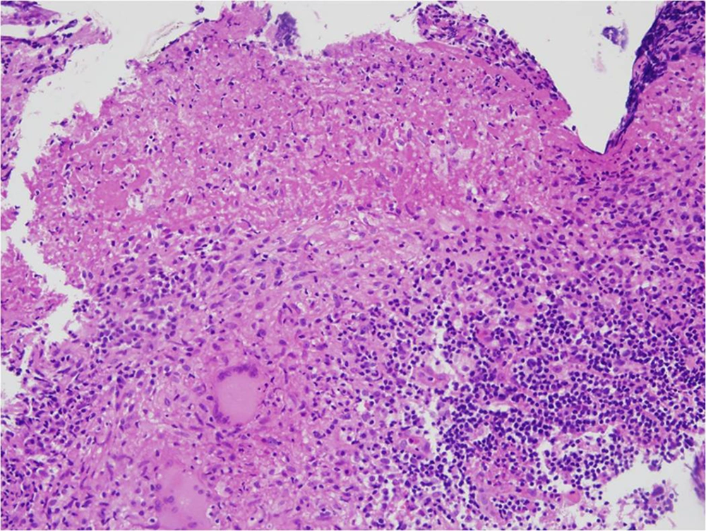
8). However, there are no definite imaging features to make an accurate diagnosis of nasopharyngeal TB and a biopsy is required to confirm the diagnosis and to differentiate it from malignancy and the other conditions described above.
It is worth noting that most nasopharyngeal TB involves the posterior roof of the nasopharynx, equivalent to the “adenoid” during childhood or early adolescence (
4). The findings lend support to a preceding report, stating that TB directly involves nasopharyngeal lymphoid tissue (
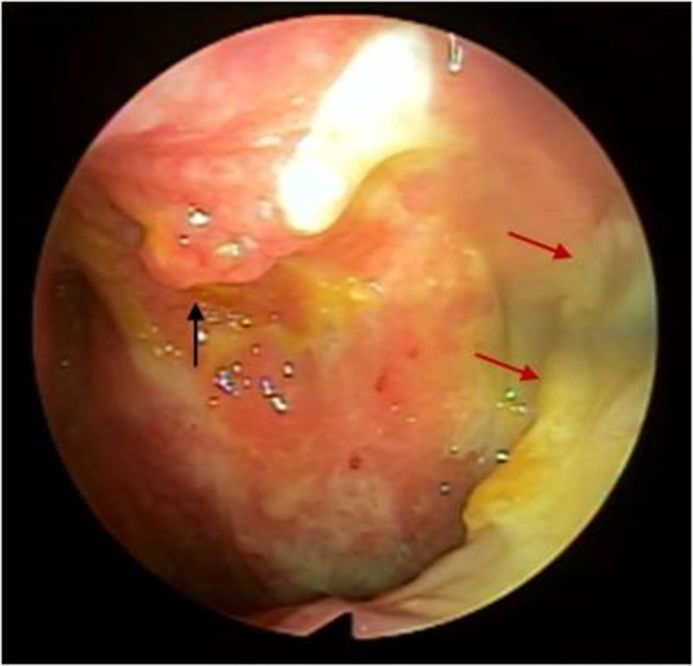
3). Regardless, our case showed the two main patterns of primary nasopharyngeal TB: the polypoid mass at the nasopharyngeal roof and, also, the diffuse soft tissue thickening of nasopharyngeal wall, consistent with previous reports and studies. However, the nasopharyngeal carcinoma is most often centered in the lateral pharyngeal recess (also called the Fossa of Rosenmuller). Sites of predilection and morphologic features can be potential diagnostic clues to differentiate nasopharyngeal TB from malignancy.
The Eustachian tube is lined with a mucous membrane, continuous with the pharynx and the mastoid air cells; therefore, the infections from nasopharynx can travel from the nasopharynx along the mucosal membrane of the Eustachian tube to the middle ear cavity. In addition, obstructing masses in the nasopharynx prevent air flow from passing through the Eustachian tube, creating a negative pressure in the middle ear cavity, followed by effusion (
10). Therefore, it is unclear which came first, the nasopharyngeal TB or TOM, in this case? However, given the physiologic anatomy of the nasopharynx and middle ear cavity, there is a high probability of secondary involvement of the nasopharyngeal TB into the middle ear and mastoid antrum, through pharyngeal opening of the Eustachian tube.
Previous studies of nasopharyngeal TB have not provided in-depth discussion of the possibility for extranasopharyngeal spread and cranial nerve involvement. There have been limited single case reports identifying extensive nasopharyngeal involvement of TB beyond the nasopharyngeal mucosa and submucosa, skull base or parapharyngeal space (
1). In this case report, besides in the nasopharyngeal roof, soft tissue masses and mucosal thickening were also noted in the pharyngeal opening of the Eustachian tube. Additional notable findings included soft tissue densities of the ipsilateral middle ear cavity, as well as along the facial nerve canal.
In conclusion, TB infection should be considered in the necrotic polypoid masses with regional mucosal lesions, centered on the posterior nasopharyngeal wall, especially in endemic areas. In addition, radiologists should keep in mind that pharyngeal orifice of the Eustachian tube can be a potential route for the spread of nasopharyngeal TB, from the nasopharynx to the middle ear cavity. Therefore, it is important to determine if obliteration of the nasopharyngeal orifice has occurred, in order to prevent acid bacilli from invading middle ear cavity.