


1. Introduction
2. Case Presentation
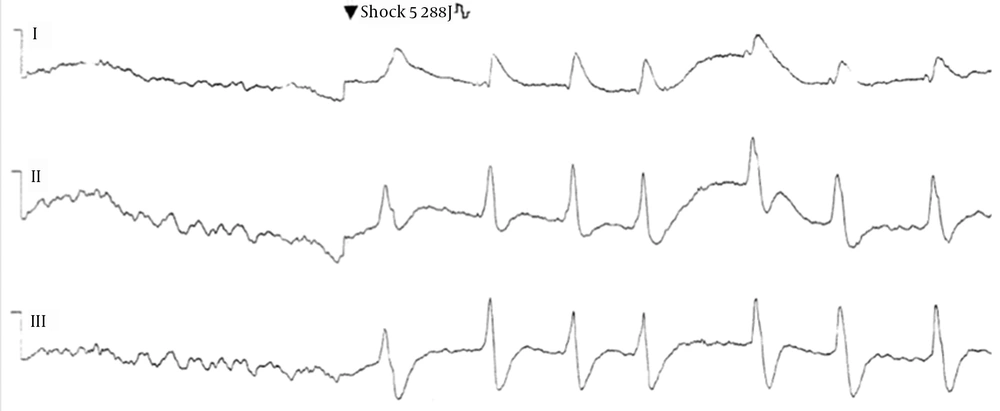
A 45-year-old man with a near drowning experience who underwent cardiopulmonary resuscitation. ECG shows conversion of ventricular fibrillation to sinus rhythm after the fifth 200-J transthoracic shock.
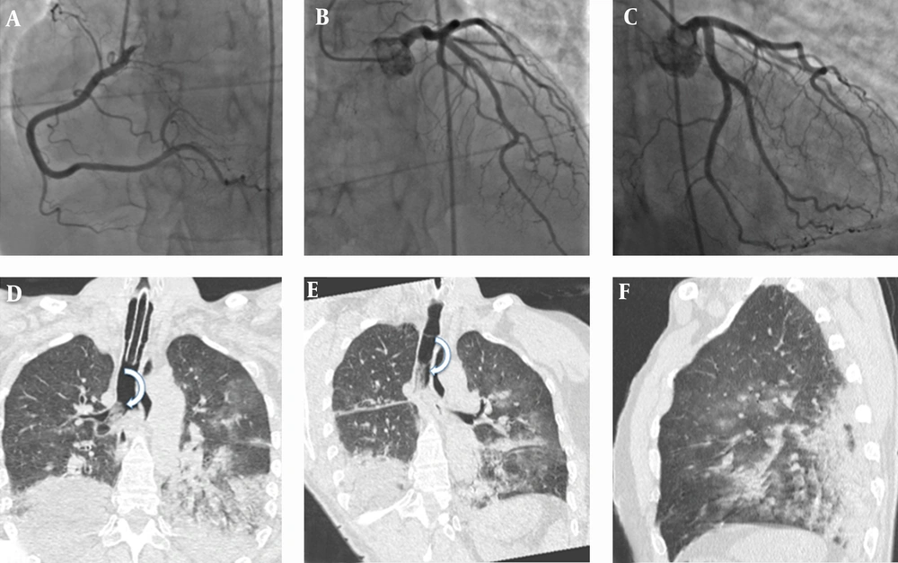
A-C, Coronary angiography performed at admission shows normal coronary flow without obvious angiographic obstruction. Coronal (D, E) and sagittal (F) thin-section chest computed tomography reveals bilateral and diffuse areas of ground-glass attenuation and airspace consolidation with air bronchogram seen mainly in lung bases resulted from lung damage and ventilation-perfusion mismatching caused by water inhalation and laryngospasm secondary to hypoxia. Curved arrows in D and E show the presence of secretions in the trachea and right main bronchus.
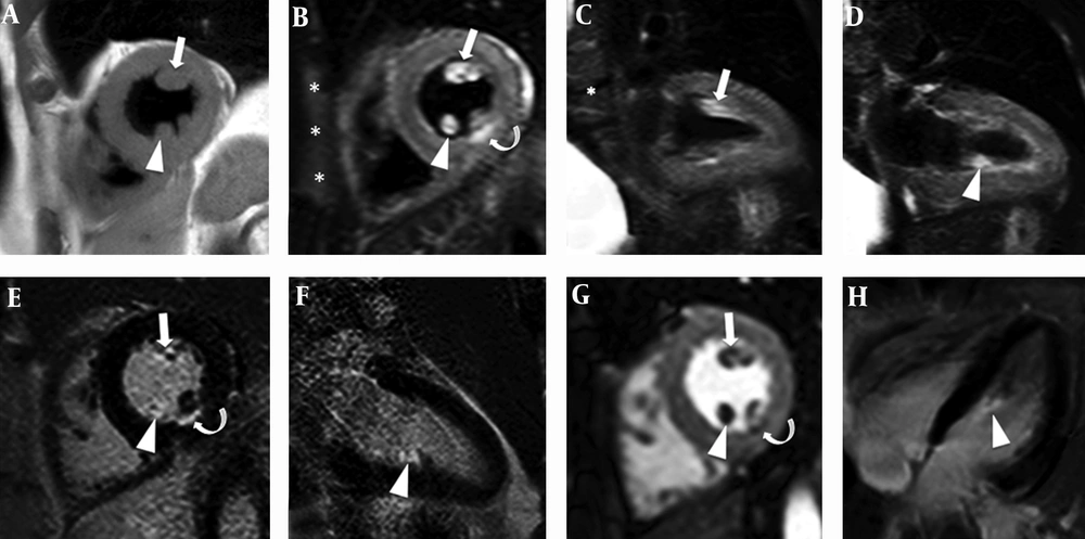
A, Short-axis T1 weighted (T1W) anatomical image shows the anterolateral (arrow) and posterior (arrowhead) muscles. In the short-axis (B) and vertical long axis (C, D) T2W-STIR images, the anterolateral (arrows in B and C) and posteromedial (arrowheads in B and D) papillary muscles have a hyperintense signal caused by increased water content (myocardial edema). On late contrast-enhanced (CE) imaging (E, F, and H), the anterolateral (arrow) and the medial portion of the posterior papillary muscles (arrowheads) are strongly enhanced. On T2W-STIR images (curved-arrow in B), a subtle hyperintensity, suggestive of edema, was detected in the middle inferior wall and CE-IR images confirmed a small (< 10 mm) correspondent subendocardial infarction (curved-arrow in E). The first-pass perfusion image (G) clearly shows a perfusion defect localized exactly at this cardiac structure. Short-axis T2W-STIR image (asterisks in B) also shows slight edema of the anterior chest wall probably due to chest compressions and transthoracic shocks.

