Fibroadenoma of the breast is a very common benign lesion, especially in younger women, and is considered more closely related to an aberrant development of the breast tissue than to a true neoplasm (
2). Although in general fibroadenoma is not considered to increase the risk of cancer, it is often associated to other pathologies such as sclerosing adenosis, duct ectasia, epithelial apocrine metaplasia, fibrocystic disease, and papillomatosis (
1). However, in women over 40 years of age with fibroadenoma, the concomitant occurrence of invasive cancers has been reported in up to 5% of cases (
2). So, the case presented here is not particularly rare, but two facts made the imaging diagnosis of this case particularly challenging: the type of tumor and its location. First, the tumor type, triple negative invasive ductal carcinoma of no special type, has been described as a tumor that often does not exhibit major suspicious findings of malignancy by mammography or US such as an irregular shape, ill-defined margins, or microcalcifications (
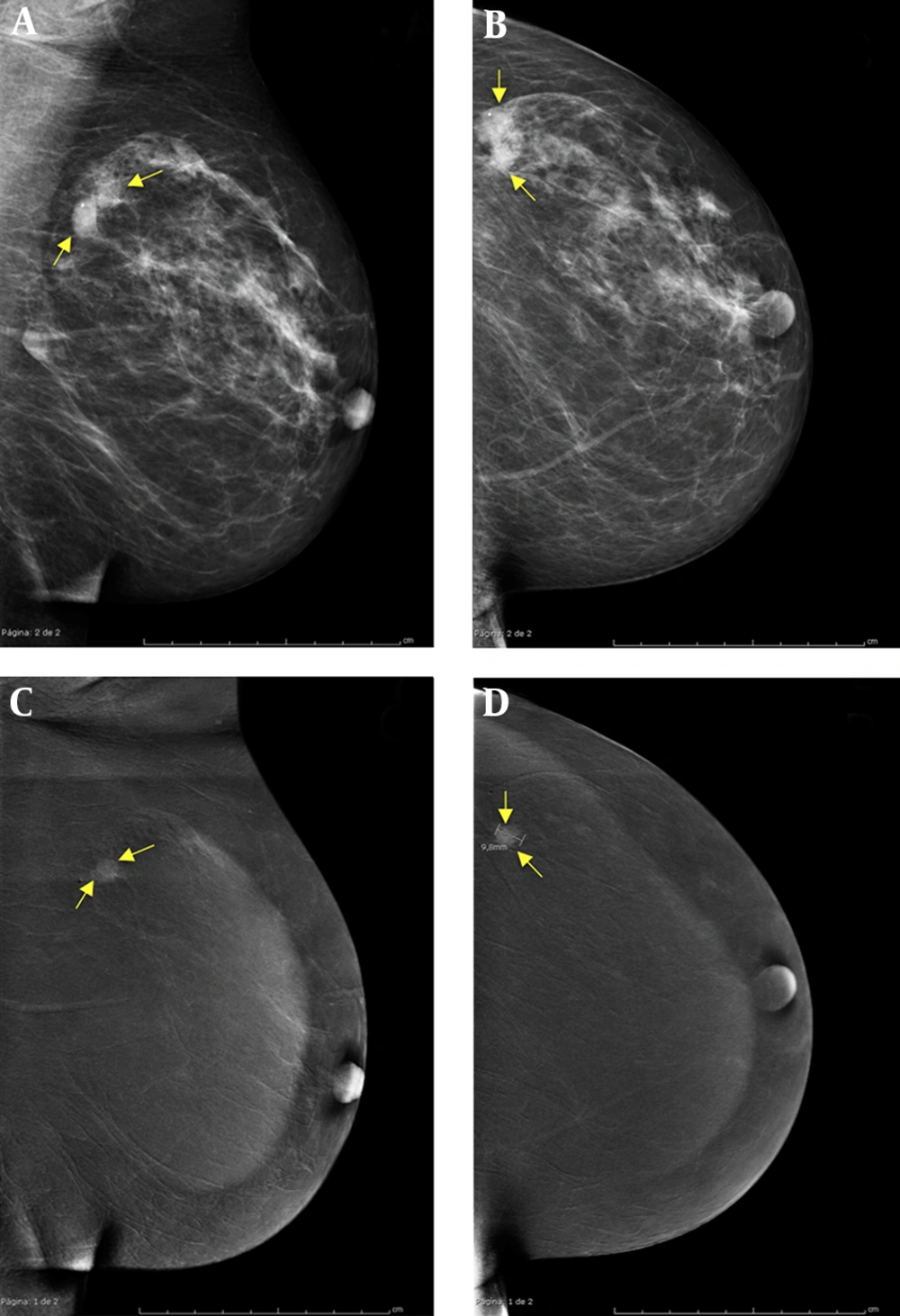
9). In this case, in the mammogram, the tumor gave an appearance indistinguishable from a fibroadenoma. Secondly, its location was adjacent to a true fibroadenoma, and the two masses appeared partially superimposed on the mammographic image, giving the appearance of either an irregularly shaped single fibroadenoma or of two adjacent rounded and smaller fibroadenomas.
It has been reported that a small percentage of fibroadenomas in older women may harbor malignant tumors inside (0.02% to 0.125%) (
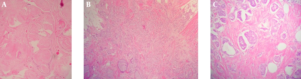
3). However, the pathology of the surgical specimen revealed that in this case, these two lesions were completely independent, although located very close to each other. For these two reasons, this case could easily have fallen within the 4- 19% of malignant breast lesions that are missed by mammography (
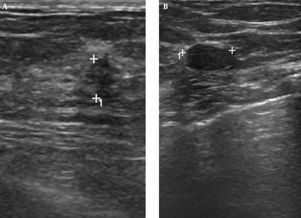
10). Moreover, in this case, although BUS gave a suspicion of malignancy in a part of the lesion, the guided biopsy resulted in a histopathological diagnosis of fibroadenoma. Probably due to the proximity of both lesions, the biopsy procedure was imprecise and incorrectly took a sample of the benign lesion instead of the carcinoma. Mainly due to the age of the patient, additional imaging studies were recommended, since it has been reported that fibroadenomas may be associated with malignant lesions with some frequency in older women (
2).
In the last decade, new methods have been developed to improve the sensitivity in the diagnosis of breast lesions, focusing on characteristics such as the neoangiogenesis of malignancies. Thus, techniques, such as contrast-enhanced breast MRI or CESM, have been developed (
4). Currently it is accepted that MRI is the most sensitive imaging technique for the diagnosis and staging of breast cancer (
5). However, MRI is an expensive technique that is not yet readily available in many developing countries. In addition, MRI also presents limitations for some patients such as overweight or claustrophobic women, or patients wearing pacemakers or metallic prostheses. CESM is a recently introduced imaging modality (2011), which is based on the attenuation of radiation as it passes through different materials, in this case iodine and soft tissue. Hence, after injecting iodinated contrast the usual mammographic views are presented, with the difference being that two consecutives images are acquired for each view: one of low energy and the other one of high energy (
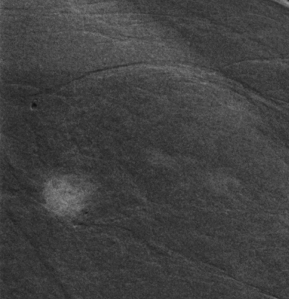
8). Post-processing allows for the creation of one combined image, where contrast uptake-areas are enhanced, and the normal tissue areas are suppressed (
8). Thus, CESM is both a routine mammographic study and a contrasted image capable of finding angiogenesis. This technique is very new, and the characterization of benign and malignant breast lesions on CESM is still a subject of research. To date, some pitfalls and disadvantages of CESM have been described such as its limited performance with malignant microcalcifications with no underlying mass (
8,
11), with the interpretation of ganglions and adenopathies (
8), or in patients with silicone breast implants (
8). In addition, the fact that CESM requires approximately a 1.5 times greater radiation dose than conventional mammography (
8,
11) has been seen as a disadvantage. However, to date, the false negative and false positive rates of CESM have been reported to be very low (
11), and recent studies have demonstrated that it could have a sensitivity and sensibility similar to that of MRI, while having the advantage of being faster, much more economical, and not presenting the aforementioned contraindications of MRI (
4,
6,
7). As such, CESM is a very promising imaging diagnostic tool, which may be useful in the diagnosis of complex breast lesions, as illustrated by this presented case report.