Male breast diseases are most often benign (
10). Gynecomastia is the most common benign breast pathology in male breasts, with a reported prevalence of approximately 4% in the age group of 10-19 years, and it has been found in up to 55% of male breasts in one autopsy specimen series (
10). Mastitis, a benign breast inflammation, is exceedingly rare in men with only three cases reported in the literature to date as a case report or original article (
9,
11).
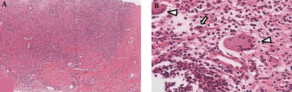
IGLM is a rare, benign inflammatory disease of the breast that mainly affects women of reproductive age (
5,
7,
12). IGLM is characterized by a chronic, lobular inflammatory process and noncaseating granulomatous inflammation without an obvious etiology. The diagnosis of IGLM can be made by excluding all known causes of granulomatous inflammation, such as mammary ductectasia, Wegener granuloma, sarcoidosis, tuberculosis, and histoplasmosis (
12).
The etiology of IGLM is unclear, but an autoimmune origin is the most widely accepted theory because of the response to steroid therapy (
5). Bani-Hani et al. (
12) identified four possible mechanisms of IGLM, and its association with lactation or hyperprolactinemia is explained by extravasated lactational secretions damaging the ductal epithelium and leading to a granulomatous inflammatory response. Additionally, oral contraceptives can cause granulomatous lobular mastitis by a chemical reaction. Our patient did not have any hyperprolactinemic condition caused by a pituitary tumor or drug-induced galactorrhea.
IGLM develops mostly in women of reproductive age (mean age: 33.1 - 39.5 years, range: 22 - 59 years) and women with children, who have a history of oral contraceptive use and/or lactation (
5-
7,
13). The most common symptom is a palpable mass, followed by pain, swelling, erythema, and axillary lymphadenopathy. In IGLM, nipple retraction rarely occurs in women, but it usually occurs in 40-50% of men with breast cancer (
14). Inflammatory signs may not always be present clinically; thus, a misdiagnosis of breast cancer can be made (
5).
Imaging features of IGLM in female patients have been reported infrequently since IGLM was first described by Kessler et al. in 1972 and recently reviewed in several reports of small series. There is a wide spectrum of radiologic findings of IGLM because of various imaging modalities, such as mammography, US, and magnetic resonance imaging (
4,
7,
15,
16). Focal asymmetric densities were the most frequent mammographic finding, followed by an irregular mass in most case series or studies of IGLM. Microcalcifications were not seen on any mammograms in our patient with IGLM. According to several reports, the most frequent sonographic finding is an irregular, heterogeneous hypoechoic mass with tubular extension (
7,
15,
16). Some studies of color Doppler US in patients with IGLM reported increased intralesional and perilesional vascularity (
15,
17). US-elastography is widely used in breast imaging. The differentiation between IGLM and breast malignancy has been studied using radiation force impulse imaging, i.e., acoustic radiation force impulse (ARFI) elastography (
13), and the authors revealed that IGLM had lower shear-wave velocity than breast malignancy. This indicated that IGLM was softer than a malignancy, and a higher diagnostic accuracy was achieved using ARFI combined with US in this retrospective study.
However, imaging findings of IGLM could not been fully differentiated from those of breast malignancy on a mammogram or ultrasonogram (
3,
4,
7,
10,
16). A definitive diagnosis of IGLM should be made histologically from biopsy results because clinical features and imaging findings are not specific and similar to those of breast cancer.
The aforementioned clinical features and representative imaging features of IGLM were obtained from studies with female patients with IGLM. These features of IGLM have not been studied in male patients so far.
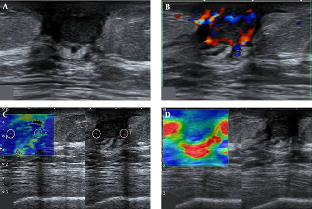
In the present case, ultrasonographic findings showed an oval, circumscribed, hypoechoic mass in the subareolar portion of the right breast. On a color Doppler ultrasonogram, perilesional and internal vascularity was noted. Furthermore, soft elasticity, according to BI-RADS Fifth Edition classification system, was observed using US-elastography. The sonographic finding of our patient is not comparable with the typical sonographic finding of IGLM reported in past studies. Two case reports of IGLM in men did not publish mammograms or sonograms. Further study of the imaging features of IGLM in male patients is needed. To the best of our knowledge, this is the first report of IGLM arising in a male breast with imaging studies of ultrasonography, including color Doppler US and US-elastography.
In conclusion, IGLM rarely occurs in male breasts, although the lobule is not developed in male breasts and the imaging features on a mammogram or ultrasonogram are not comparable to that of female IGLM. When a palpable breast mass occurs in a man, physicians must consider male breast diseases ranging from benign diseases, such as gynecomastia, to breast cancer. Moreover, inflammatory breast diseases such as IGLM should be considered as a differential diagnosis, although the imaging findings may not be comparable with typical IGLM.