A 29-year-old woman was diagnosed with SLE three months ago complicated by end-stage renal disease presented with melena. Physical examination revealed anemic features with a temperature of 38.4°C, blood pressure of 130/90 mmHg, and pulse of 90 beats per minute. Laboratory studies revealed severe anemia (hemoglobin 5.8 g/dL, normal range 12 - 16 g/dL), thrombocytopenia (platelet count 79,000/mm
3, normal range 150,000 - 450,000/mm
3), and leukopenia (white blood cell count 1,050/mm
3, normal range 5,000 - 10,000/mm
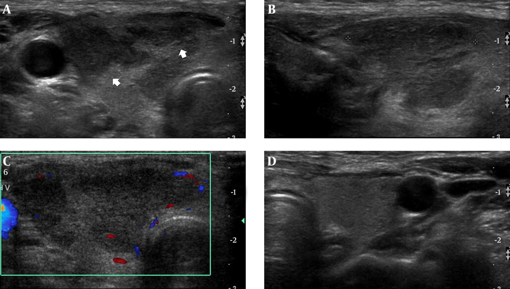
3). Chest X-ray was normal. We could not found the focus of infection. Emergency gastroduodenoscopy revealed several ulcers with hematoma in the stomach and duodenum. There was no evidence of fungal infection around the ulcer. Hemoclip was applied for bleeding control and the bleeding stopped. She was hospitalized for observation while receiving blood transfusion. After four days, she complained of right neck mass and palpitation. Sonography (iU-22; Philips Medical System, Bothwell, WA, USA, 5 - 12 MHz high frequency linear-array transducer) of the thyroid showed diffuse enlargement of the right thyroid gland and the anterior-posterior (AP) diameter was 2.1 cm. An ill-defined hypoechoic lesion involving the middle and upper portion of the right thyroid gland was detected. In this area, sonographic tenderness was present. Referentially, the echogenicity of a normal thyroid gland is slightly higher than the neck muscle and it is homogeneous. The size of a normal thyroid lobe is 1.3 - 1.8 cm in the anteroposterior dimension (
6). Color Doppler images revealed poor vascularity within this hypoechoic lesion. The left lobe was unremarkable. There was no evidence of enlarged cervical lymph nodes (
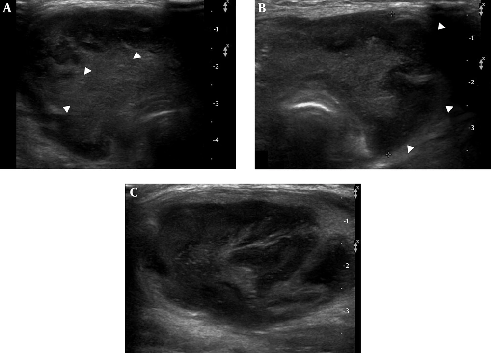
Figure 1). Thyroid function tests revealed a thyrotropin (TSH) level of less than 0.02 μIU/mL (normal range: 0.38 - 4.94 μIU/mL) and free thyroxine (FT4) level of 4.05 ng/dL (normal range: 0.71 - 1.85 ng/dL), indicating thyrotoxicosis. We considered subacute thyroiditis with hyperthyroidism. She was empirically treated with beta blocker and steroid for subacute thyroiditis. However, the patient continued to complain of palpitation with a growing neck mass. Repeat thyroid function showed a higher increase of free thyroxine (FT4) level to 7.83 ng/dL with serum TSH level of 0.02 μIU/mL. Propylthiouracil was added to the treatment strategy, but there was no significant effect and the patient complained of progressive neck swelling and palpitation. After two weeks from the initial sonography, follow up sonography revealed a significant increase in the size of both thyroid glands. The AP diameter was 4cm with lobulating contour. The entire thyroid gland showed irregular hypoechogenicity along the subcapsular region with isoechogenicity in the central portion (
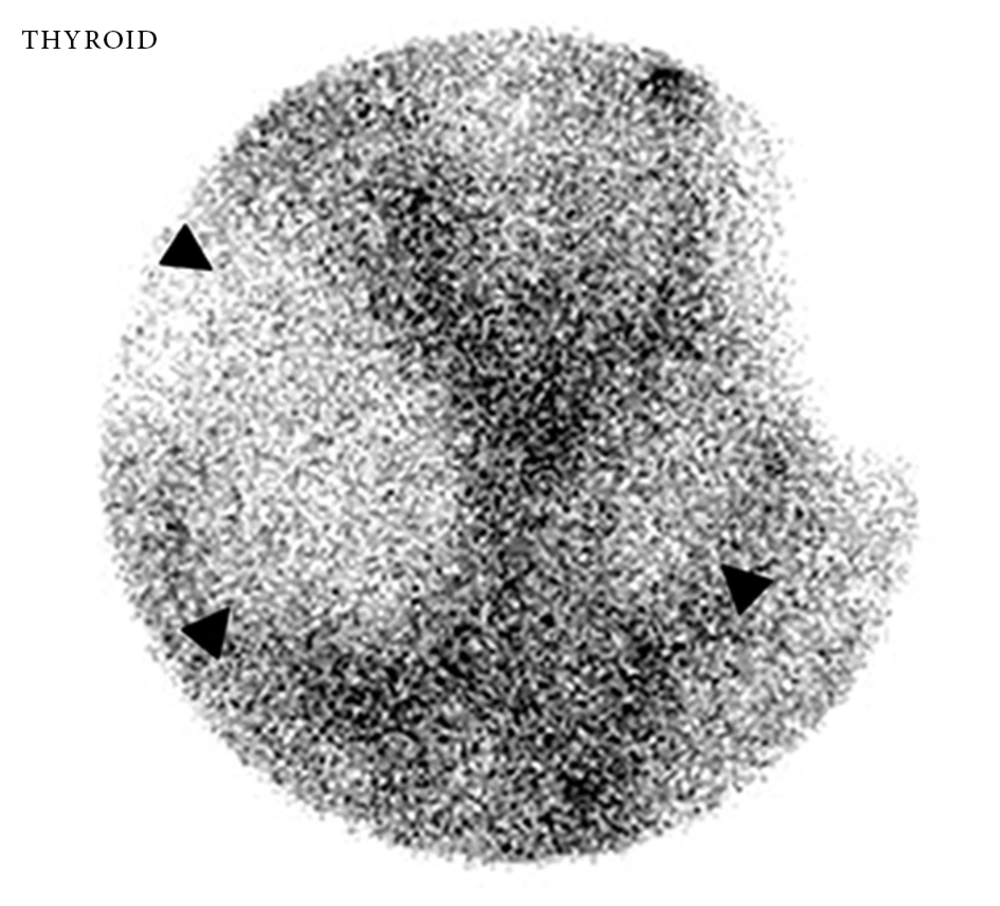
Figure 2). Sonographic findings suggested marked progression of thyroiditis. On scintigraphy, both thyroid glands were noted as large photon defect (
Figure 3). We suspected other diseases such as fungal or suppurative thyroiditis rather than subacute thyroiditis. There was no evidence of fungal infection involving the lung in chest computerized tomography (CT). She had no symptoms of sinusitis. There was no evidence of sinusitis on the Waters’ and Caldwell views; therefore, we did not perform paranasal sinus CT. The next day, the patient was found to have severe dyspnea and hypoxemia. She was endotracheally intubated. After stabilizing her thyroid hormone levels in order to prevent thyrotoxic crisis (thyroid storm), the patient underwent a total thyroidectomy. During the surgery, we noted diffuse enlargement of both thyroid glands. Papillary-like inflammatory infiltration was seen in the peripheral portion of both thyroid lobes with internal hemorrhagic necrosis. Histopathological examination and staining of the surgical specimen with methenamine silver revealed extensive necrotic tissue with many fungal yeast and hyphae characteristic of
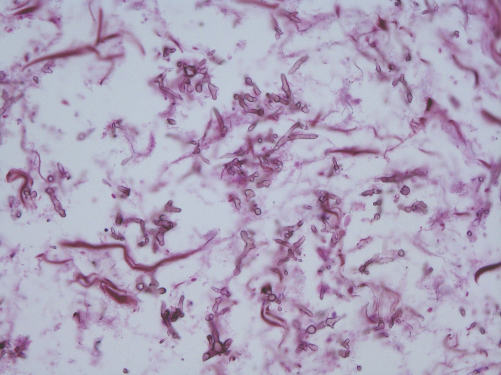
Aspergillus species (
Figure 4). The patient was started on intravenous Amphotericin B. The remainder of the postoperative days were uneventful. The patient was discharged on the 20th postoperative day. The patient is on follow up and has been free of the disease for two years.
All procedures were in accordance with the ethical standards of the responsible committee on human experimentation (institutional and national) and with the Declaration of Helsinki of 1975, as revised in 2008 (
5). Informed consent was obtained from the patient.