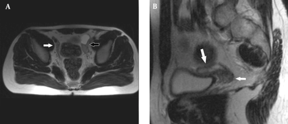
An 18-year-old male patient was referred to the hospital for nonpalpable testes and urination abnormalities. Penoscrotal hypospadias with nonpalpable testes were found on physical examination, The laboratory tests such as testosterone, luteinizing hormone (LH), lactate dehydrogenase (LDH), alpha fetoprotein (AFP), and beta human chorionic gonadotropin (ßHCG) levels were normal. On the other hand, there was a three-fold increase (36.38 mIU/ml) in the follicle stimulating hormone (FSH) level. Karyotype analysis was reported as 80% 46 XY and 20% 45X chromosomal pattern. Contrast-enhanced abdominal MRI and diffusion weighted imaging were performed to demonstrate associated anomalies. There was a left undescended testis with heterogeneous signal near the bladder. Epididymal cyst was found as a cystic lesion associated with left undescended testis. A linear structure like fallopian tube and a heterogeneous nodular lesion like ovary were seen on the right side on T2 weighted images (
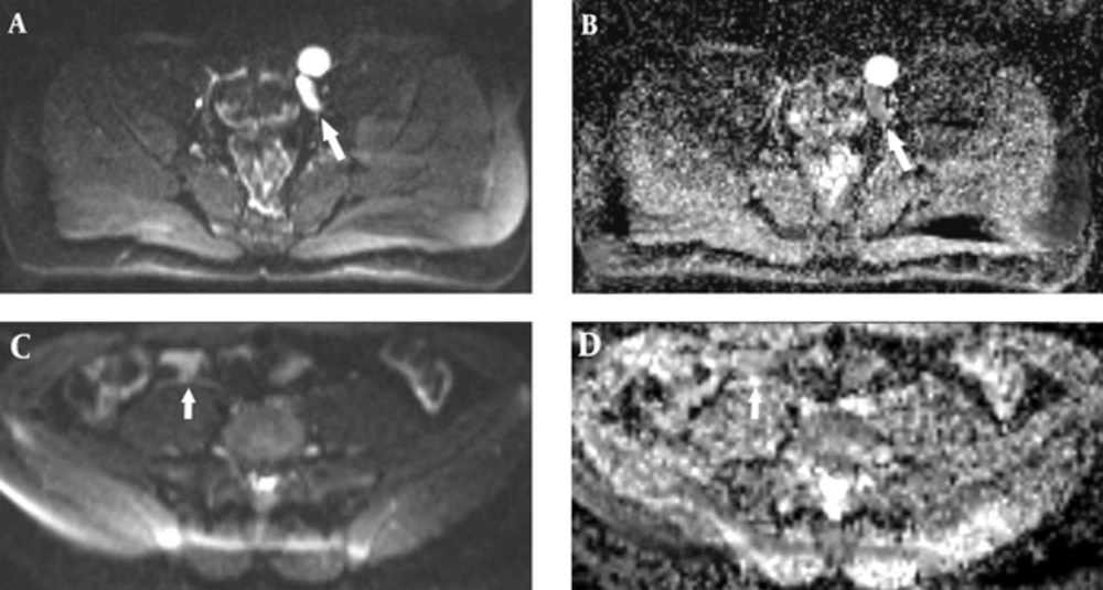
Figure 1). The right side gonad was thought to be an ovary on T2 sequences because of configuration and location on the distal end of the fallopian tube. The right seminal vesicle was normal, but there was no left seminal vesicle. Prostate and prostatic urethra were normally seen, but penile urethra was not visualized. Müllerian duct remnant as a linear cystic lesion was shown between the prostate and rectum. The fusion of müllerian duct remnant and prostatic urethra opened in the skin at the level of perineum. The restriction of diffusion which indicates increased cellularity was seen in both the right ovary and left testis (
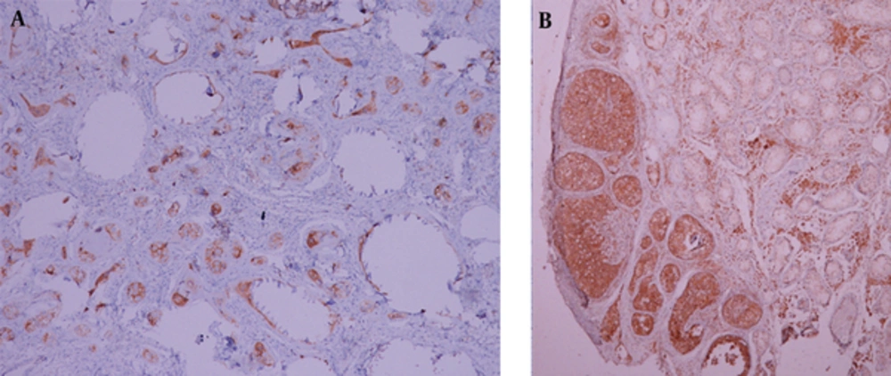
Figure 2). Diagnostic laparoscopy, hysterosalpingo-oophorectomy and abdominal orchiectomy were performed because of high suspicion of malignancy. Histopathologically, gonadoblastomas (in situ malignant germ cell tumors) were found in both the right ovary and left testis (
Figure 3).