


1. Background
2. Objectives
3. Materials and Methods
3.1. Livewire


3.2. LVness

3.3. Gray Space Map
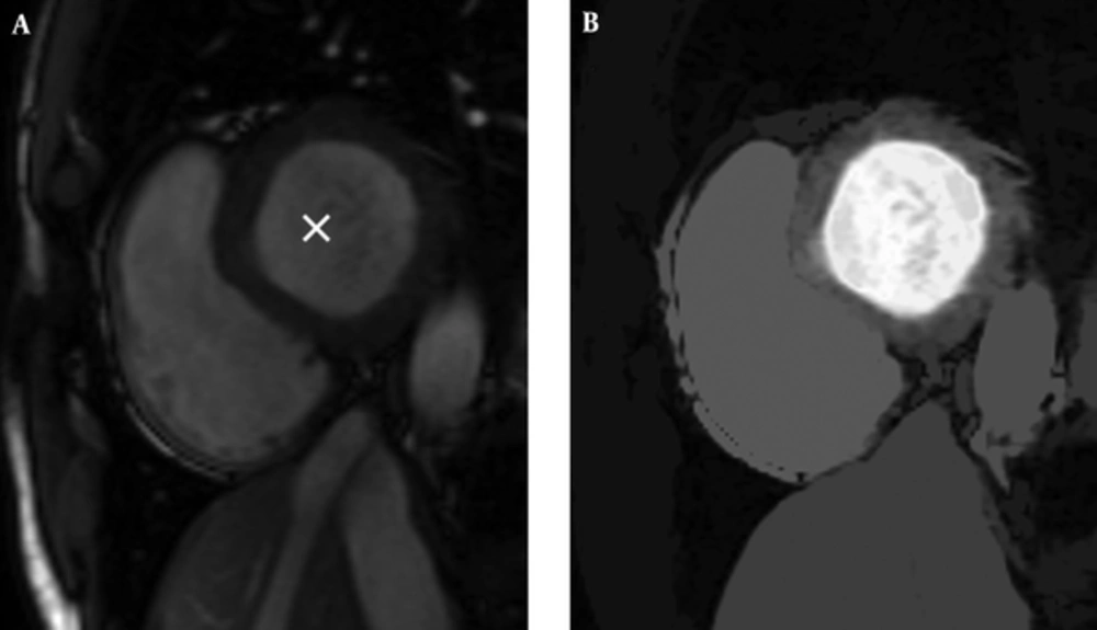
Gray space map with a seed pixel inside the left ventricle. A, The original image with the seed displayed by a cross mark; B, Resulted gray space map.
3.4. LVness Generation


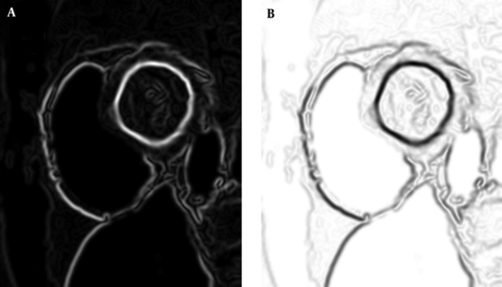
A, Result of third step (non maximal suppression: NMS) of canny edge detector algorithm. B, fLV is the complement of NMS.


3.5. Graph Searching Algorithm
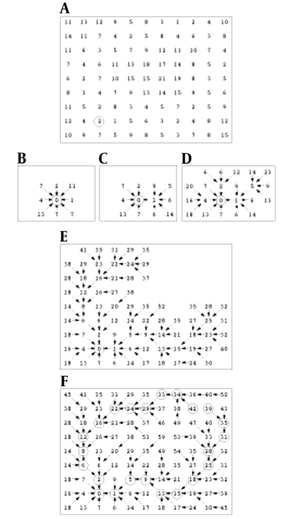
Cost expansion. A, Local cost map. B, Seed point expanded. C, Two points expanded. D, Five points expanded. E, 47 points expanded. F, Completed total cost path/pointer map with optimal paths
3.6. Ethical Approval
3.7. Informed Consent
4. Results
4.1. LVness Generation
4.2. Quantitative and Qualitative Evaluation

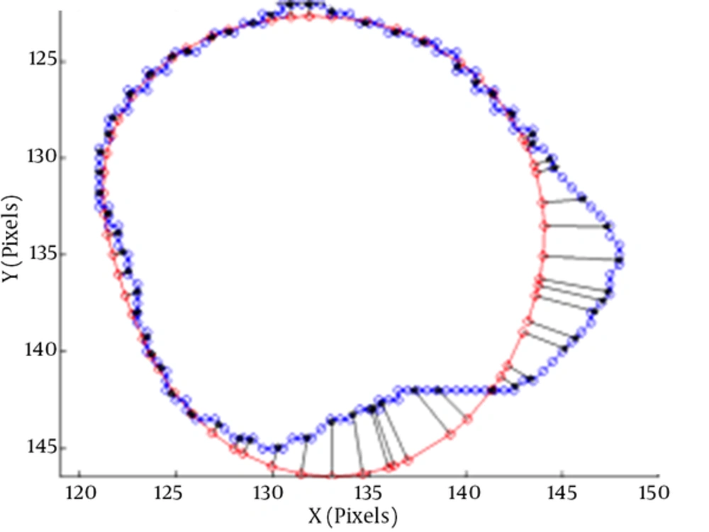
The average perpendicular distance APD between two contours (3)
| Dataset | DM | APD | |
|---|---|---|---|
| Training | ED | 0.95 ± 0.26 | 1.48 ± 1.32 |
| ES | 0.89 ± 0.11 | 6.23 ± 4.33 | |
| Validation | ED | 0.92 ± 0.19 | 4.11 ± 1.19 |
| ES | 0.90 ± 0.12 | 4.77 ± 0.12 | |
| Online | ED | 0.88 ± 0.34 | 3.45 ± 0.79 |
| ES | 0.86 ± 0.29 | 5.83 ± 2.35 |
Abbreviations: APD, average perpendicular distance; DM, dice metric; ED, end diastole; ES, end systole.
aValues are expressed as mean ± standard deviation.
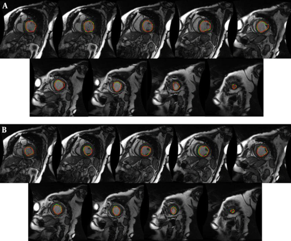
Manual (red) and proposed approach (green) segmentation of the left ventricle from base to apex for an instance case of MICCAI 2009 challenge database (3). A, End diastole; B, End systole.
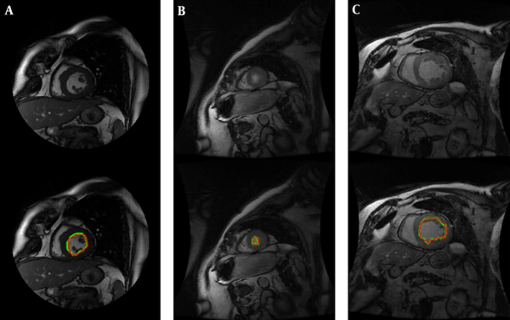
A, Manual (red) and proposed approach (green) segmentation of the left ventricle in some challenging slices containing presence of papillary muscles; B, Apical slices; C, Unclear borders. Top row: original image and bottom row: segmentation result.
5. Discussion
| Method | DM | APD |
|---|---|---|
| Proposed method | 0.95 ± 0.26 | 1.48 ± 1.32 |
| Classic livewire (22) | 0. 88 ± 0.22 | 8.13 ± 3.87 |
| Oghli et al. 2013 (31) | 0.79 ± 0.33 | 9.12 ± 1.23 |
| Oghli et al. 2012 (32) | 0.73 ± 0.56 | 9.58 ± 2.19 |
| (Queiros et al. 2014) (33) | 92.7 ± 0.23 | 10.51 ± 9.17 |
| (Ngo and Carneiro, 2014) (34) | 93.23 ± 0.34 | 22.20 ± 21.74 |
| (Hu et al. 2013) (35) | 0.61 ± 0.29 | 15.08 ± 8.91 |
| (Constantinides et al. 2012) (36) | 0.80 ± 0.19 | 9.79 ± 5.38 |
| (Jolly et al. 2009) (37) | 0.88 ± 0.04 | 2.97 ± 0.38 |
| (Liu et al. 2012) (38) | 0.78 ± 0.20 | 9.26 ± 4.93 |
| (Huang et al. 2011) (39) | 0.81 ± 0.16 | 7.28 ± 3.58 |
| (Schaerer et al. 2010) (40) | 0.77 ± 0.16 | 9.64 ± 4.15 |
Abbreviations: APD, average perpendicular distance; DM, dice metric.
aValues are expressed as mean ± standard deviation.
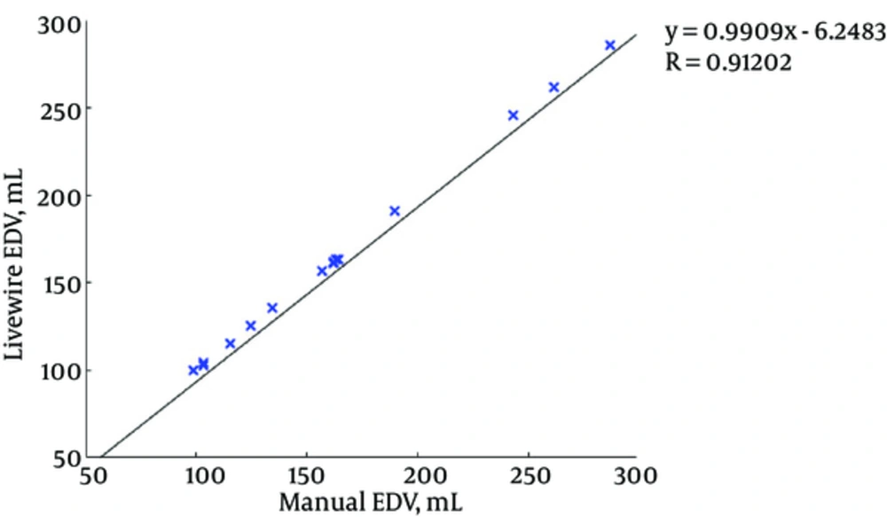
Correlation graph for end-diastolic volume (EDV)
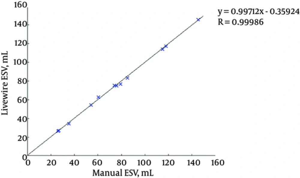
Correlation graph for end-systolic volume (ESV)
