A 36-year-old woman, gravida 2, para 2, had a palpable mass in the mid inner portion of the right breast. The mass had been present for several months and was painful. The patient had a history of focal excision for fibroadenoma in the upper inner quadrant of the right breast in another hospital. The patient had no family history of breast cancer. Skin retraction, nipple discharge or palpable lymph nodes were not observed on clinical examination. On mammography (
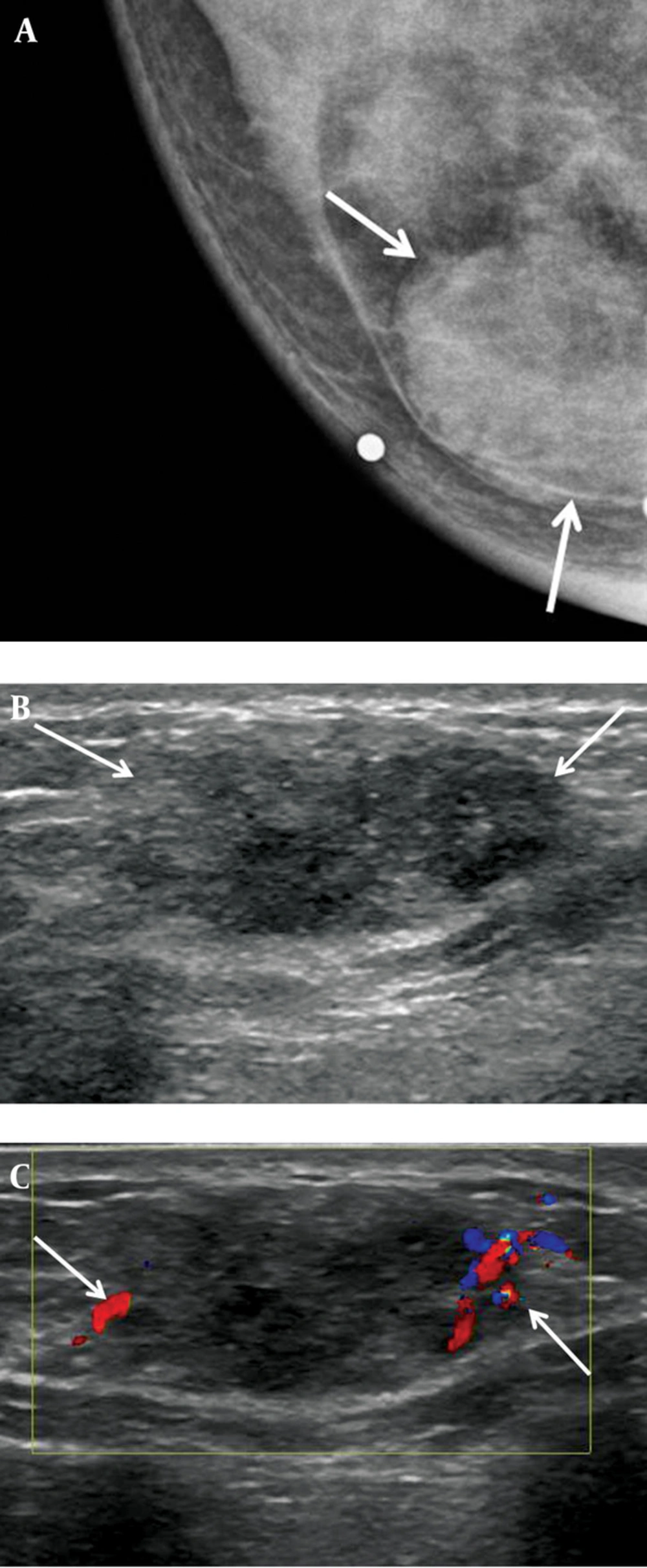
Figure 1 A), a 2-3 cm in diameter, circumscribed, oval-shaped, isodense mass was seen in the right inner breast on the craniocaudal view. This mass was not detected on the mediolateral oblique view. Microcalcifications were not detected on mammography. Ultrasonography (
Figure 1 B) showed an indistinct, 2.3 × 1.1 × 1.7 cm, oval shaped, heterogeneous and mild hypoechoic mass in the palpable area. On power Doppler study, a blood vessel was seen in the periphery of the mass (
Figure 1 C). Breast MRI was performed using a 3.0 T -MR imaging system (Achieva 3.0 T TX; Philips Healthcare) with a dedicated phased-array breast coil, and the patient in the prone position. DWI was acquired in the transverse plane and covered both breasts. A spin-echo single-shot echo-planar-imaging sequence with diffusion-sensitizing gradients was applied along orthogonal direction. These images were used to synthesize isotopic transverse images (repetition time ms/echo time ms, 5417/72; b values, 0, 600 and 1000 s/mm
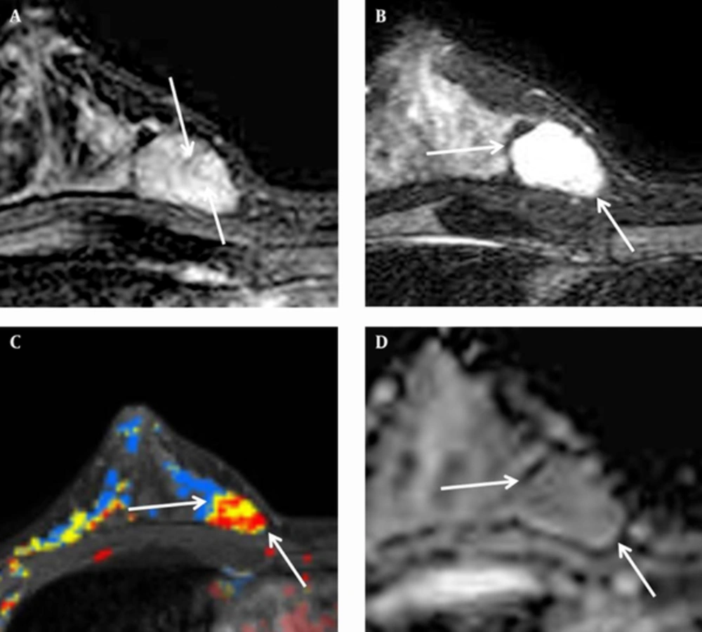
2; image matrix, 96 × 126; field of view, 320 × 320 mm; section thickness, 3 mm; section gap, 0 mm; three signal acquired; acquisition time, 80 s). After DWI, T2-weighted fast spin-echo transverse images were also obtained. The following image parameters were used: 5727/70; flip angle, 90°, image matrix, 620 × 309; field of view, 581 × 342 mm; section thickness, 3 mm; section gap, 0 mm. A three-dimensional T1-weighted fast spoiled gradient-echo sequence was also performed with transverse imaging using one pre-contrast and six post-contrast dynamic series, immediately after contrast injection and after 60, 120, 180, 240, and 300 seconds. The image parameters were as follows: 6/3; flip angle, 0°; image matrix, 436 × 436; field of view, 330 × 340 mm; section thickness, 3 mm; section gap, 1.5 mm. Gadoterate meglumine (Dotarem; Guerbet, Villepinte, France) was injected per kilogram of body weight into an antecubital vein with a power injector (Spectris; Medrad, Indianola, PA, USA) at a rate of 2 mL/s. DCE-bMRI revealed a smooth-marginated, heterogeneously well-enhanced mass (
Figure 2 A), which demonstrated high signal intensity on T2-weighted imaging (
Figure 2 B) and low signal intensity on T1-weighted imaging. This showed early rapid enhancement and delayed washout enhancement (
Figure 2 C) on the kinetic analysis in the majority of the mass. This mass shows heterogeneous high signal intensity on DWI with high b-value (b = 1000) without definite diffusion restriction (
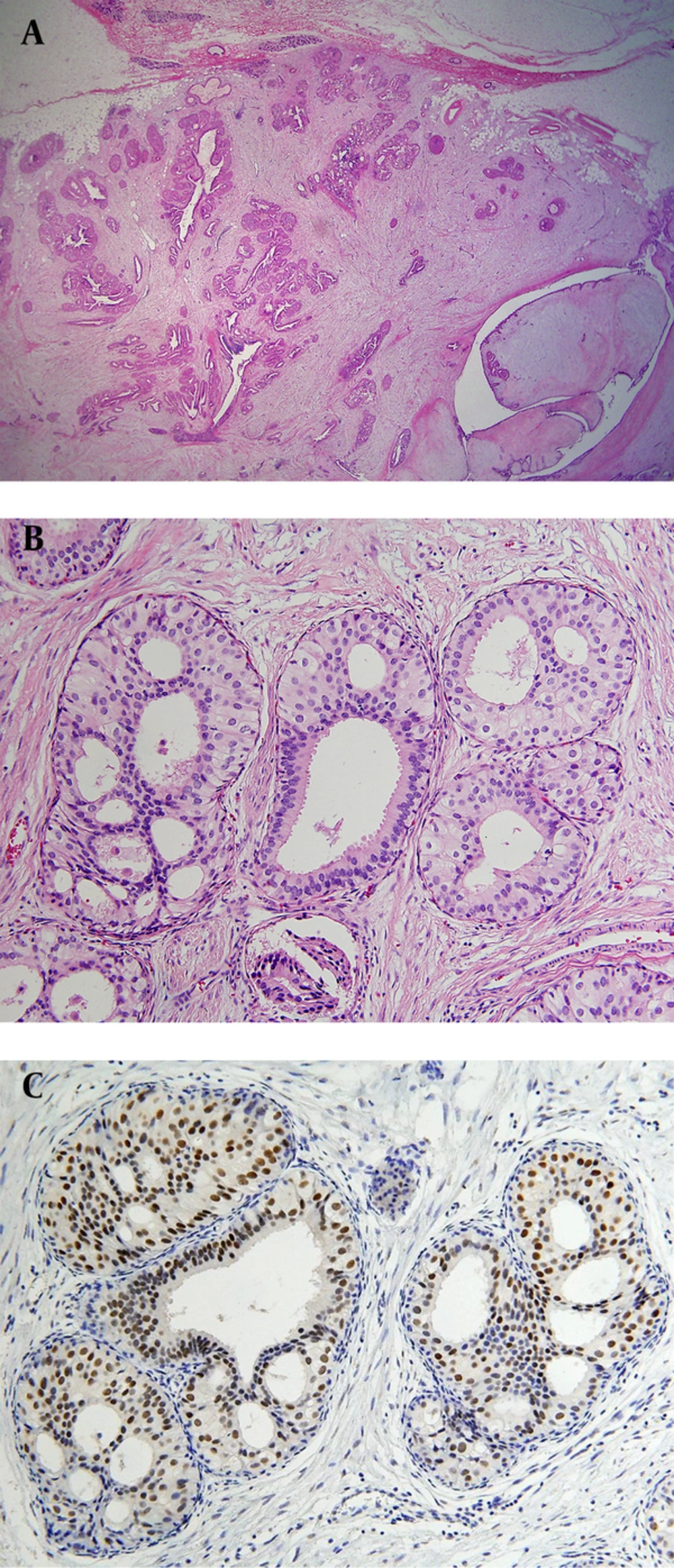
Figure 2 D). The patient underwent breast-conserving surgery for the palpable mass. Histopathological examinations (
Figure 3) showed multiple foci of atypical ductal hyperplasia and ductal carcinoma in situ, with a cribriform or solid pattern, within a juvenile fibroadenoma. In the DCIS areas, most ductal epithelial cells showed overexpression of estrogen receptor (ER) immunostaining, and p63-positive myoepithelial cells were preserved only in the periphery. There was no evidence of metastasis in sentinel lymph nodes. Radiation therapy was performed in the right breast after surgery. The patient had no evidence of recurrence 12 months after the surgery.