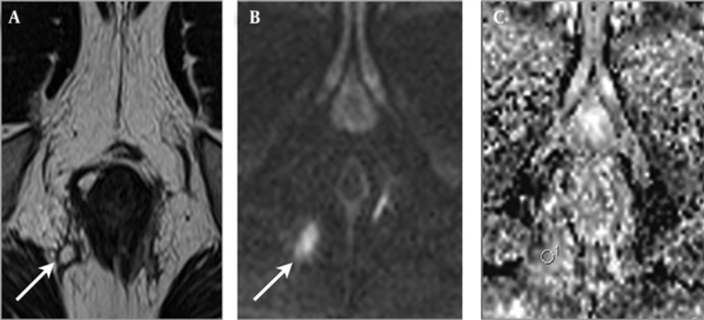
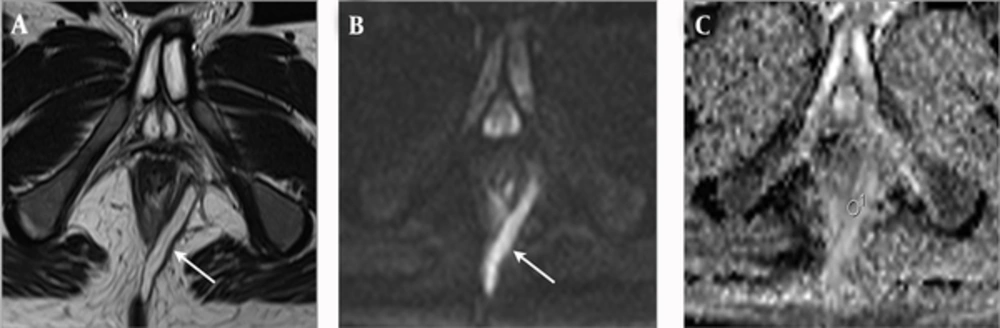
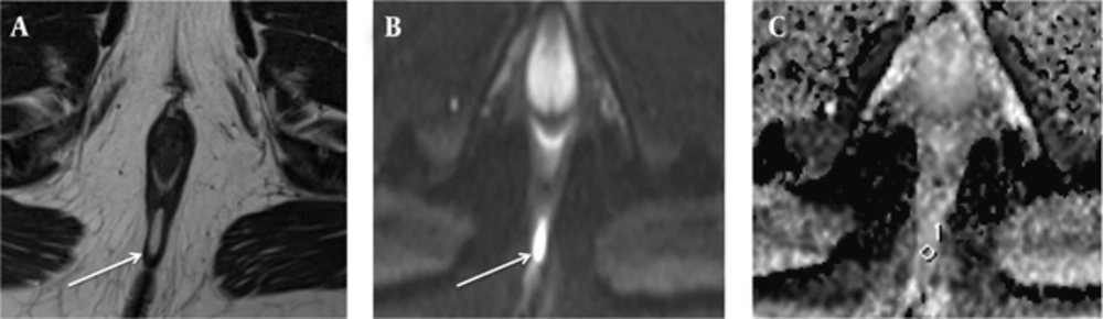
Dynamic-contrast enhanced MRI is often used for the assessment of perianal fistulas and the increased conspicuity of fistula is the most important advantage of this technique (
6). Additionally, several studies have shown that dynamic contrast-enhanced MRI can help determine perianal Crohn’s disease activity (
7,
8). However, it has been shown that contrast-enhanced T1WI may exaggerate the degree of perianal disease activity and the number of active fistulas (
15). Additionally, clinically inactive fistulas may enhance after intravenous gadolinium administration and may be incorrectly diagnosed as an active fistula (
15). Because of the known contraindications of contrast agents that may lead to nephrogenic systemic fibrosis and hypersensitivity (
16), alternative MRI methods that provide similar information, as contrast agents, are needed. The DWMRI is an alternative MRI technique that is cost-effective, with a very short imaging time, compared to contrast-enhanced imaging.
Perianal abscesses are quite common in patients with active fistulizing diseases (
17). Abscesses are usually manifested by acute onset of pain and require immediate surgical treatment (
18). In addition, several symptoms of positive inflammation (i.e., pain at defecation or pain at rest) more frequently occur with perianal abscesses, compared to active and inactive fistulas (
19). Previous studies showed that clinical findings and disease activity increase when an abscess accompanies a fistula (
17-
19). In the present study, the mean ADC value of the PIA fistulas was significantly lower than the mean ADC value of NIA fistulas, in patients with abscess. Conversely, we did not find statistically significant differences between the mean ADC values of the PIA and NIA groups, in patients with only perianal fistulas. Our results indicate that the presence of an abscess causes restricted diffusion and low ADC values in perianal fistulas, which are correlated with fistula activity.
Restricted diffusion has been shown in many different inflammatory processes, including encephalitis, pyelonephritis, and abscesses formation (
20,
21). In addition, inflammation and fibrosis have been shown to cause decreased ADC values and restricted diffusion in the liver (
22). The characteristic histological findings of active Crohn’s disease are infiltration of the lamina propria and submucosa of the small bowel, by inflammatory cells, and presence of lymphoid aggregates. Oto et al. (
23) reported that these histological findings are characterized by a brighter signal on DWMRI and lower ADC values, in the inflamed bowel wall. In our study, the mean ADC value of the PIA fistulas was found significantly different from the mean ADC value of NIA fistulas, in the presence of an abscess. We suggest that the low ADC values of PIA fistulas, in the present study, could be secondary to increased cellularity, due to perianal inflammation. An increased cell density can narrow the extracellular space and restrict the diffusion of the water molecules in a PIA fistula. Thus, allowing DWMRI discriminating between active and inactive perianal fistulas that are accompanied by an abscess.
Hori et al. (
11) and Dohan et al. (
12) examined the role of DWMRI in the detection of perianal fistulas. The most important limitation of these studies was their small sample size. Although our study included more fistulas (n = 56) than these studies, we did not find any statistically significant difference between the number of fistulas, determined by T2WI alone and by DWMRI alone. In the current study, visibility of fistula tracks was better on the combined evaluation of T2WI and DWMRI, compared to the visibility on T2-weighted alone, which is consistent with the results of Hori et al. (
11) and Dohan et al. (
12).
In an another study, Yoshizako et al. assessed the value of DWMRI in evaluating perianal fistula activity. Yoshizako et al. (
3) found a significant difference in ADCs between PIA and NIA fistulas (P = 0.0019) and concluded that DWMRI can be helpful for evaluating perianal fistula activity. Although 30 abscesses were evaluated in their study, Yoshizako et al. (
3) did not classify the fistulas based on their association with abscess. In the current study, when we measured ADC values of all fistulas, regardless of the presence of an abscess, we did not find a statistically significant difference between ADC values of PIA and NIA fistulas (P = 0.636).
In a study that examined the role of DWMRI in determining the fistula activity in perianal fistula, Dohan et al. (
12) did not find a correlation between ADC values and fistula activity, which is similar to our findings. However, we found a statistically significant correlation between disease activity and ADC values in abscess accompanied fistulas (P = 0.036) and suggested that increased fistula activity may be related to the presence of abscesses.
Our study has several limitations. First, a series of subjective clinical findings (pain or restriction of daily or sexual activity) were used, rather than objective surgical, inflammatory, and anatomic parameters. In addition, although the CRP level is the most widely used biochemical marker of inflammation, it is not specific to perianal fistulas and can reflect other inflammatory processes, occurring at the time of the investigation. Second, the visibility of fistulas was evaluated on a 3-point scale, rather than a 5-point scale, which might have affected the findings. Third, the present study was a retrospective and single-center study. Although our sample size for abscess associated fistulas was small, the number of perianal fistulas not accompanied by abscess, evaluated by DWMRI in the current study, is the largest in the published literature. In the future, a prospective study should be performed with a larger number of patients, to clarify the efficacy of DWMRI for determining activity and visibility of perianal fistulas. Finally, in the current study, we did not compare DWMRI with contrast enhanced T1WI and fat-suppressed T2WI, regarding their effectiveness in evaluating disease activity and detecting fistulas.
In conclusion, we found decreased ADC values and restricted diffusion in perianal fistulas, accompanied by abscesses, which show disease activity. Also, in our study, the visibility of fistula tracks was better when DWMRI and T2WI were combined, compared to T2WI alone. Therefore, we concluded that DWMRI is a valuable tool for evaluating fistula activity in patients with perianal abscess and provides better visibility of fistulas compared to T2WI.