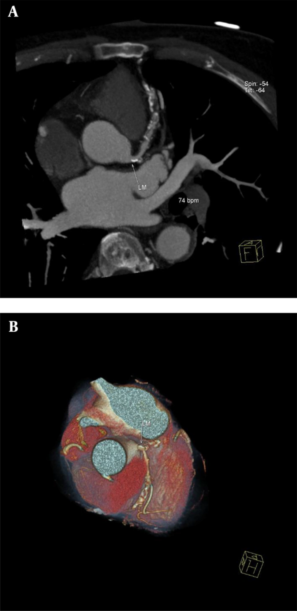
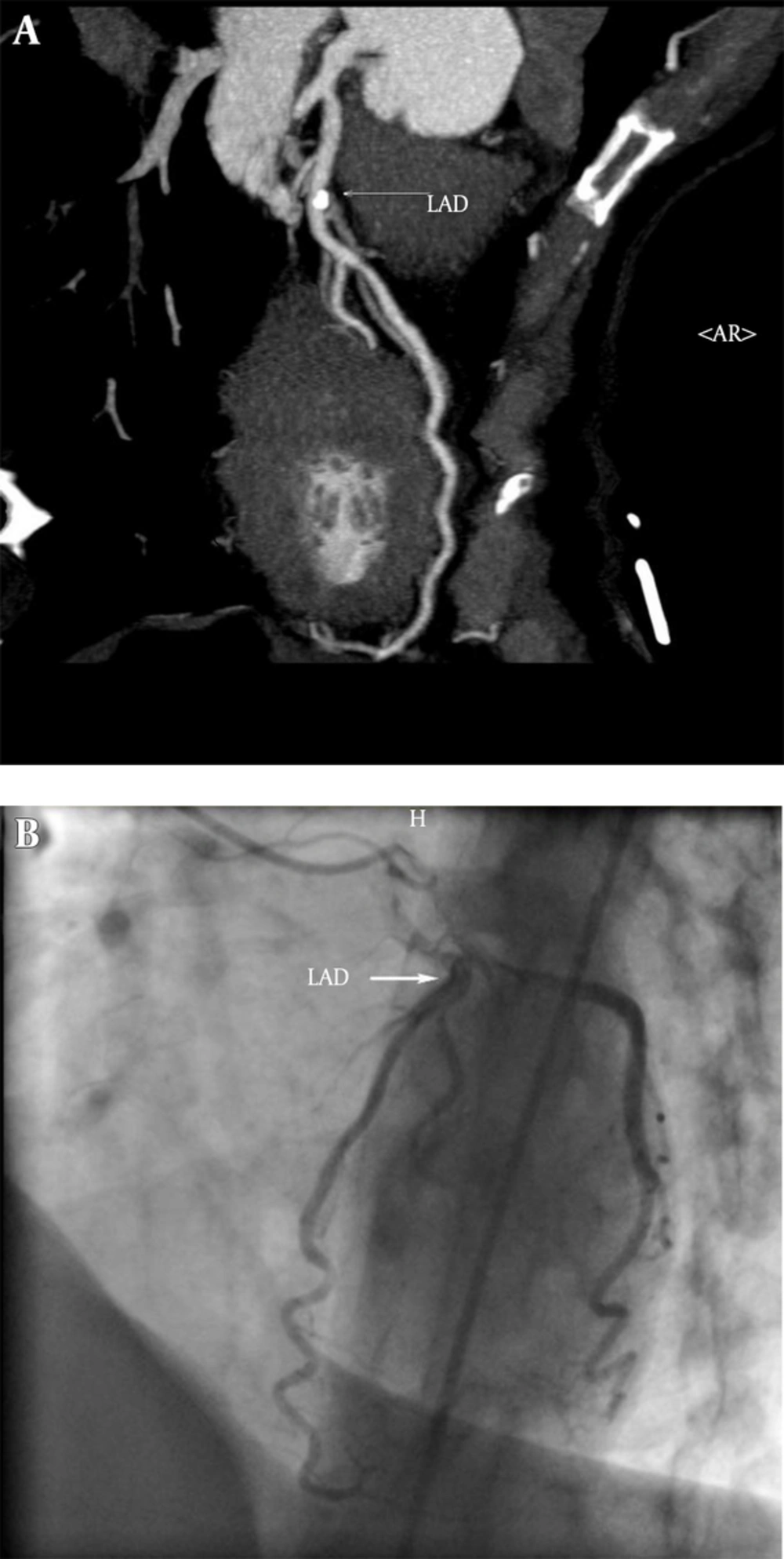
The results of the present study showed that DSCT coronary angiography has clinically acceptable accuracy in the diagnosis of coronary artery stenosis among patients who were referred to our center during daily clinical practice.
Likelihood ratio (LR) is a useful index of any diagnostic test. Unlike sensitivity and specificity, LR can be used at the individual patient level and it individualizes the probability of a diagnosis. Furthermore, these measures are based on a ratio of sensitivity and specificity, hence not varying in different populations or settings (
22). For a given diagnostic test, LRs greater than 1 indicate that the test result is associated with the disease, and values closer to zero indicate that the finding decreases the probability of the disease. Generally, LRs above 10 and below 0.1 are considered to provide strong evidence to rule in or rule out diagnosis (
22,
23). Good LR values of current study confirmed that DSCT coronary angiography provides a high diagnostic accuracy for evaluation of CAD in daily clinical practice.
Our findings are consistent with several other studies that have previously investigated the diagnostic performance of DSCT coronary angiography. In a study by Scheffel and colleagues, per segment analysis of DSCT coronary angiography findings showed a sensitivity of 96.5% and specificity of 97.5%. Also PPV and NPV of DSCT coronary angiography were reported as 85.7% and 99.4%, respectively (
10). The results of our study showed a lower accuracy for DSCT coronary angiography compared to the study conducted by Scheffel et al. This could be explained by the difference in the setting of the study and recruitment of the subjects. While in the study by Scheffel et al., all the subjects were recruited with specific inclusion and exclusion criteria, we selected our subjects from patients who were referred to our center during daily clinical practice and aimed to show the accuracy of DSCT coronary angiography while it is used in a real clinical setting and not a research setting. Cademartiri et al. also tested the accuracy of CTCA in the real world setting and they have reached a similar conclusion (
24).
In a recent meta-analysis (
25) on the diagnostic performance of DSCT coronary angiography, the pooled diagnostic parameters of per segment analysis were reported as follows: sensitivity, 95%; specificity, 97%; PLR, 35.2; and NLR , 0.05, which are in accordance with the current study. In addition, per vessel analysis of the DSCT findings showed a sensitivity, specificity, PLR, and NLR of 97%, 94%, 16.4, and 0.03, respectively which are in line with our findings.
In total, 14 segments (2.2%) were non-assessable in our study. This value is comparable to the systematic review by Salavati et al. (
14) that evaluated 21 studies and reported that 2% of the segments which were studied by CTCA were non-assessable.
The current study was performed in a daily routine clinical setting, which makes it different in some aspects compared to other studies. In routine practice, a high number of diagnostic studies should be performed in the radiology department every day. This limits the evaluation time that is spent for each DSCT coronary angiography study and might lead to the decrease of the diagnostic accuracy of DSCT compared to the studies that performed DSCT coronary angiography in a research setting. Also, considering the time limitation in a daily routine clinical setting, all the evaluations were done visually and this could also be considered as another cause of discrepancy between DSCT and CCA.
In a systematic review by Mowatt G. et al. it was found that the diagnostic accuracy of CTCA for the evaluation of stenosis of LCX artery is lower than other major coronary arteries (
15). It was hypothesized that this might be due to the anatomical position of the LCX artery, which is close to the atrium, and thereby is more affected by atrial contraction (
26). Similarly, in our study, it was found that the diagnostic accuracy of LCX is slightly lower than LM-LAD and RCA, although the difference was not significant. The fact that no significant difference in the diagnostic accuracy of DSCT findings between RCA, LM-LAD, and LCX artery territories were found, might imply that the diagnostic performance of DSCT is not much influenced by temporal resolution (
27).
Conventional coronary angiography still remains the gold standard for diagnosing coronary aneurysm and ectasia. However, CCA provides no information about the vessel wall (
28). Therefore, CCA may underestimate the size of the aneurysm, and when it is occluded or contains thrombosis, the aneurysm may not even be seen on CCA (
29). Excellent correlation between MDCT coronary angiography and CCA has been reported in the detection of coronary aneurysm and stenoses in patients after childhood Kawasaki syndrome (
30). However in our study, normal segments of the artery between multiple aneurysms were reported as significant stenosis, in one patient, ensuing in false positive results for DSCT imaging study. Therefore, interpretation of luminal stenosis in patients with coronary aneurysms needs special consideration.
The capability of depicting additional findings is one of the unique features of the CT angiography technique. According to the American college of radiology guidelines, interpretation of non-cardiac findings are necessary in CTCA imaging studies (
31). Lazoura et al. demonstrated incidental non-cardiac findings in as high as 56% of the patients undergoing CTCA (
32). Similar to our study, others have reported findings such as pleural effusion, pulmonary embolus, and pulmonary malignancy (
33,
34). This feature might even help to determine the cause of chest pain or respiratory symptoms in patients with normal coronary arteries (
35).
The retrospective nature of the study confined our control over the use of daily beta-blockers by the subjects, although an additional beta-blocker was not prescribed for heart rate control before CTCA study. While this is a retrospective study, we did not ask patients to do both of the examinations. As a substitute, we chose patients who underwent both DSCT coronary angiography and CCA in a 60-day period. So, this could lead to a high prevalence of CAD in our patient population. PPV and NPV are influenced by disease prevalence; consequently, the ability of DSCT to detect and to rule out stenosis could be overestimated. However, this limitation could be balanced with the use of LR in our study. Moreover, we should state that current results are reached in a population with a high prevalence of CAD, which is the favorable population presented to a referral cardiovascular center. The small number of patients was another limitation of the present study.
In conclusion, our results indicate that DSCT coronary angiography provides a high diagnostic accuracy for evaluation of CAD in the daily routine practice of a referral cardiovascular setting. These results are comparable to studies performed in a research setting. Moreover, acceptable LR values of the current study demonstrate that second-generation DSCT coronary angiography is a reliable tool that could be used as a non-invasive method for assessment of CAD in the clinical setting.