Iliotibial band friction syndrome (ITBFS) is a misuse damage triggered by repeated abrasion of the iliotibial band through the lateral femoral epicondyle (
1,
2). ITBFS is commonly observed in bicyclists, marathon runners, commandos, or rangers among the relatively young aged active population (
2). Diagnosis is habitually established from a complete medical anamnesis and physical examination (
6). Treatment is typically conservative consisting of rest, anti-inflammatory drugs, physiotherapy, steroid injection, and surgical treatment in selected cases (
1,
7,
8).
The etiopathogenesis of this syndrome is controversial and it is still unclear which tissue is responsible for the inflammatory progression: the bursa or the synovial recess of the knee joint. Ekman et al. acknowledged ITBFS as a problem in the bursa underneath the iliotibial band using magnetic resonance imaging (
2,
6). Histologically Nemeth et al. revealed that the inflamed tissue is the lateral synovial recess of the knee joint (
7). It was also speculated that the combination of extrinsic factors and intrinsic or anatomical factors may predispose to develop overused injuries including ITBFS. Malalignment of the lower extremity has been implicated in iliotibial band syndrome (
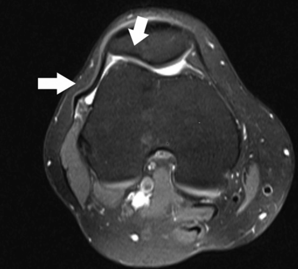
3). In our study, we also found an association between the anatomic factors including facet shape and height of the patella in our study. We used Wiberg classification for patellar facet shape.
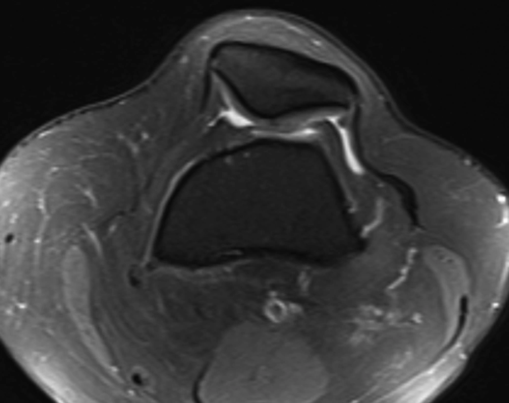
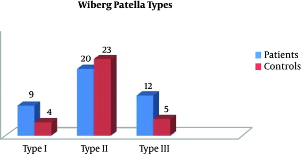
Wiberg classification of patella was based on differences in facet shape and lengths (
4). In Wiberg classification, three types of patella were termed. These shapes can be well-defined in axial MRI planes. In 1941, Wiberg described three types of patella based on lengths of the medial and lateral facets. In Wiberg type I patella has same sized facets medially and laterally, whereas in type II, the medial facet is slightly shorter than the lateral facet, and in type III the medial facet is markedly smaller and has a concave shape. In large-scale anatomic studies, 10% of the normal population has type I, 65% has type II and 25% has type III (
4,
9).
Stoller et al. (
9) proposed that an inexplicably short medial facet predisposed the patella to chondromalacia patellae. They described that the small area of contact between the medial facet of the patella and the medial femoral condyle led to increased stress on the medial articular cartilage of the patella during physiologic motion. In our study Wiberg type I and III patella were significantly higher in ITBFS group whereas type II was significantly higher in control group. Thus we thought that facet shape of patella could be associated with ITBFS.
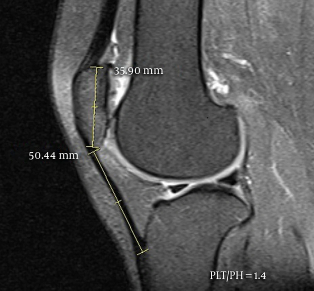
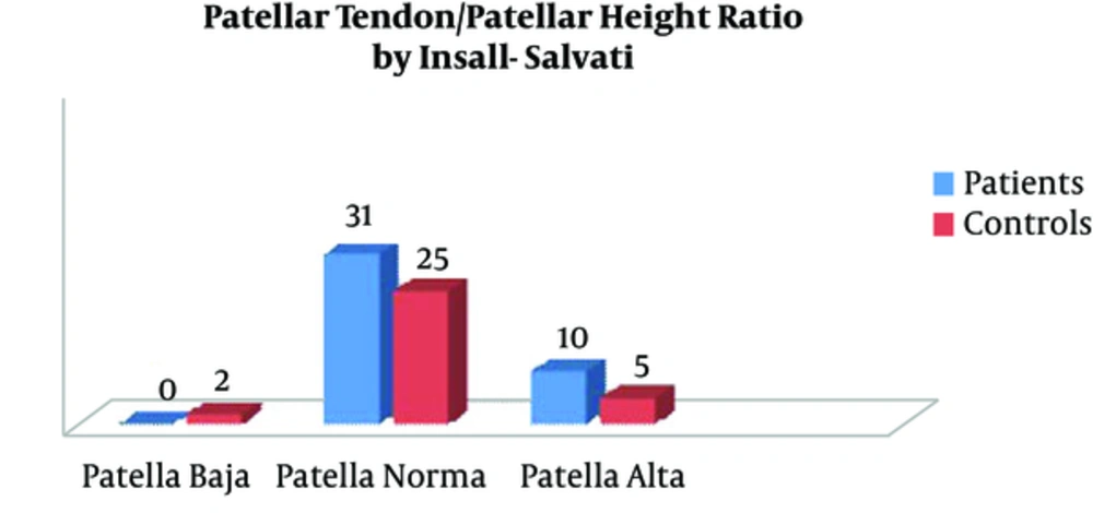
In our study we used Insall - Salvati ratio for detection of patella height. Insall and Salvati described the patellar tendon - patella ratio as the ratio of maximal length of the patella to the distance from the distal tip of patella and the tibial tuberosity, which is normally between 0.8 and 1.2. Patellar positions above and below these ratios are called patella alta or patella baja, respectively (
9). Shabshin et al. (
5) used MRIs of extended knees to measure the PLR and suggested that PLR of > 1.50 or < 0.74 defined patella alta and baja, respectively. Barbier-Brion et al. (
10) showed that patients with patella lateral femoral friction syndrome (PLFFS) have anatomical predisposition for instability with patella alta (P < 0.0001). Patella baja has been related with quadriceps tendon rupture, neuromuscular disorders, and post-surgical advancement of the tibial tuberosity (
11). Patella alta is associated with lateral patellar dislocation, chondromalacia patellae, patellar ligament rupture, patellar and quadriceps tendonitis.
In our study, there was a strong association between patella alta and ITBFS suggesting that patella alta may become provocative and take role improving pathology in the knee.
The diagnosis of ITBFS usually depends on clinical signs and symptoms. Anterior knee pain and pain in motion are the major symptoms (
1,
2). Magnetic resonance imaging is necessary to show pathological findings and to exclude other reasons that may cause similar symptoms (
6). Ultrasound has minor role in imaging but can be useful for follow up once the diagnosis is made (
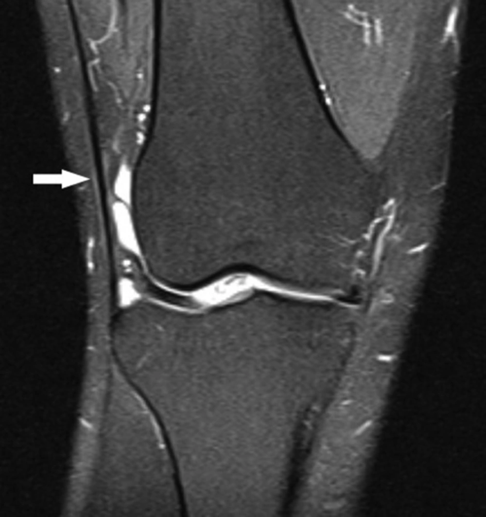
12). Vasilevska et al. (
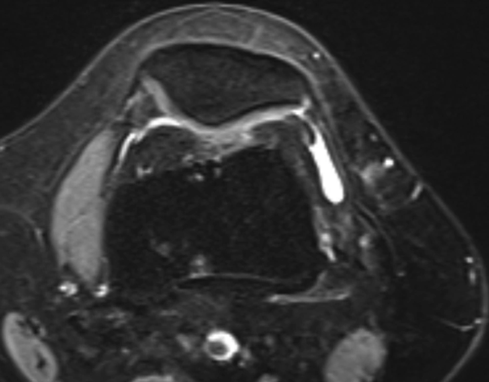
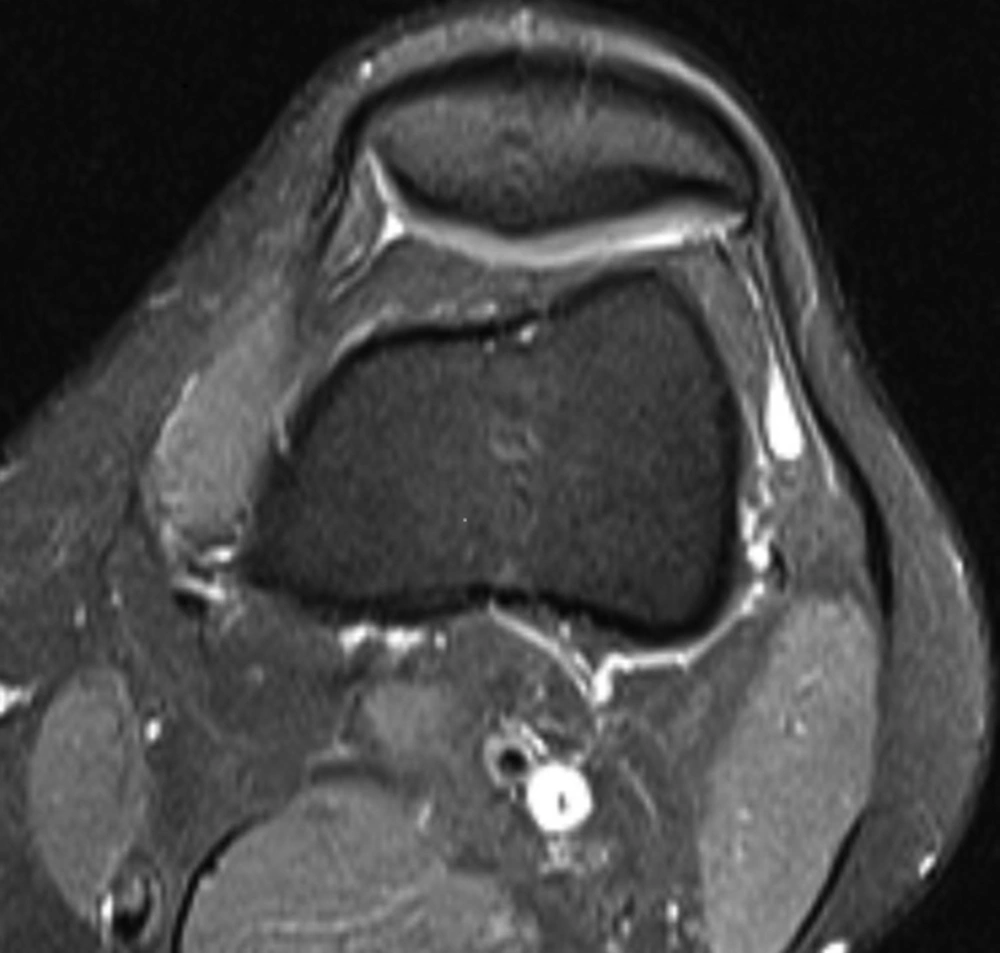
13) studied 128 patients to assess the frequency of MRI signs of ITBFS in patients with advanced medial compartment knee osteoarthritis and stated that ITBFS frequently (74.2%) accompanies osteoarthritis of the knee. Their model was that decreased medial joint space created a varus knee deformation, hence putting additional tension to the iliotibial band. They defined MRI findings of ITBFS including poorly defined abnormalities of signal intensity and localized fluid collection lateral, distal or proximal to the lateral epicondyle, and signal intensity abnormalities superficial to or deep by the iliotibial band. We used MRI findings confirming ITBFS including localized fluid collection, thickening of the iliotibial band, lateral patellofemoral joint distance reduction in axial plane and lateral movement of the patella.
As far as we know there is no data regarding size criteria for iliotibial band thickness. Thus, we evaluated the thickness of iliotibial band subjectively. This was the first limitation of our study. In addition, our study had other limitations including the small sample size, and retrospective structure.
In conclusion, ITBFS has some objective imaging criterions in MRI and may be associated with internal or anatomic factors including patellar shape and height.
ITBFS is more frequently accompanied with patella alta and type I and III patella according to Wiberg classification.