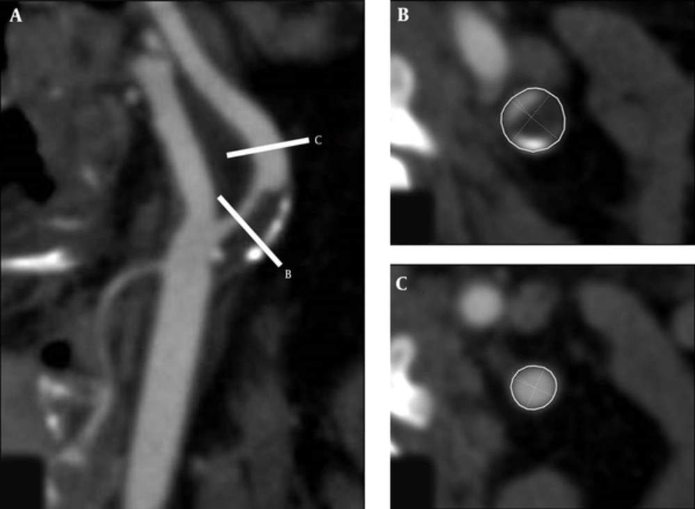
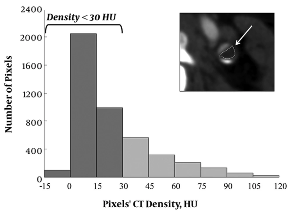
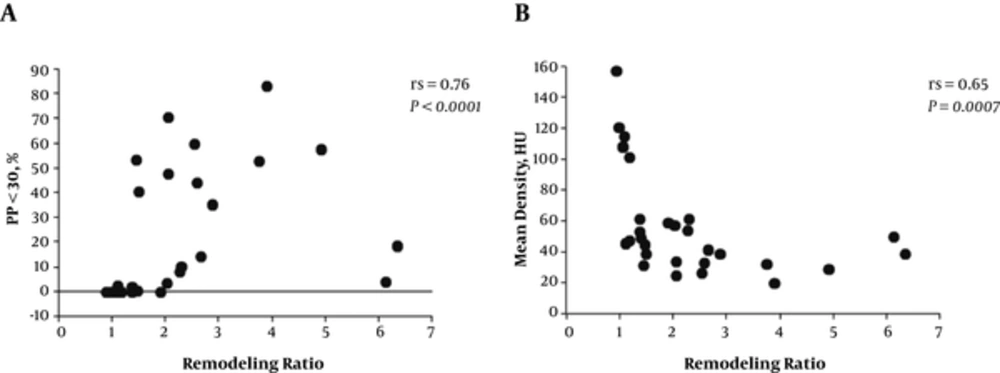
We demonstrated that the percentage of the low density plaques of less than 30 HU calculated by histogram analysis and the remodeling ratio were correlated with the AHA type VI lesions, indicating vulnerable and complicated plaques. Of note, these complex plaques are associated with a higher incidence of cerebrovascular events (
4-
6).
Recently, the advanced imaging modalities have enabled visualizing atherosclerotic plaques. US, MRI, and CTA were non-invasive imaging modalities compared with other modalities such as intravascular ultrasound (IVUS) (
8). CTA can rapidly provide data on the whole carotid artery with high spatial resolution, making it a good candidate for routine imaging modality of carotid artery disease (
3,
9). The assessment of percent luminal narrowing using CTA has been established in carotid artery disease in previous studies like NASCET (
3,
5,
8). Also, CTA enables visualization of plaque calcification, and morphology; smooth, irregular, or ulcerated surfaces (
8). Previous studies demonstrated that diagnostic accuracy of plaque ulceration by CTA was significantly higher than by US (
8,
15). Recently, an additional new objective index, the remodeling ratio, has been considered useful for assessment of positive remodeling secondary to atherosclerotic plaque inflammation (
3,
5,
7,
8). CT has a disadvantage that measurement of plaque density may be inferior to MRI for determining the characteristics of plaques, as described in the later section (
5). MRI could detect characteristics of the vulnerable plaque with high sensitivity and specificity. However, the resolution was not high enough to detect small structures or components, and a long scan time was needed, causing partial volume effects and motion artifacts. The resolution, cardiac motion artifacts, and signal intensity were strongly influenced by hardware, vender-specific software, and scan parameters (
8,
16). US is an easy and reproducible investigation tool and allows assessment of the degree of stenosis and morphology with high spatial resolution. However, the accuracy is operator dependent and only the cervical portion of the extracranial carotid is accurately imaged. Although the thickness of carotid intima media obtained using US has been shown to be correlated with the risk of stroke, the recent study in the general population showed no such correlation (
8).
In previous studies, carotid plaque density was associated with recent ischemic neurologic events (
9). It was reported that lipids, hemorrhage, and necrotic debris (called “soft tissue” by the authors) had the lowest tissue densities on CT and intraplaque hemorrhage was found in AHA type VI (
4). However, the density of other tissue components (fibrosis or calcifications) and contrast agent or calcified portions in the adjacent lumen influenced the plaque densities, which affected the mean plaque densities and decreased the accuracy of plaque classification (
4,
10,
11). For the coronary atherosclerotic plaque characterization, a few studies have reported the potential role of a quantitative histogram analysis for distribution of pixels showing low density (
10,
11). For the carotid atherosclerotic plaques, the correlation between MDCT images for measurement of lipid core area and histological sections was poor for calcified plaques in a previous study (
2). In our carotid artery study, histogram analysis showed significant differences between the type VI and non-type VI groups, but mean plaque densities showed no significant differences. Also, histogram analysis showed a more significant correlation with the remodeling ratio than the analysis of mean plaque density. As the extent of expansive remodeling may indicate underlying atherosclerotic plaque vulnerability of the ICA (
3,
5), histogram analysis may be helpful for the carotid atherosclerotic plaque evaluation on the CTA. Furthermore, the extent of the remodeling ratio correlated well with the plaque evaluation as previously reported (
5).
This study had several potential limitations. First, the presence of neurological symptoms showed no significant correlation with the remodeling ratio, PP < 30, or AHA plaque types. As we selected patients treated by CEA, there were only four (14%) asymptomatic patients, leading to the small sample bias. Second, as we have already mentioned in the “Patients and Methods”, we selected the cutoff for low-density plaques based on the previous coronary CTA histogram study (
11) using a similar scanner to ours. However, the cutoff may be less because the HU criteria of coronary plaques were reported to be significantly higher than those of carotid plaques, which are caused by more partial volume effects from lumen contrast enhancement and the surrounding tissue due to the smaller plaque size (
12). In addition, the cutoff may be different according to CT vendor or number of detector rows (
12). Thus, further evaluation is necessary.
In conclusion, histogram analysis of the carotid artery plaque density may be more useful for the evaluation of atherosclerosis on CTA compared with the conventional analysis of mean plaque density, and the combined analysis of histogram analysis and remodeling ratio may help to predict future stroke events.