

Full Text
Full text is available in PDF
IJ Radiology
Posterior longitudinal ligament (PLL) originates at the basiocciput, extends along the posterior aspect of the vertebral bodies and intervertebral discs and inserts into the sacrum. When hypertrophied (HPLL) and ossified (OPLL), the ligament encroaches upon the spinal canal. The OPLL is diagnosed when ossification originates within or spans the space between two discs to involve the ligament overlying the centrum of the vertebral body in between. The HPLL is defined by thickening of the PLL that compresses the dural tube. The differential diagnosis should be made based on the existence of ossification either on the X-rays or the histological findings. The relation between HPLL and OPLL is still controversial, and there is no universal consensus as to whether HPLL evolves into OPLL. For diagnosis, plain films may be useful. MRI should be the initial study for a patient with non-traumatic myelopathy. OPLL appears as an area of low signal and CT-myelogram provides better resolution of osseous anatomy and longitudinal retrovertebral opacity. Etiology of OPLL is unknown, but high incidence of diffuse idiopathic skeletal hyperostosis (DISH) among OPLL patients suggests a hereditary diathesis of spinal ligament ossification. The diagnostic features of DISH are: calcifications and ossifications along the anterolateral aspect of vertebral bodies, osteophytes, preservation of disk height, an absence of excessive disk disease, bony ankylosis, and sacroiliac erosion. Radiography is sufficient for diagnosing DISH. Occasionally, computed tomography (CT) scanning may be performed to evaluate complications. Bone scanning and MRI do not play a significant role.
© 2014, Author(s). This open-access article is available under the Creative Commons Attribution 4.0 (CC BY 4.0) International License (https://creativecommons.org/licenses/by/4.0/), which allows for unrestricted use, distribution, and reproduction in any medium, provided that the original work is properly cited.
Bahrami Motlagh H, Zare Mehrjardi M. Imaging Presentations of Spinal Instability. I J Radiol. 2014;11(30th Iranian Congress of Radiology):e21297. doi: https://doi.org/10.5812/iranjradiol.21297
Bakhshandehpour G, Khaksar P, Rakhsha S, Dalili S, Movahhedi MR. MR Myelogram: Is There any Role for Diagnosis of Spinal and Extra-Spinal Disorders?. I J Radiol. 2014;11(30th Iranian Congress of Radiology):e21318. doi: https://doi.org/10.5812/iranjradiol.21318
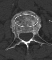
Liu D, Zhong D, Cao A. Use of Computed Tomography Compression Ratio in Diagnosis of Disc Injuries and Posterior Ligamentous Complex Injuries in Osteoporotic Thoracolumbar Compression Fractures. I J Radiol. 2021;18(4):e111779. doi: https://doi.org/10.5812/iranjradiol.111779
Emami Razavi SZ, Aryan A, Kazemi S, Rostamian A, Jahangiri A, et al. Prevalence of Hip Ossification and Related Clinical Factors in Cases With Spinal Cord Injury. Arch Neurosci. 2015;2(4):e25395. doi: https://doi.org/10.5812/archneurosci.25395
Mehrian P, Karimi MA, Kahkuee S, Bakhshayeshkaram M, Ghasemikhah R. Solitary Osteochondroma of the Thoracic Spine with Compressive Myelopathy; A Rare Presentation. I J Radiol. 2013;10(2):77-80. doi: https://doi.org/10.5812/iranjradiol.12015

Checking
Last Update: 2 weeks ago
Last Update: 2 weeks ago
Last Update: 2 weeks ago
Last Update: 4 days ago
Ordering Reprints
Articles are published under the Creative Commons license stated on each article. No permission or royalty fee is required for uses permitted by that license. CCC handles optional bulk and customized reprint orders. Any quotation covers production and delivery services only, not copyright permission. > Request Reprints from CCC