


1. Background
2. Objectives
3. Patients and Methods
3.1. Study Population
3.2. Ethics Issues
3.3. MRI Imaging Acquisition
| Sequence | Plane | FOV, mm | Matrix | Thickness, mm | Phase direction | TR, ms | TE, ms | TI, ms | No. of signals acquired | Acquisition time, min:sec |
|---|---|---|---|---|---|---|---|---|---|---|
| PSIR | Sagittal | 240 | 320 × 256 | 3 | A-P | 2200 | 10 | 400 | 2 | 2:53 |
| STIR | Sagittal | 240 | 320 × 256 | 3 | A-P | 3000 | 13 | 150 | 2 | 1:39 |
| T2W TSE | Sagittal | 240 | 320 × 256 | 3 | A-P | 2800 | 120 | - | 3 | 2:03 |
| PSIR | Axial | 220 | 320 × 256 | 4 | A-P | 2900 | 10 | 400 | 2 | 3:40 |
| T2W TSE | Axial | 220 | 320 × 256 | 4 | A-P | 3500 | 120 | - | 3 | 2:37 |
Abbreviations: A, anterior; FOV, field of view; P, posterior; PSIR, phase sensitive inversion recovery; STIR, short tau inversion recovery; TE, echo time; TI, inversion time; TR, repetition time; TSE, turbo spin echo
3.4. Image Analysis
3.4.1. Number of Lesions
3.4.2. Lesion Conspicuity
3.5. Statistical Analysis
4. Results
4.1. Sagittal View
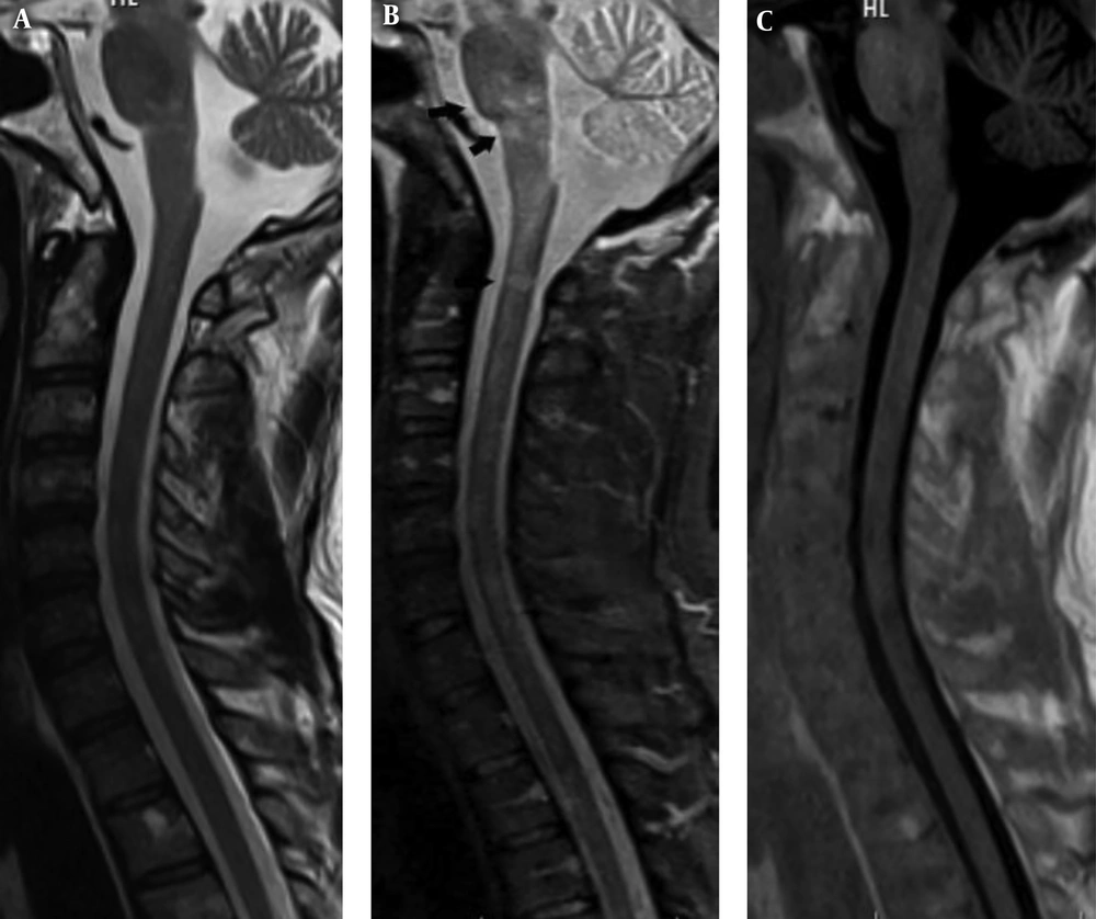
A 32-year-old woman with relapsing remitting multiple sclerosis (RRMS) who had a neurological attack. Sagittal images in T2 weighted turbo spin echo (T2W TSE) (A), short tau inversion recovery (STIR) (B) and phase sensitive inversion recovery (PSIR) (C) sequences have been shown. The patient has plaques in the pons, medulla oblongata and at the level of C2 that are not visible on PSIR and T2W TSE images, while it is delineated in STIR sequence (black arrows). Note the better lesion count in STIR compared to other sequences in this case.
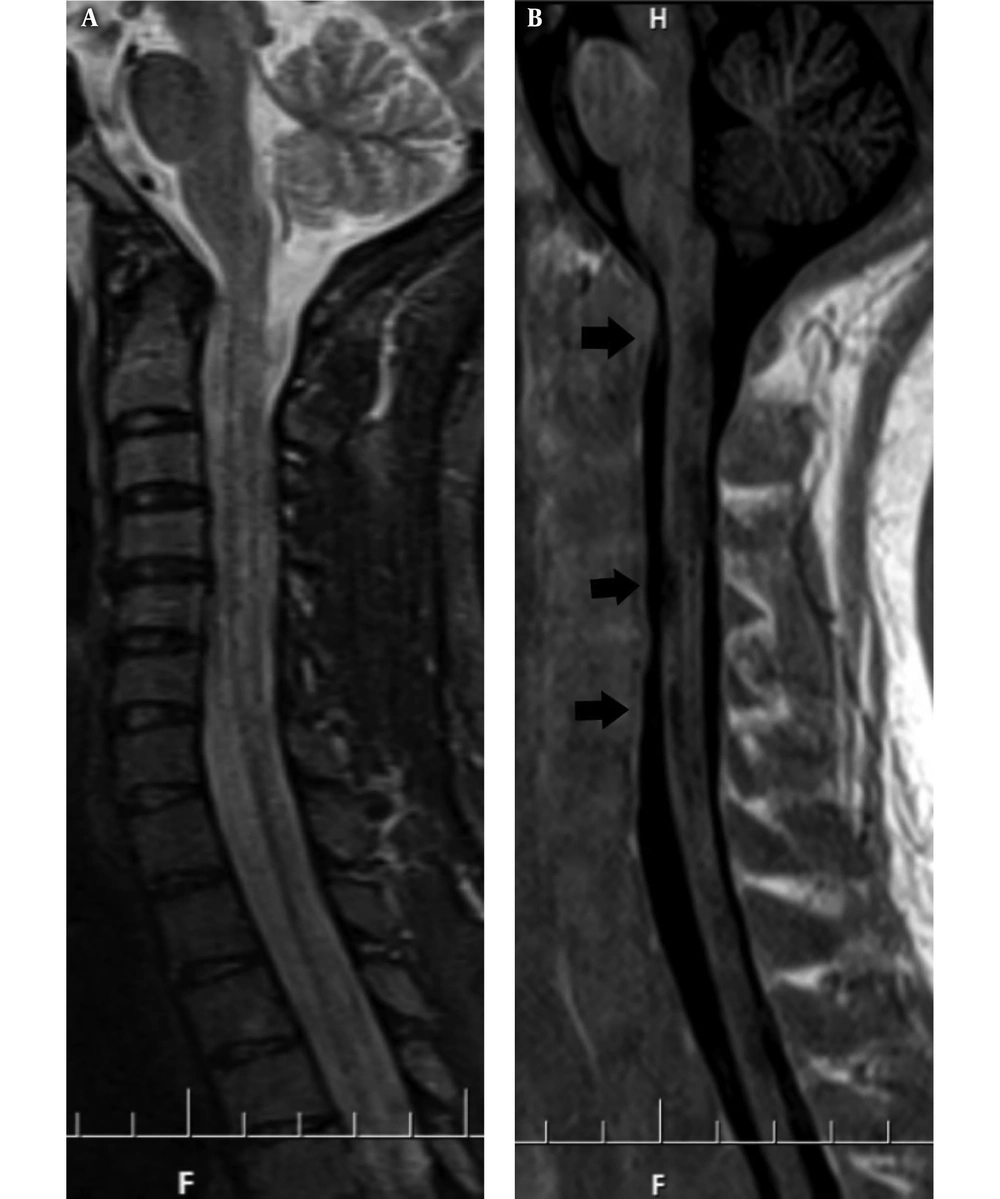
A 28-year-old female patient who is a known case of relapsing remitting multiple sclerosis (RRMS) on follow up. Sagittal short tau inversion recovery (STIR) image shows subtle high signal intensity (dirty white matter) without delineation local lesions at the cord (A). Sagittal phase sensitive inversion recovery (PSIR) shows multiple local hypointense lesions at the level of C2 - T2 (black arrows) (B). The lesions could be seen in a poor conspicuity in STIR, while in PSIR, it is better detected.
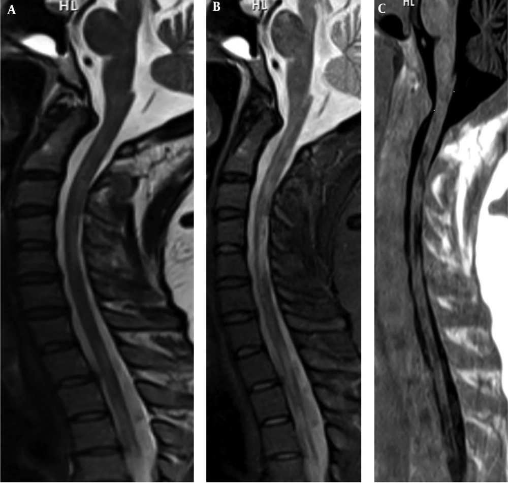
A 32-year-old male patient with secondary progressive multiple sclerosis (SPMS) who presented with tingling, numbness and weakness of both lower limbs. Sagittal T2 weighted turbo spin echo (T2W TSE) (A), short tau inversion recovery (STIR) (B) and phase sensitive inversion recovery (PSIR) (C) images show multiple MS plaques extending from C2 to T3. They display bright signal intensity (SI) on T2W TSE and STIR and low SI on PSIR. Note that lesions have more conspicuity in PSIR than STIR and T2 W images.
| Plaque number | Mean ± SD of plaque No. | Range | Total plaque counts in all patients | Comparison of plaque number | ||||||
|---|---|---|---|---|---|---|---|---|---|---|
| All three sequences | PSIR vs. T2W TSE | STIR vs. T2W TSE | PSIR vs. STIR | |||||||
| P | Mean difference | P | Mean difference | P | Mean difference | P | ||||
| T2W TSE | 1.7 ± 1.1 | 0 - 4 | 59 | < 0.001 | 1.7 | < 0.001 | 1.9 | < 0.001 | 0.2 | 0.3 |
| STIR | 3.5 ± 1.4 | 1 - 8 | 124 | |||||||
| PSIR | 3.4 ± 1.4 | 1 - 7 | 118 | |||||||
Abbreviations: PSIR, phase sensitive inversion recovery; SD, standard deviation; STIR, short tau inversion recovery; TSE, turbo spin echo.
| Imaging conspicuity | No. (%) | Comparison of plaque imaging conspicuity (P values) | |||
|---|---|---|---|---|---|
| All three sequences | PSIR vs. T2W TSE | STIR vs. T2W TSE | PSIR vs. STIR | ||
| T2W TSE | |||||
| Poor | 29 (90.6) | < 0.001 | < 0.001 | < 0.001 | < 0.001 |
| Moderate | 3 (9.4) | ||||
| Good | 0 | ||||
| STIR | |||||
| Poor | 3 (9.4) | ||||
| Moderate | 23 (71.9) | ||||
| Good | 6 (18.8) | ||||
| PSIR | |||||
| Poor | 1 (3.1) | ||||
| Moderate | 7 (21.9) | ||||
| Good | 24 (75) | ||||
Abbreviations: PSIR, phase sensitive inversion recovery; SD, standard deviation; STIR, short tau inversion recovery; TSE, turbo spin echo.
aPSIR conspicuity better than STIR and T2W TSE, STIR conspicuity better than T2W TSE
4.2. Axial View
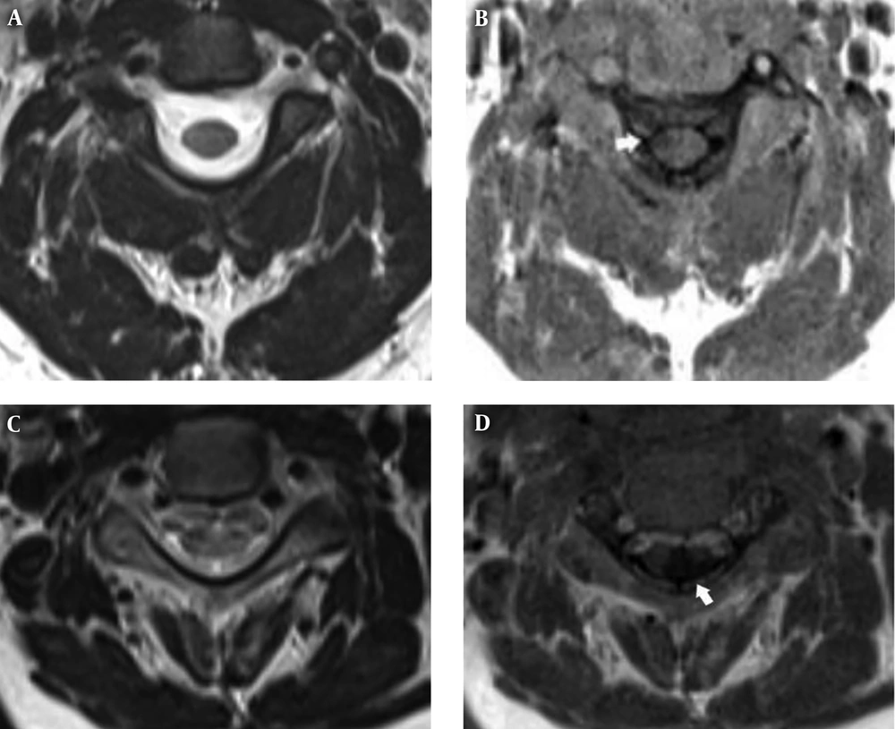
A 36-year-old woman with relapsing-remitting multiple sclerosis (RRMS) and upper extremity weakness (A and B). A 32-year-old man with secondary-progressive MS (SPMS) and upper and lower extremity paresthesia (C and D). Axial T2 weighted turbo spin echo (T2W TSE) (A and C). Axial phase sensitive inversion recovery (PSIR) (B and D). MS plaques are seen in PSIR sequence (white arrows) (B and D). No sign of plaques is detected in T2W TSE (A and C).
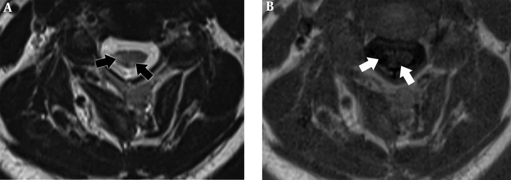
A 42-year-old patient with relapsing remitting multiple sclerosis (RRMS). Axial T2 weighted turbo spin echo (T2W TSE) (A). Axial phase sensitive inversion recovery (PSIR) (B). In both images, two plaques are seen in the cervical/spine. The lesion is clearer, the border is clear and differentiation between the lesion and the normal tissue is better in the PSIR sequence (white arrows) compared to T2W TSE (black arrows).
| Plaque number | Imaging conspicuity, No. (%) | ||||||||
|---|---|---|---|---|---|---|---|---|---|
| Mean ± SD | Range | Total plaque counts in all patients | Mean difference | P value | Poor | Moderate | Good | P value | |
| T2W TSE | 2.4 ± 2.1 | 1 - 10 | 83 | 1.3 ± 1 | 0.004 | 23 (65.7) | 12 (34.3) | 0 | < 0.001a |
| PSIR | 3.7 ± 2.5 | 1 - 12 | 126 | 0 | 13 (37.1) | 22 (62.9) | |||
Abbreviations: PSIR, phase sensitive inversion recovery; SD, standard deviation; TSE, turbo spin echo.
aPSIR conspicuity better than T2W TSE.


