Iranian Journal of Radiology

Image Credit:Iranian Journal of Radiology

An Alternative Loop Snare Technique with Many Loops Using a Simple Guide-Wire for Removal of a Double J Stent: A Case Series

Author(s):
Su Ho KimSu Ho Kim1, Byung Gil ChoiByung Gil Choi2,*, Oh Jung SukOh Jung Suk2, Ho Jong ChunHo Jong Chun1, Hae Giu LeeHae Giu Lee2
1Department of Radiology, Eunpyeong St. Mary’s Hospital College of Medicine, The Catholic University of Korea, Seoul, Korea
2Department of Radiology, Seoul St. Mary’s Hospital College of Medicine, The Catholic University of Korea, Seoul, Korea
*Corresponding Author: Department of Radiology, Seoul St. Mary’s Hospital College of Medicine, The Catholic University of Korea, Seoul, Korea. Tel: 82-222581485, Email: [email protected]

IJ Radiology:Vol. 17, issue 3; e98787
Published online:Jul 13, 2020
Article type:Brief Report
Received:Dec 04, 2019
Accepted:Jun 15, 2020
How to Cite:Kim SH, Choi BG, Jung Suk O, Chun HJ, Lee HG. An Alternative Loop Snare Technique with Many Loops Using a Simple Guide-Wire for Removal of a Double J Stent: A Case Series. I J Radiol. 2020;17(3):e98787. doi: https://doi.org/10.5812/iranjradiol.98787

Abstract

Background:

Removal of a double J (DJ) stent has been conventionally performed with conventional snare wires, tissue forceps, or stone baskets under fluoroscopic guidance. However, if the lumen is collapsed, is very wide, or if the tip of the DJ stent is lodged in the bladder wall, the procedure becomes difficult.

Patients and Methods:

We retrospectively reviewed four patients who had undergone retrieval or replacement of their DJ stents by interventional radiologists with an alternative loop snare technique using a guide-wire after conventional attempts using a snare catheter had failed.

Results:

Five DJ stents were completely removed without any complications.

Conclusion:

The loop snare technique using a guide-wire could be an alternative and inexpensive approach for difficult DJ stent removal.

1. Background

The placement of a double J (DJ) ureteral stent is usually performed by a urologist through the bladder via cystoscopy. However, if bladder access is difficult or the urethral stricture is severe, placement may be carried out using a percutaneous renal nephrostomy (PCN). The removal and exchange of DJ stents are also typically performed using a retrograde approach, but retrieval procedures become difficult when DJ stents have migrated to the renal pelvis or become lodged in the bladder wall. In these cases, patients are referred to an interventional radiologist and removal of the DJ stent is attempted under fluoroscopic guidance with conventional snare wires, tissue forceps, or stone baskets (1). However, if the lumen has collapsed, is very wide, or if the tip of the DJ stent is lodged in the bladder wall, there is a limit to the amount of time that could be spent handling these devices. In addition, tissue forceps and stone baskets could cause physical damage to the bladder wall. If the use of any one device is difficult, various other devices may be tried, which could increase the cost of the procedure.

2. Objectives

Therefore, we report an alternative loop snare technique using a guide-wire.

3. Patients and Methods

3.1. Patients

This retrospective study was approved by our Institutional Review Board. Between 2017 and 2018, 17 patients were referred to interventional radiology for exchange or removal of the DJ stent. DJ stents were successfully removed using a conventional technique in 13 patients. Four patients underwent retrieval or change of the DJ stent with an alternative loop snare technique by an interventional radiologist using a guide-wire. A patient underwent the same procedure twice. There were two males and two females, with an age range from 43 to 57 years (mean: 48.6 years) (Table 1).
Table 1.Summary of Patient Demographics
PatientAge/sexUnderlying diseaseProcedureLocation of tip of stentTP, minTC, minTA, min
143/MCystitis cystica1Renal pelvis20164
245/FCervical cancer2Bladder26215
3Renal pelvis25178
357/MBladder cancer4Renal pelvis433211
453/FOvarian cancer5Bladder21165

Abbreviations: TA, time for alternative loop snare technique; TC, the elapsed time for the conventional technique; TP, total procedure time.

3.2. Technique
All procedures were performed with patients under moderate sedation by two certified attending interventional radiologists with 7 years and 21 years of experience, respectively.
Prior to starting the procedure, both the size and position of the DJ stents were evaluated under fluoroscopy to determine where to perform the procedure. When the tips of the three migrated DJ stents were located in the renal pelvis, the procedure was carried out by an antegrade approach using the PCN route. When the tips of the two stents were in the bladder, the procedure was performed using a retrograde approach via the transurethral route. Performing a retrograde approach, a 7-F latex nelaton catheter (Sewoon, Chingdao, China) was first inserted into their urinary bladders for passage of the guide wire.
After determining the best route, an 8-Fr. introducer sheath (Radiofocus, Terumo Corp., Tokyo, Japan) was inserted over the 0.035-inch guide-wire. Then a 6-Fr conventional snare catheter (Multi-Snare® set, pfm medical ag, Koln, Germany) with a loop diameter of 20 mm was inserted into the introducer sheath. The loop of the snare catheter was then advanced between the tip and the wall. If it failed to capture the tip despite several attempts, the procedure was changed to the alternative loop snare technique.
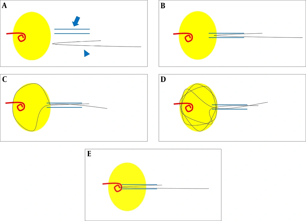
The alternative loop snare was fabricated using a 0.018-inch, 180-cm long hydrophilic guide-wire (Radiofocus, Terumo Corp., Tokyo, Japan). The end of the guide-wire was bent to the length of the sheath and inserted into the introducer sheath with the bent end first. The reason for bending as much as the length of the sheath is that if the short string is held, the bent end does not exit the sheath. By advancing the guide-wire loop to the introducer sheath tip, the short string could be fixed with one hand, which allowed for the radiologist to push the long string of the wire slightly. The reason for this motion is to prevent tissue damage when the bent section is sharp. If the radiologist continues to push the long string part, multiple loops will be created in the lumen. Due to the elasticity of the guide-wire, these loops are formed along the wall of the lumen. Even if the tip of the DJ stent is lodged in the lumen, the loop could still be inserted between the DJ stent tip and the wall. If the tip of the DJ stent is expected to come into one of the multiple loops, the operator could pull both the short string and the long string at the same time and check if the DJ stent is caught. If not, another attempt should be made to push the long string and the short string at the same time. If the DJ stent is caught, the radiologist should pull the wire and guide the DJ stent into the sheath to remove it. These steps are demonstrated using a flask and a straw (Figure 1) to represent the lumen and the sheath.
A, Prepare the introducer sheath (blue arrow) and a 0.018-inch guide-wire (blue arrowhead). Bend the end of the guide-wire as much as the sheath length. The yellow circle represents the lumen, such as a bladder or a renal pelvis; B, Insert the introducer sheath in the lumen and push the bent end of the guide-wire through the sheath; C, Create a loop by pushing the long string. The short string should be held to prevent the folded end from entering the lumen; D, When the long string is inserted continuously, the loop touches the lumen wall, and multiple loops are created due to the elasticity of the guide-wire; E, If a foreign body enters the loops, pull the short string and the long string simultaneously and guide both strings into the sheath. Finally, remove the foreign body along with the sheath.
Figure 1.

A, Prepare the introducer sheath (blue arrow) and a 0.018-inch guide-wire (blue arrowhead). Bend the end of the guide-wire as much as the sheath length. The yellow circle represents the lumen, such as a bladder or a renal pelvis; B, Insert the introducer sheath in the lumen and push the bent end of the guide-wire through the sheath; C, Create a loop by pushing the long string. The short string should be held to prevent the folded end from entering the lumen; D, When the long string is inserted continuously, the loop touches the lumen wall, and multiple loops are created due to the elasticity of the guide-wire; E, If a foreign body enters the loops, pull the short string and the long string simultaneously and guide both strings into the sheath. Finally, remove the foreign body along with the sheath.

Technical success for retrieval of the DJ stent was defined as removal of the entire DJ stent from the body. The total procedure time was defined as the total time from insertion of the introducer sheath to removal of the DJ stent. The elapsed time for the conventional technique was measured as the time from insertion of the introducer sheath to conversion of the procedure. The time length of the alternative loop snare technique was calculated as the time from conversion of the procedure to removal of the introducing sheath.

4. Results

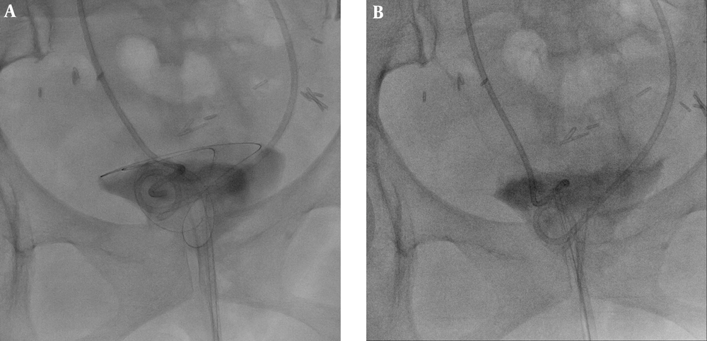
The overall success rate for retrieval of the DJ stent in this study was 100% (5/5). The tips of three DJ stents were placed in the renal pelvis, while the tips of two stents were located in the bladder (Figure 2).
A 53-year-old woman had two DJ stents due to ovarian cancer with bladder metastasis. The patient was referred to our department to replace the DJ stents, which were functioning poorly. A, After the conventional technique using a snare catheter failed, an 8-Fr introducer sheath was inserted through the urethra into the bladder. Then, a bent, 0.018-inch guide-wire was inserted through the sheath. Multiple loops were made by pushing the long string; B, When the DJ stent entered one of the loops, the short and long strings were pulled out simultaneously to introduce both strings into the sheath.

Figure 2.

A 53-year-old woman had two DJ stents due to ovarian cancer with bladder metastasis. The patient was referred to our department to replace the DJ stents, which were functioning poorly. A, After the conventional technique using a snare catheter failed, an 8-Fr introducer sheath was inserted through the urethra into the bladder. Then, a bent, 0.018-inch guide-wire was inserted through the sheath. Multiple loops were made by pushing the long string; B, When the DJ stent entered one of the loops, the short and long strings were pulled out simultaneously to introduce both strings into the sheath.


All DJ stents were examined after retrieval and were removed in their entire length without any evidence of component disruption. All DJ stents were 7-Fr ureteral stents (Percuflex®; Boston Scientific, Natick, MA). The total procedure time ranged from 20 to 43 minutes. The elapsed time for the conventional technique ranged from 16 to 32 minutes, while the total operative time for the alternative loop snare technique ranged from 4 to 11 minutes. Additional commercially available retrieving devices or procedures were not required. There were no significant complications related to these procedures. Follow-up plain films showed appropriate stent placement in all patients.

5. Discussion

Radiologic techniques are routinely used to guide the percutaneous retrieval of intravascular foreign bodies (1). Occasionally, these techniques are applied to the percutaneous retrieval of foreign bodies in the genitourinary tract and the pleural space (2, 3).
When a DJ stent is removed, the advantage of an alternative loop snare technique, compared to existing techniques, is that the loop can easily advance between the wall and the tip of the stent, which facilitates capture. Another advantage is that this technique uses only introducer sheaths and a 0.018 inch-guide-wire. As a result, this technique is cost effective because no additional device is needed. This approach also caused no damage to the surrounding tissues in any of our five procedures because it used a soft guide-wire to make the loop. No complications occurred in any patients.
Previous studies have shown that intravascular foreign bodies can successfully be removed using a self-made snare catheter (4, 5). The alternative loop snare technique using a guide-wire creates many loops, and the loop size is expanded to the maximum size in accordance with the lumen space, making it easy to ensnare the tip of the DJ stent. One disadvantage of this study is that there are only a few procedures that have led to a small sample size. However, this technique is very easy to reproduce and could be applied in any situation.
In conclusion, the loop snare technique using a guide-wire can be an alternative, relatively safe, inexpensive, and effective approach for removing foreign bodies that are difficult to retrieve with conventional methods.

Footnotes

  • Authors' Contributions: Study concept and design: Su Ho Kim, Byung Gil Choi, and Hae Giu Lee. Acquisition of data: Su Ho Kim. Analysis and interpretation of data: Su Ho Kim. Drafting of the manuscript: Su Ho Kim. Critical revision of the manuscript for important intellectual content: Byung Gil Choi. Statistical analysis: Su Ho Kim. Administrative, technical, and material support: Byung Gil Choi, Jung Suk Oh, and Ho Jong Chun. Study supervision: Byung Gil Choi.

  • Conflict of Interests: All authors declare there is no conflict of interest.

  • Ethical Approval: This retrospective study was approved by our Institutional Review Board. The ethical approval code is KC18RESI0577.

  • Funding/Support: The authors received no specific funding for this work.

References

Copyright

Copyright © 2020, Author(s). This is an open-access article distributed under the terms of the Creative Commons Attribution-NonCommercial 4.0 International License (http://creativecommons.org/licenses/by-nc/4.0/) which permits copy and redistribute the material just in noncommercial usages, provided the original work is properly cited.

Similar Articles

22
Jul
2015

Fluoroscopy Guided Transurethral Placement of Ureteral Metallic Stents

Song Myung Gyu,
Tae-Seok Seo,
Cheol Min Park,
Jae Woong Choi,
Jong Mee Lee,
Yang shin Park

Myung Gyu S, Seo T, Park CM, Choi JW, Lee JM, et al. Fluoroscopy Guided Transurethral Placement of Ureteral Metallic Stents. I J Radiol. 2015;12(3):e91315. doi: https://doi.org/10.5812/iranjradiol.20269v2

2
Oct
2019
Iranian Journal of Radiology

A Modified Wire-Loop Snare Technique to Retrieve Inferior Vena Cava Filters with Severe Tilt and Embedded Hooks

Yuewei Wang,
Haofu Wang,
Hai Xin,
Jun Li

Wang Y, Wang H, Xin H, Li J. A Modified Wire-Loop Snare Technique to Retrieve Inferior Vena Cava Filters with Severe Tilt and Embedded Hooks. I J Radiol. 2019;16(4):e84404. doi: https://doi.org/10.5812/iranjradiol.84404

16
Apr
2016

An Extremely Rare Complication of Ureteral Pigtail Stent Placement: A Case Report

Davood Arab,
Arash Ardestani Zadeh,
Rahimeh Eskandarian,
Mehrshad Asaadi,
Kamran Ghods

Arab D, Ardestani Zadeh A, Eskandarian R, Asaadi M, Ghods K. An Extremely Rare Complication of Ureteral Pigtail Stent Placement: A Case Report. Nephro-Urol Mon. 2016;8(3):e36527. doi: https://doi.org/10.5812/numonthly.36527

30
Jun
2010

Percutaneous Retrieval of a Dislodged Guide Wire by Dormia Basket in a 98-year-old Patient, Still a Safe and Feasible Approach

N Zirak,
Gh Soltani,
A Maraghe moghaddam,
MH Nezafati,
M Abbasi,
H Javan

Zirak N, Soltani G, Maraghe moghaddam A, Nezafati M, Abbasi M, et al. Percutaneous Retrieval of a Dislodged Guide Wire by Dormia Basket in a 98-year-old Patient, Still a Safe and Feasible Approach. Int Cardiovasc Res J. 2010;4(2):e65020. doi:

29
Jul
2015

Double-J Versus External Ureteral Stents in Kidney Transplantation: A Retrospective Analysis

Thomas Vogel,
Markus Utech,
Fabian Schmidt,
Wiebke Holscher Keplin,
Ricarda Diller,
Jens Brockmann
,et al.

Vogel T, Utech M, Schmidt F, Holscher Keplin W, Diller R, et al. Double-J Versus External Ureteral Stents in Kidney Transplantation: A Retrospective Analysis. Nephro-Urol Mon. 2015;7(4):e27820. doi: https://doi.org/10.5812/numonthly.27820

Download PDF1.52 MB

Crossmark

Crossmark

Checking

Share on
Cited by
Metrics

Ordering Reprints

Articles are published under the Creative Commons license stated on each article. No permission or royalty fee is required for uses permitted by that license. CCC handles optional bulk and customized reprint orders. Any quotation covers production and delivery services only, not copyright permission. > Request Reprints from CCC 

Search Relations

Author(s):

Related Articles