
1. Introduction
First branchial cleft anomalies constitute a rare subset of congenital malformations affecting the head and neck region, accounting for less than 10% of all branchial apparatus defects (1). These anomalies are typically classified into three structural forms based on their embryological development: Cysts, which are enclosed epithelial-lined sacs; sinuses, with a single external opening; and fistulas, which exhibit both internal and external tracts.
Embryologically, the branchial arches appear bilaterally in five distinct pairs around the fourth week of gestation. The first and second arches play crucial roles in craniofacial development, giving rise to structures such as the mandible and hyoid, respectively. Between these arches lies the first branchial cleft, which deepens dorsally to form the external auditory canal. Failure in the complete regression or closure of its ventral portion may result in a spectrum of anomalies collectively referred to as first branchial cleft malformations (2).
The extent and location of the developmental defect determine the resulting anatomical presentation. These lesions typically originate near the cartilaginous or osseocartilaginous junction of the external auditory canal and may extend inferiorly toward the mandibular or submandibular region, depending on the severity (1).
Multiple classification systems have been proposed, with the work classification being the most widely accepted. Type I anomalies are purely ectodermal in origin and typically present as cystic or sinus-like lesions located medial to the conchal bowl, often terminating blindly within the parotid region. In contrast, type II anomalies have both ectodermal and mesodermal components, often containing skin adnexa and cartilage. These are more complex and may traverse the parotid gland and lie in proximity to, or intersect with, branches of the facial nerve (1, 3).
Among these malformations, external auditory canal duplication is an extremely rare congenital anomaly, with an annual incidence of approximately 1 per 1,000,000 live births (4). This anomaly is characterized by the presence of an accessory canal — either rudimentary or partially formed — adjacent to a normally developed external canal. It may involve only the soft tissue portion or extend into the cartilaginous segment. Clinical manifestations range from preauricular pits and recurrent otorrhea to more serious complications such as persistent otitis externa or varying degrees of conductive or mixed hearing loss (3).
2. Case Presentation
We report the clinical case of a 1-year-and-6-month-old male infant presenting with duplication of the external auditory canal (Figure 1). Informed written consent for the publication of medical data and images was obtained from the patient’s legal guardians.
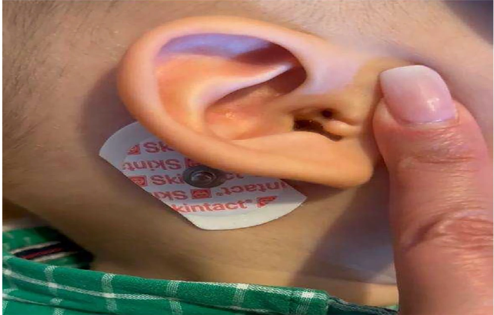
Figure 1.
Duplicating ear canal with ear tag at the entrance of ear canal in the right ear
2.1. Auditory System Assessment
A comprehensive audiological evaluation was conducted for the patient, including otoscopic inspection, tympanometry, acoustic reflex testing, and auditory brainstem response (ABR). These diagnostic measures were employed to assess auditory function in the presence of the duplicated canal anomaly. Parental consent for the case documentation and publication was formally secured.
2.2. Auditory Brainstem Response
The ABR is a non-invasive, objective method used to evaluate the integrity of the auditory pathway from the cochlea to the brainstem. In this procedure, surface electrodes are positioned on the scalp and earlobes to capture the brain’s electrical responses to auditory stimuli such as brief clicks or tone bursts. It is especially valuable for hearing screening in infants and for the diagnosis of retrocochlear or central auditory disorders (5-8). The ABR test generates five key waveform peaks.
A. Wave I: Arises from the distal portion of the auditory nerve.
B. Wave II: Originates from the proximal segment of the auditory nerve.
C. Wave III: Reflects activity in the cochlear nucleus.
D. Wave IV: Corresponds to responses from the superior olivary complex.
E. Wave V: Represents neural activity in the lateral lemniscus and inferior colliculus.
The evaluation focuses on the latency (in milliseconds) and amplitude (in microvolts) of these waves. Notably, the presence of Wave V at low stimulus intensities (5 - 20 dB) is a clinical indicator of near-normal auditory thresholds. In this patient, the ABR findings were:
A. Right ear: Mild conductive hearing loss.
B. Left ear: Moderate to severe mixed hearing loss.
2.3. Diagnostic Flow Summary
2.3.1. Overview of the Condition
1. Congenital anomaly involving external auditory canal duplication.
2. Frequently associated with sensorineural or mixed hearing loss.
2.3.2. Patient Overview
1. Male infant, 1 year and 6 months old.
2. Clinical signs: Accessory ear canal; ear tag located at the right external canal entrance.
2.3.3. Audiological Evaluation
1. Test performed: The ABR.
2. Findings:
A. Right ear: Mild conductive hearing impairment.
B. Left ear: Moderate to severe mixed-type hearing impairment.
3. Discussion
The presence of a duplicated external auditory canal is a rare anatomical anomaly resulting from incomplete obliteration of the ventral portion of the first branchial cleft during embryogenesis. This malformation may involve only the membranous part of the external auditory canal or extend into the cartilaginous component (3). Its clinical presentation is variable and may include recurrent ear discharge (otorrhea), preauricular pits or depressions, palpable periauricular masses, and in some cases, conductive or mixed hearing loss (9).
In the current case, ABR testing revealed mild conductive hearing loss in the right ear and moderate to severe mixed hearing loss in the left ear. These findings are consistent with previous literature describing duplicated auditory canals as potential causes of conductive auditory deficits due to malformed external structures that disrupt sound transmission pathways (9-11). The mixed hearing loss observed in the left ear suggests additional sensorineural involvement, possibly implicating the cochlea or auditory nerve.
3.1. Epidemiology
Such anomalies of the external auditory canal are uncommon and warrant early functional evaluation, and the true prevalence is difficult to establish due to the limited number of documented cases. The literature suggests no clear gender predilection (9). The scarcity of large-scale epidemiological studies likely stems from underdiagnosis and clinical overlap with other first branchial cleft anomalies.
3.2. Pathophysiological Correlation and Auditory Brainstem Response Utility
The ABR serves as a valuable diagnostic modality in such congenital anomalies, as it allows for early, non-invasive assessment of auditory pathway function. Conductive hearing loss, as seen in the right ear, may be attributed to anatomical blockage or sound conduction inefficiency through the duplicated canal. The mixed hearing loss on the contralateral side implies a more complex pathology involving both conductive and sensorineural elements, which could be due to associated inner ear maldevelopment or secondary inflammation (6).
3.3. Management Strategies
Management of duplicated ear canals must be individualized based on the severity of symptoms and hearing impairment. In symptomatic cases, surgical intervention is often warranted. Standard approaches include complete surgical excision of the duplicated tract or canaloplasty, depending on the anatomical complexity and involvement of surrounding structures. Canaloplasty may be preferred when preservation or reconstruction of the external auditory canal is desired. Surgical planning must consider the proximity of the tract to the facial nerve, especially in type II first branchial cleft anomalies, to avoid iatrogenic nerve injury (12). In asymptomatic or mild cases, conservative monitoring may be sufficient.
For patients with hearing deficits, early amplification with hearing aids or other rehabilitative strategies should be implemented to optimize speech and cognitive development during critical periods of language acquisition (6).
3.4. Prognosis and Long-Term Considerations
Prognosis largely depends on the extent of the structural anomaly and the severity of the associated hearing loss. Timely diagnosis, appropriate audiological monitoring, and targeted intervention can significantly improve outcomes. In this case, the right ear’s conductive component may respond well to medical or surgical correction, while the mixed hearing loss in the left ear may require ongoing audiological and possibly neurologic evaluation (10).
Clinical outcomes reported in the literature are generally favorable when early intervention is provided. For example, a retrospective review by Triglia et al. demonstrated that most patients with first branchial cleft anomalies who underwent surgical excision remained asymptomatic at follow-up, with no recurrence and preserved facial nerve function (13). Furthermore, Laccourreye et al. reported improved hearing thresholds in children undergoing corrective surgery for external auditory canal malformations, particularly when conducted before the age of 3 years (14).
These findings emphasize the importance of multidisciplinary care and early treatment to optimize auditory and developmental outcomes in patients with duplicated external auditory canals.
3.5. Advances in Imaging and Diagnostic Accuracy
The use of sophisticated imaging methods, such as high-resolution CT and MRI, has dramatically transformed the assessment of first branchial cleft anomalies, including rare presentations like duplicated external auditory canals. These advanced imaging techniques provide an unparalleled view of the anatomical details, enabling visualization not only of the duplication itself but also of its relationship with adjacent soft tissues and, in some cases, cartilage. Obtaining such accurate preoperative information is essential, as it helps clinicians avoid complications such as facial nerve injury, a known risk during surgical correction of these anomalies (15, 16). However, in the present case, imaging studies such as CT or MRI were not performed due to financial constraints faced by the patient’s family. This limitation reflects challenges in resource-limited settings and emphasizes the need for careful clinical and audiological evaluation to guide diagnosis and management.
3.6. Emerging Rehabilitative Approaches and Multidisciplinary Management
Surgery is not the only treatment option for individuals experiencing hearing loss due to these rare ear anomalies. Recent advancements in hearing technologies, such as bone conduction hearing aids and cochlear implants, have significantly expanded the rehabilitative possibilities, particularly for patients with combined conductive and sensorineural hearing loss. These devices can effectively improve hearing, enabling better speech and communication development. Comprehensive management typically involves a multidisciplinary team including otolaryngologists, audiologists, and speech therapists to address both the anatomical and functional aspects of the condition (17).
3.7. Key Findings
3.7.1. Auditory Brainstem Response Evaluation
In this case, ABR testing identified two distinct patterns of hearing impairment: A mild conductive hearing loss in the right ear and a moderate to severe mixed hearing loss in the left ear (Figure 2) (7). These findings underscore the importance of comprehensive auditory assessment in infants with congenital anomalies such as duplicated external auditory canals. Recognizing the specific type and degree of hearing loss enables clinicians to implement timely and appropriate management strategies, including surgical, medical, or rehabilitative interventions. Early identification is essential for minimizing developmental delays in speech, language, and cognitive skills and for improving the patient’s overall quality of life.
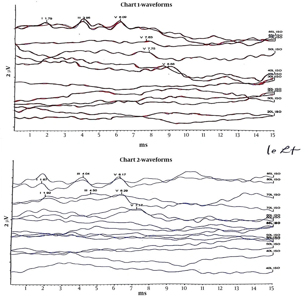
Figure 2.
Auditory brainstem response (ABR) test (chart 1: Right ear shows mild conductive hearing loss; chart 2: Left ear shows moderate to severe mixed hearing loss).