1. Introduction
Ehlers-Danlos syndrome (EDS) is a relatively rare condition involving a group of patients with inherited connective tissue disorder. This syndrome is caused by 3 fundamental mechanisms, including deficiencies of collagen-processing enzymes, dominant negative effects of mutant collagen, chains, and haploinsufficiency. The main findings of EDS cases include articular hypermobility, skin extensibility, and tissue fragility (1-3). The prevalence of EDS has been reported as 1 in 5000 to 100000 live births in different communities; however, the epidemiology of the specific types is largely unknown. EDS affects both sexes and has no racial predisposition (4-6). EDS has a wide range of expressing pattern depending on the type of collagen being affected. Villefranche introduced a classification system for EDS, which contained 6 major types. Variations were mainly based on the clinical, biochemical, and molecular differences (2, 3) (Table 1). The classical type characterized with hypermobility is known to be the most common form of EDS having type V collagen deficiency in patients with classic EDS, which demonstrates marked skin hyperextensibility, wide atrophic scarring, and significant joint hypermobility (1). Other findings comprised smooth velvety skin, molluscoid pseudotumors over pressure points, subcutaneous spheroids on the forearms or chins, muscle hypotonia, ecchymosis, and tissue fragility (5). Surgical or traumatic bruising can vary from mild to moderate and can be accompanied by delicate hyperextensible skin (7). Repeated dislocation of TMJ could be reported along with epicanthus, strabismus, narrow nasal bridge, and shaggy hair in facial area (8).
Intraoral manifestations include highly fragile mucosa and frequent periodontal tissue injuries. This is usually seen following minor oral surgeries such as a simple tooth extraction (9). Bleeding tendency is higher in cases of EDS compared to normal cases.Early-onset of generalized periodontitis has been reported in some cases (10, 11). The tongue is very soft with Gorlin sign being visible in almost 50% of the patients with EDS (9). The palate is usually deep and dome-shaped (9, 12). Dental anomalies include enamel hypoplasia, some degrees of root deformity, pulp stones, missing and supernumerary teeth (8, 13-16). This article aimed to focus on various clinical complications of EDS with more emphasis on oral and dental findings and management of a 6.5 year-old-female with EDS.
Table 1.
Different Types of EDS With Varying Degrees of Tissue Involvements
| Villefranche Types | Collagen Defects | Main Features |
|---|---|---|
| Classical | Type V | Skin hyperextensibility, wide atrophic scarring, significant joint hyper mobility |
| Hypermobility | Unknown | Generalized joint hyper mobility and musculoskeletal pains |
| Vascular | Type III | Ecchymoses, smooth, muscle wall fragility, small joint hyperextensibility, translucent skin, easy and excessive bruising |
| Kyphoscoliosis | Lysyl hydoxylase | Hypotonia, joint laxity, congenital scoliosis, ocular fragility |
| Arthrochalasia | Type I | Severe generalized joint hypermobility with recurrent subluxations, congenital bilateral hip dislocation |
| Dermato-sparaxis | Procollagen N-peptidase | Skin fragility leading to sever ecchymoses |
2. Case Presentation
A 6.5-year-old Iranian female was presented to the pediatric dental clinic at Shahid Beheshti University of Medical Sciences in September 2010. Patient had a familial history of EDS with parents being cousins. Patient was reported to have been born with asphyxia after full term pregnancy, which led to a period of incubation for 11 days. A history of urinary tract infections (UTI) was also noted with moderate degrees of kidney problem (hydronephrosis) and a prolonged respiratory problem. Several different drugs are administered to tackle organ problems, including salbutamol inhalation aerosol (Ltd co., China) and the cap of pamidronate (Bioadvantex Pharma, USA). An informed consent form was signed by parents for the GA (general anesthesia) dental procedure and the report preparation. Complete Blood Count (CBC) was evaluated with other vital signs as a routine part of the case investigation. CBC test results indicated that all measures were within the normal range.
Extra oral examination revealed severe hyperelasticity of the skin in various parts (Figure 1). Hypermobility of the joints was noted (Figure 2), which was detected soon after birth. Therefore, the patient had to be controlled for several joint dislocations while skin examination showed slight bruises and dystrophic scars. Intraoral examination revealed no dental abnormality in shape or number of the teeth as expected. However, dental caries was present with poor oral hygiene (Figure 3A) with an unusual tongue posture (Figure 3B). These findings were further confirmed by an orthopantomograph view (Figure 4).For treatment and management of the case, several teeth were extracted with the rest being restored in one visit under general anesthesia (Figure 5).
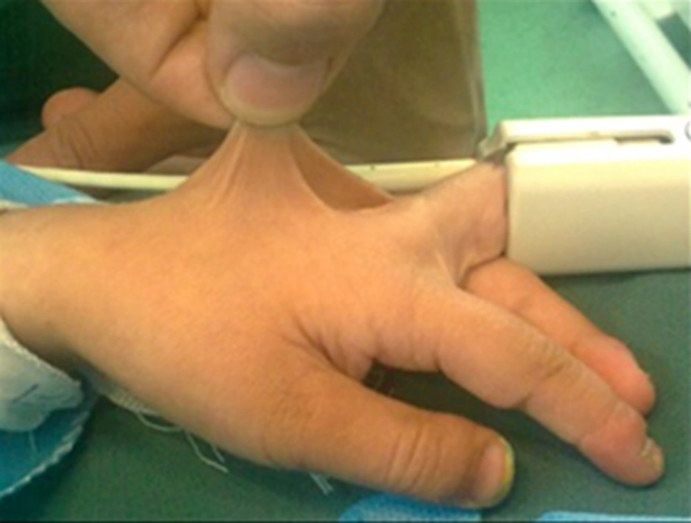
Figure 1.
Severe Hyperelasticity of the Skin on Hand
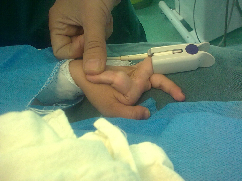
Figure 2.
Hypermobility and Hyperextensibility of Joints in Fingers
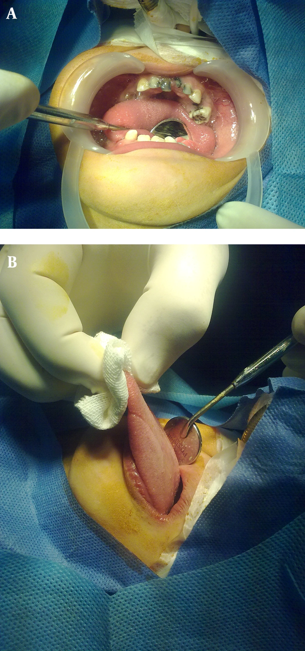
Figure 3.
Intraoral View Showing: A) Extensive Dental Caries Along With B) Extendable Tongue
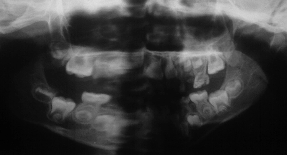
Figure 4.
Panoramic View of the Teeth and the Jaws Showing Preoperative Status
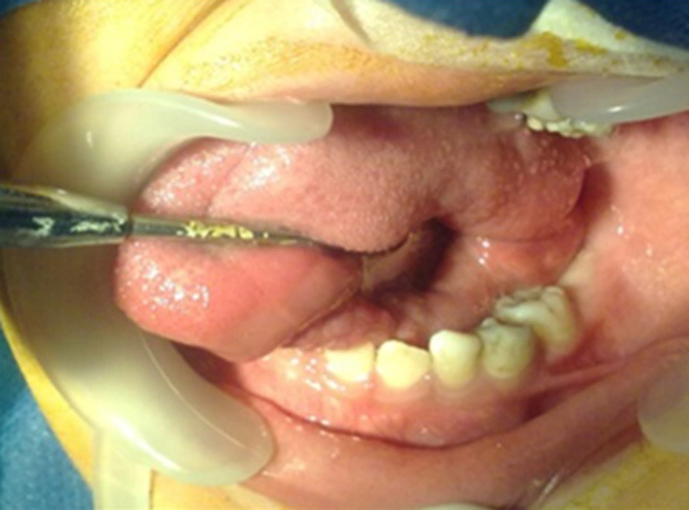
Figure 5.
A Sample of Intraoral View (Mandibular Left Side) Following Dental Treatment Under G.A
3. Discussion
Widely known cases of EDS suffer from varying degrees of extra movements and these clinical signs play an important role in identifying their specific type. Further laboratory investigations were performed on skin biopsy with results showing disturbed or abnormal collagen structure in classical type (5).
Major diagnostic criteria for classical type include skin hyperextensibility, wide atrophic scares, and significant joint hypermobility. Similar clinical signs were observed in the presented case. Several other reported diagnostic criteria include recurrent joint dislocation, positive family history, bleeding tendency, prolapsed mitral or tricuspid valves (5). This patient manifested all 3 major criteria as well as recurrent joint dislocations and positive family history. Absence of any musculoskeletal pain is considered as a major sign in hypermobile type of EDS, also the absence of ecchymosis and or scoliosis. Interestingly, patients with EDS are reported to carry several dental anomalies such as hypoplasia of enamel and dentin with short dilacerated roots, calcified pulp (pulp stone), congenital missing, and even presence of supernumerary teeth. However, the present case was free from any dental anomalies with only caries as the major dental problem (8, 13-16). Several active caries associated with hypoplastic enamel may somehow been related to the collagen defects associated with EDS (17).
Based on the age and the number of caries teeth, it is recommended to treat such children under general anesthesia as there is a high risk of temporomandibular joint (TMJ) dislocation in cases seen on chairside. No complication has been reported for dental treatment under general anesthesia from intubation, medication, or dental works. Overall, cases with EDS d, require pre-operative cardiac consultation and antibiotic prophylaxis cover prior to any major dental operations due to the presence of the mitral valve prolapsed (4).
Pillsbury showed that fragility of skin and blood vessels could lead to scar and hematoma formation following any traumatic event in EDS patients (18). Due to the possibility of complications following any treatment within the oral cavity, care should be taken as even low traumatic forces could lead to severe bleeding. Excessive gingival hemorrhage is reported as a common finding following teeth brushing (5, 8, 16). Surprisingly, such finding was not reported in the present case. Prolonged and repeated bleeding is reported following teeth extractions (5, 19, 20). It is critical to control laboratory blood test results, including prothrombin time (PT), partial thromboplastin time (PTT), and international normalized ratio blood test (INR) as a routine part of evaluation in patients with EDS. Blood count elements were found to be in normal range for the present case. Teeth were planned to be treated in one visit under general anesthesia. Care should be taken when suturing the socket walls of an extracted tooth in order to ensure a minimally traumatizing procedure for treatment under general anesthesia and to reduce the risk of extra trauma from local anesthetic administration. Reasonable care should be implemented when suturing the extraction sites in such individuals as any excess pressure and tension could further damage the already fragile and elastic tissues (16, 19, 21).An acrylic removable appliance is suggested to be used as a replacement for stitches to cover the surgical site (16, 21, 22). Inferior alveolar nerve block injection is banned because of its high risk of tissue damage (8, 23). Surgical flaps involving mucoperiosteom need maximum care as it is associated with high risk of tissue collapses. TMJ hypermobility and high dislocation should also be considered when undergoing dental treatment in patients with EDS. It is also recommended to arrange appointments in short periods. No orthodontic treatment is recommended to be scheduled until the full eruption of the first molars.
All initial orthodontic procedures are best provided along with routine dental treatments under general anesthesia in a single visit course for those suffering from EDS. As collagen defects influence periodontal ligament (PDL) and gingival fibers, light orthodontic forces are suggested along with a longer retention period (24).Higher frequency of root resorption and soft tissue defects are among the problems reported to be associated with orthodontic treatment of patients with EDS. Jone reported a high risk of mucosal ulceration related to bracket positioning in a case of EDS (25). Pourshahidi et al. had spotted similar findings in another similar case under dental observation (26). Fragility of gingival tissue and blood vessels could make the performance of any routine dental procedure highly sensitive. Therefore, special care should be taken when there are any interties for small dental works for chair side circumstances of patients with EDS.
The diagnosis of this case was EDS with the typical defects of collagen structure reflected in different tissue damages, including the tongue, fingers and toes. Medical and dental professionals are encouraged to see these patients willingly but carefully in order to deliver atraumatic, gentle care with a more preventive approach.