
1. Introduction
Alveolar hypoventilation is defined as an insufficient ventilation. The primary feature of this disorder is insufficient sleep-related ventilation, resulting in an abnormal increase in the arterial partial pressure of carbon dioxide (PaCO2) during sleep. Awake hypoventilation is defined as PaCO2 greater than 45 mmHg. Daytime hypoventilation may or may not occur concomitant with sleep-related hypoventilation disorders other than obesity hypoventilation syndrome (OHS). Evidence suggests that hypoventilation during wakefulness is associated with an even worse hypoventilation during sleep (1).
2. Case Presentation
In this article, we report two cases of hypoventilation in the first decade of life with abnormal presentations (e.g., arterial and venous thrombosis). We also present a case of hypoventilation with an uncommon etiology and compare the therapeutic strategies for these patients.
2.1. Case 1
A 10-year-old girl was hospitalized with complaints of headache, daytime sleepiness, and night-time fever, along with frequent thrombosis in the lower extremities, sagittal sinus, and cavernous sinus. Her growth and development were normal at the time of birth, which continued until the age of three years when she started to experience increased appetite and progressive weight gain. Gradually, severe daytime sleepiness and night-time fever occurred. At the age of seven, she showed signs of early puberty, such as pubic and axillary hair growth and breast bud formation. Acanthosis nigricans also appeared in the axillary regions and behind the neck.
The patient had an obese body stature. In the examinations, her vital signs were within the normal range, except for oxygen saturation (73%), weight (30 kg), and height (113 cm). The head and neck examinations indicated a large tongue and a long uvula. No abnormal findings were reported in the chest and abdominal examinations. Due to respiratory failure and hypoxemia, a full polysomnography (PSG) was performed as a diagnostic test, which confirmed chronic hypoventilation and obstructive sleep apnea hypopnea syndrome. The PSG results are:
Apnea hypop index (AHI) = 23.9/h
Desaturation index = 78.3/h
Oxygen desaturation < 90% = 97.8%
Following the PSG test, the patient was subjected to titration with bilevel positive airway pressure (BiPAP). BiPAP (S/T mode) was applied (14/4/0/14) along with oxygen at 2 L/min. After the application of BiPAP, her sleepiness and oxygen saturation improved, although her weight gain persisted. With continued weight gain and advancing age, higher BiPAPs were required for management.
The patient was hospitalized again at the age of 10 years with visual impairment and headache. Studies indicated superior sagittal sinus and left transverse sinus thrombosis; however, no etiology was identified, and the patient was treated with anticoagulants. Four months later thrombosis occurred in the posterior tibial veins, which extended to the left popliteal veins and common femoral vein; nevertheless, no evidence of thrombosis extending to the inferior vena cava was reported.
In the laboratory studies, the complement C3, complement C4, erythrocyte sedimentation rate (ESR), antinuclear antibody (ANA), anti-dsDNA, and thyroid function tests were in the normal range. The increased level of 8-am cortisol was not inhibited in response to the dexamethasone test. Similarly, the serum prolactin level increased. The patient’s coagulation tests and levels of protein C, protein S, and factor 5 were normal. Spiral Chest CT scan was normal. On the brain magnetic resonance imaging (MRI), the hypothalamic pituitary region showed a normal appearance. The abdominal ultrasonography showed fatty liver disease grade II, while other parameters were normal. The laboratory examination of PHOX2B gene mutation was negative.
2.2. Case 2
An eight-year-old boy was hospitalized for PSG due to daytime sleepiness, hypoxemia, and increased PaCO2. According to his mother, he had normal growth and development at birth. His normal growth continued until the age of two years when he started to experience increased appetite and progressive weight gain. In addition, cyanosis and several episodes of reduced consciousness associated with PaCO2 increase were reported. At the age of eight, a full PSG test was requested due to severe daytime hypoxemia, hypercapnia, and lack of academic achievement; the findings revealed chronic hypoventilation syndrome. The results of PSG are:
AHI = 4/h
Desaturation index = 96.5/h
Oxygen desaturation < 90% = 90%
Following the test, the patient underwent titration with BiPAP (S/T mode), which was administered (14/4/0/12) with an oronasal mask. He was hospitalized in the intensive care unit (ICU) after one year due to the reemergence of cyanosis, hypoxemia, severe daytime drowsiness, and decreased consciousness. The examinations indicated abnormal results: PCO2 = 140 mmHg and Na = 154 meq/L.
The patient underwent intubation and treatment with a mechanical ventilator. Treatment of hypernatremia was performed simultaneously, although its exact cause remained unknown, and the patient did not have any complaints of polydipsia or polyuria. After improvement and stabilization of his general condition, a PSG test was requested, which indicated the following results: AHI = 53.3/h; desaturation index = 96.5/h; mean desaturation = 61%; and oxygen saturation < 90% = 90%
Next, titration was performed, and BiPAP (25/5/0/14) was applied along with 3 L/min of oxygen. Following the application of BiPAP, the patient’s drowsiness and cyanosis were corrected, while his weight gain continued, which interfered with the BiPAP control of hypoventilation and necessitated BiPAP changes. Finally, due to incomplete hypoventilation control, the patient underwent intelligent volume-assured non-invasive ventilation (iVAPS).
According to our examinations, the level of growth hormone was lower than normal, due to obesity and delayed puberty, and failed to increase in response to the stimulation test. Therefore, after controlling the respiratory events, administration of growth hormones was initiated, although weight gain and obesity continued. The patient was re-hospitalized at the age of 16 with severe headache and reduced consciousness. The examinations indicated thrombosis in the superior sagittal sinus, however, no exact etiology was identified, and the patient was treated with anticoagulants.
2.3. Case 3
An eight-year-old boy was hospitalized due to head trauma. He was incubated and mechanically ventilated due to oxygen deprivation and increased PaCO2. To investigate the head trauma, a brain MRI was performed, which indicated no pathological abnormalities. The patient was intubated for 20 days. Hypercapnia and hypoxemia continued after extubation, and he was discharged with BiPAP application. The BiPAP application continued until he was readmitted to the ICU at the age of 16 due to pneumonia and reduced consciousness. PaCO2 increased to over 100 mmHg and oxygen reduction was detected. Accordingly, he was intubated again and underwent mechanical ventilation. Nevertheless, hypoventilation persisted following extubation.
According to the patient’s mother, he had a normal growth and development at birth. He also had no history of cardiac or respiratory problems and did not use any particular medications. In order to investigate the cause of hypoventilation, thyroid function tests (TFT), anti-HIV, C3, C4, anti-dsDNA, anti-cyclic citrullinated peptide (Anti-ccp), VDRL, ESR, and biochemical tests were performed, which indicated normal results. In the blood gas analysis, PaCO2 was consistently high and hypoxia was identified.
2.4. Spiral Chest CT Scan
Both lungs were found to be normal. No evidence of mass in the anterior, central, or posterior mediastinum was found. The hilar region was unremarkable on each side, and the main bronchi appeared normal. The thoracic skeleton and soft tissues showed no abnormalities. The pulmonary structure was also normal.
2.5. Lung High-Resolution Computed Tomography (HRCT)
A normal parenchymal architecture was found in the inspiration phase. No evidence of bronchiectasis was found. Brain MRI was normal.
A full PSG was requested for the patient due to hypoventilation. The results of PSG are: AHI = 10/h, Desaturation index = 5.2/h, Oxygen desaturation index < 90% = 61.7%, PaCO2 (pre-titration) = 63.2 mmHg, PaCO2 (post-titration) = 53 mmHg.
The patient was subjected to titration with BiPAP (S/T mode), and BiPAP (18/8/14) was applied in the S/T mode. Finally, the PHOX-2B gene mutation test was requested, which indicated negative results. The patient received BiPAP (S/T mode) in the new titration. Hypoxemia and hypercapnia were controlled, and no academic or developmental problems were reported in the follow-ups.
3. Discussion
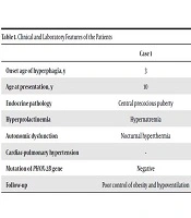
In this study, we presented three cases of pediatric hypoventilation with uncommon manifestations and etiologies. The differential diagnoses of pediatric hypoventilation include congenital central hypoventilation syndrome (CCHS), ROHHAD syndrome, Chiari malformation, Prader-Willi syndrome (PWS), and neuromuscular disorders (e.g. congenital myopathy, brain trauma, and central nervous system tumors) (Table 1).
Table 1.
Clinical and Laboratory Features of the Patients
| Case 1 | Case 2 | Case 3 | |
|---|---|---|---|
| Onset age of hyperphagia, y | 3 | 2 | - |
| Age at presentation, y | 10 | 8 | 8 |
| Endocrine pathology | Central precocious puberty | Growth hormone deficiency | - |
| Hyperprolactinemia | Hypernatremia | - | - |
| Autonomic dysfunction | Nocturnal hyperthermia | - | - |
| Cardiac-pulmonary hypertension | - | + | - |
| Mutation of PHOX-2B gene | Negative | Negative | Negative |
| Follow-up | Poor control of obesity and hypoventilation | Poor control of obesity and hypoventilation | Good control |
Congenital central hypoventilation syndrome (CCHS) with autonomic dysfunction, hypoventilation, and gastrointestinal motility disorders commonly occur in the neonatal period. However, some cases of late-onset CCHS may be misdiagnosed with ROHHAD syndrome. Considering the negative results of PHOX-2B genetic mutations, this diagnosis was ruled out in all our three patients (1-4). Another differential diagnosis of hypoventilation is Chiari malformation, which is characterized by hypoventilation associated with herniation of cerebellar vermis and caudal brainstem with disruption of blood flow. However, in our three patients, the normal results of brain MRI ruled out this diagnosis (1).
PWS is also a differential diagnosis of pediatric hypoventilation, which occurs due to the deletion of the long arm of chromosome 15. The signs of PWS include early-onset obesity, mental retardation, and small hands and feet. PWS is usually ruled out genetically. However, since the onset age of hypoventilation and its clinical signs in our patients were not compatible with this syndrome, no genetic study was carried out, and the disease was ruled out based on the clinical signs (2).
Another differential diagnosis of pediatric hypoventilation is ROHHAD syndrome. A diagnostic criterion of this syndrome, based on the international classification of sleep disorders (ICSD-3), is the sudden and progressive weight gain after the age of two years, followed by hypoxemia and hypercapnia. In other studies that are done on patients with ROHHAD syndrome, the prevalence of increased appetite and progressive weight gain was 83% after the age of two years (1, 5). It has been reported that 75% of patients with this syndrome experience hypoventilation (5); two of our patients were presented with one or two of the symptoms.
Autonomic dysregulation is another sign of ROHHAD syndrome with different manifestations, including blurred vision in 25% of cases, pain reduction in 13% of cases, and excessive sweating in 10% of cases (5). Our first patient was presented with night-time hyperthermia, which is an autonomic dysregulation. Another diagnostic criterion for ROHHAD syndrome is disruption of the hypothalamus axis, which has various manifestations, including hyperprolactinemia, central hypothyroidism, growth hormone deficiency, growth hormone insensitivity to stimulation tests, delayed or precocious puberty, and ACTH deficiency (1, 2, 5). Our first patient experienced hypothalamic deficiency, which manifested as premature central puberty and hyperprolactinemia. The second patient showed a reduction in the growth hormone level, which did not respond well to the stimulation test.
Based on the above-mentioned findings, diagnosis of ROHHAD syndrome was suggested for two of our patients (first and second cases). After full PSG and titration, they were treated with BiPAP (S/T). One important finding in these two patients was arterial and venous thrombosis without a known etiology, despite normal coagulation tests and absence of known genetic mutations. It should be noted that arterial or venous thrombosis has not been reported as one of the signs of ROHHAD syndrome (6). Considering the unknown etiology of the disease, variety of clinical symptoms, and course of ROHHAD syndrome (due to the low prevalence of this syndrome), it is suggested to consider this finding in other studies on ROHHAD syndrome in order to select the most appropriate treatment or prophylaxis.
As the above discussion attests, other causes of hypoventilation were ruled out, including CCHS (considering the onset age and negative results of PHOX-2B genetic mutation), ROHHAD syndrome (due to other symptoms such as sudden weight gain after 2 - 3 years of age, hyperphagia, and HPA axis disorder), Chiari malformation (due to normal brain MRI), and PWS (considering the onset age of symptoms and absence of other symptoms of the disease). After a careful review of the patient’s history, which indicated the onset of hypoventilation after a head trauma, absence of other clinical symptoms, and normal mental and physical development, head trauma was described as the cause of hypoventilation, as reported in rare cases (1).
Another important finding was the difference between the first two patients and the third one in terms of response to treatment and control of hypoventilation after a full PSG and titration. The first two patients experienced a progressive increase in weight gain and obesity. Also, hypoventilation management was challenging, as we needed to repeat titration and readjust the BiPAP device; even in one case, we used the iVAPS device alternatively. However, there was no such problem in the third patient, and in the subsequent follow-ups (every 6 - 12 months), normal physical and mental development, as well as clinical signs, were reported; furthermore, paraclinical findings for hypoventilation were within an acceptable range. We compared clinical features, laboratory findings, and follow ups of three cases in Table 1.