Jentashapir J Cell Mol Biol

Image Credit:Jentashapir J Cell Mol Biol

In Silico Study of Analysing the Capability of Vinblastine and Vincristine as Inhibitory Agents for Targeting the Cathepsin B Enzyme by Molecular Docking

Author(s):
Ahmad HafeziAhmad HafeziAhmad Hafezi ORCID1, Zahra KhamarZahra KhamarZahra Khamar ORCID1,*
1Department of Biology, Faculty of Science, Ferdowsi University of Mashhad, Mashhad, Iran

Jentashapir Journal of Cellular and Molecular Biology:Vol. 16, issue 3; e165042
Published online:Sep 06, 2025
Article type:Research Article
Received:Aug 02, 2025
Accepted:Aug 26, 2025
How to Cite:Hafezi A, Khamar Z. In Silico Study of Analysing the Capability of Vinblastine and Vincristine as Inhibitory Agents for Targeting the Cathepsin B Enzyme by Molecular Docking. Jentashapir J Cell Mol Biol. 2025;16(3):e165042. doi: https://doi.org/10.5812/jjcmb-165042

Abstract

Background:

Cathepsin B (CatB) is a lysosomal cysteine protease belonging to the papain family, involved in antigen processing during the immune response, hormone activation, and circulation, as well as in the pathology of chronic inflammatory diseases and cancer. The expression levels of the CatB enzyme are elevated in numerous tumor cells, serving as both a prognostic and therapeutic marker for various cancers. Considering the specific functional role of CatB within cancer cells, it is possible to inhibit cancer progression by either reducing the activity of the CatB proteolytic enzyme or enhancing lysosomal permeability. The alkaloids vincristine and vinblastine have been recognized for their significant contributions to the category of antitumor medications, owing to their strong anticancer effects, functioning as effective mitotic inhibitors.

Objectives:

Investigate the potential of vinblastine and vincristine molecules to function as ligands that interact with the CatB enzyme through the process of molecular docking.

Methods:

In this study, the first two ligands were sourced from the PubChem database. Subsequently, the structure of the CatB enzyme was acquired from the RCSB Protein Data Bank website. After optimizing the molecular structures, they were saved in PDBQT format, which included the addition of Gasteiger and Kollman charges to both the ligands and the target enzyme, respectively. Finally, the molecular docking process was carried out using the Melligro Virtual Docker software, specifically targeting the cavity where the primary ligands bind to the CatB enzyme.

Results:

The results obtained from the molecular docking analysis demonstrated that vinblastine can bind to the active site of the CatB enzyme, forming hydrogen bonds with Met 196. The estimated bond length was 3.16 angstroms. The total energy measured was -218.647 kcal/mol for the MolDock score and -129.661 kcal/mol for the Rerank score. Vincristine demonstrated significant potential for hydrogen bonding with the Ser 175 residues of both chains A and B. The scores derived from the docking results suggest that vincristine possesses a relatively strong capacity to bind to this site, with a MolDock score of -184.221 kcal/mol and a Rerank score of -96.239 kcal/mol. Additionally, the positioning of both ligands with the primary ligands of the cathepsin enzyme was compared using the Discovery Studio software.

Conclusions:

Both vincristine and vinblastine demonstrated considerable potential for interacting with the enzyme CatB, which may facilitate additional research into the in vitro activity of these ligands.

1. Background

1.1. Cathepsins

Cathepsins are a type of endopeptidase present in the majority of cells, leading to cell autolysis and the autodigestion of tissues. They are categorized into serine (cathepsins A and G), aspartic (cathepsins D and E), and cysteine cathepsins (cathepsins B, C, F, H, K, L, O, S, V, W, and X) based on their structural characteristics and catalytic mechanisms (1-3). Among the various cathepsins, cathepsin B (CatB) holds significant relevance due to its critical involvement in numerous pathologies and carcinogenic processes (2). The CatB is a lysosomal cysteine protease belonging to the papain family, which is essential for intracellular protein catabolism (1, 4, 5). It is produced in the rough endoplasmic reticulum (RER) as a proenzyme consisting of 339 amino acids, accompanied by a 17 amino acid signal peptide. This enzyme plays a crucial role in degrading excess organelles and proteins within the acidic environment of lysosomes, thereby facilitating recycling processes (3, 6). Additionally, it participates in various physiological functions, including antigen processing during the immune response, hormone activation, and the regulation of blood circulation (1, 4, 7). The CatB is implicated in the pathogenesis of chronic inflammatory diseases (3,5) and is also associated with cancer (7, 8). The CatB enzyme exhibits activity outside the cell in tumors and plasma (1, 4, 6). Cathepsins serve a dual function in tumor progression, with this process being contingent upon the equilibrium between proteases and their inhibitors (5). In numerous tumor cells, the expression of the enzyme CatB is elevated, serving as a prognostic and therapeutic marker for various cancers. Lysosomal cathepsins are frequently present in cancer cells to meet metabolic demands linked to heightened invasion and metastasis (1, 5, 8, 9). The increased expression of CatB in many human cancers at both the mRNA and protein levels indicates that this enzyme may possess pro-apoptotic characteristics (5, 8, 9). The CatB is located on chromosome 8p22, which is recognized as a tumor suppressor. Alternative splicing plays a significant role in the oncogenic potential of CatB (2).
Considering the specific functional role of CatB in cancer cells, there are presently two methods to directly or indirectly influence cancer cell death by targeting it: (A) Inhibition of the proteolytic enzyme CatB's activity. As CatB is involved in cancer metastasis by modifying extracellular matrix remodeling and promoting angiogenesis, its inhibition leads to a decrease in the migration, invasion, and proliferation of cancer cells. RNA interference can be employed to inhibit CatB, thus diminishing the invasion, growth, and angiogenesis of gliomas (1, 5); (B) Enhanced lysosomal permeability, which results in the release of CatB into the cytoplasm and subsequent apoptosis of tumor cells, has been extensively studied in therapeutic strategies targeting CatB for various cancer types. Nevertheless, since increased lysosomal permeability can induce cancer cell death, potential side effects must be assessed when utilizing these drugs (1).
The application of CatB inhibitors in vitro has demonstrated a reduction in both the motility and invasion of tumor cells. The inhibitors employed encompass a range of protein inhibitors, some of which are of endogenous origin and serve as regulators of CatB activity within the cell, such as cystatins. Conversely, certain exogenous protein inhibitors have been extracted from various natural sources. The utilization of X-ray crystal structures of CatB in complex with these protein inhibitors has facilitated the design and synthesis of numerous new small molecular weight compounds that act as CatB inhibitors. Typically, these compounds feature an electrophilic moiety that interacts with CatB (4).

1.2. Vinblastine and Vincristine

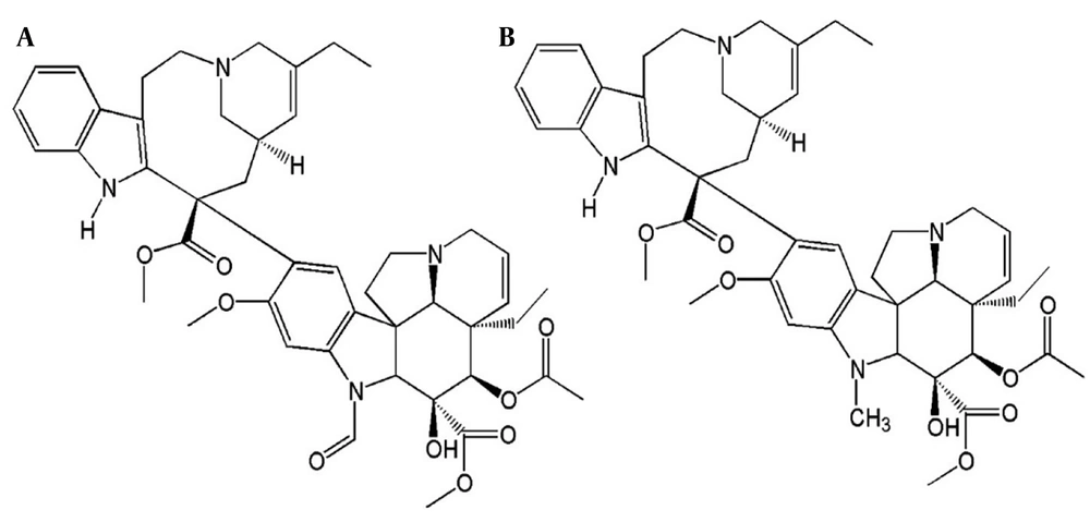
The alkaloids vincristine and vinblastine (Figure 1), both obtained from the Madagascar periwinkle (10), Vinca rosea L., have secured a significant position within the antitumor drug category due to their anticancer properties (11-13). Vincristine and vinblastine function as inhibitors by attaching to microtubules and the mitotic spindle during the metaphase stage of the cell cycle (12, 14, 15). Microtubules, which form the cytoskeleton, consist of dimers of α- and β-tubulin, and are essential for the transport and positioning of intracellular organelles, as well as for the separation of chromatids during anaphase in mitosis (16-18). Both vincristine and vinblastine are effective mitotic inhibitors, with their molecular structures differing solely by one carbonyl group. The primary sites of toxicity for vincristine are the nervous system, whereas for vinblastine, it is the hematopoietic system (10, 15). Vinblastine has a methyl group, while vincristine has a formyl group on the indole nitrogen of the vinblastine skeleton (10, 13).
The structure of vincristine (A) and vinblastine (B) (<a href="#A165042REF19">19</a>)
Figure 1.

The structure of vincristine (A) and vinblastine (B) (19)

Vincristine, a naturally occurring vinca alkaloid, attaches to the vinca domain within the h-tubulin subunit, leading to microtubule destabilization at different concentrations (14, 16, 18, 20). At elevated concentrations, this results in the total disintegration of the microtubule network, which in turn facilitates the formation of the mitotic spindle (16, 18). Vincristine is an antimitotic medication that has been utilized in the treatment of cancer for more than 40 years (18, 20). Studies indicate that both vincristine and vinblastine lead to significant alterations in the lysosomal compartment, making cells more susceptible to lysosomal membrane permeability. This results in an increase in lysosomal volume and leakage, which subsequently activates the intrinsic apoptotic pathway. For instance, in HeLa cervical carcinoma cells, vincristine induces mitotic arrest and considerable cell death, initially characterized by an increase in lysosomal volume, ultimately leading to apoptosis (18, 21). Apoptosis is regarded as the most effective cell death mechanism for inhibiting tumor growth (16, 18, 21).

2. Objectives

The main aim of this study is to investigate the potential of vinblastine and vincristine molecules to function as ligands that attach to the CatB enzyme through molecular docking, thus offering a further approach for employing these medications to target different locations in cancer cells.

3. Methods

In this research, the Molegro Virtual Docker (MVD) software version 6 was initially installed on the Windows operating system. Subsequently, the structures of Vinblastine and Vincristine, identified by CID: 13342 and CID: 5978 respectively, were retrieved from the www.pubchem.ncbi.nlm.nih.gov website in the form of an SDF file. Both structures were optimized utilizing the Avogadro software and stored in the Mol2 format. Subsequently, the Gasteiger charge was applied to these ligands using the AutoDock Tools 1.5.7 software and saved in PDBQT format. In order to prepare the CatB molecule for the docking procedure, the PDB structure was first downloaded from the www.rcsb.org website (ID = 1gmy). Subsequently, this molecule was imported into AutoDock Tools, where it was saved as a PDBQT file by eliminating water molecules, adding polar hydrogens, and incorporating Kollman charges. Subsequently, these molecules were imported into the MVD software, and in the preparation section of the molecule, the option 'if missing' was chosen. Following the optimization of the selected peptides by the software, the docking process for these ligands was conducted at the binding site of the primary CatB ligands within cavity 998, with coordinates X = 37.60, Y = 36.70, and Z = 31.82. In the course of this procedure, the MolDock score (GRID) mode was chosen, and the MolDock simplex evolution (SE) algorithm was implemented as well. Subsequently, the software was instructed to conduct 10 distinct runs for each ligand at the designated position. Ultimately, the outcomes of this procedure were assessed (22-24).

4. Results

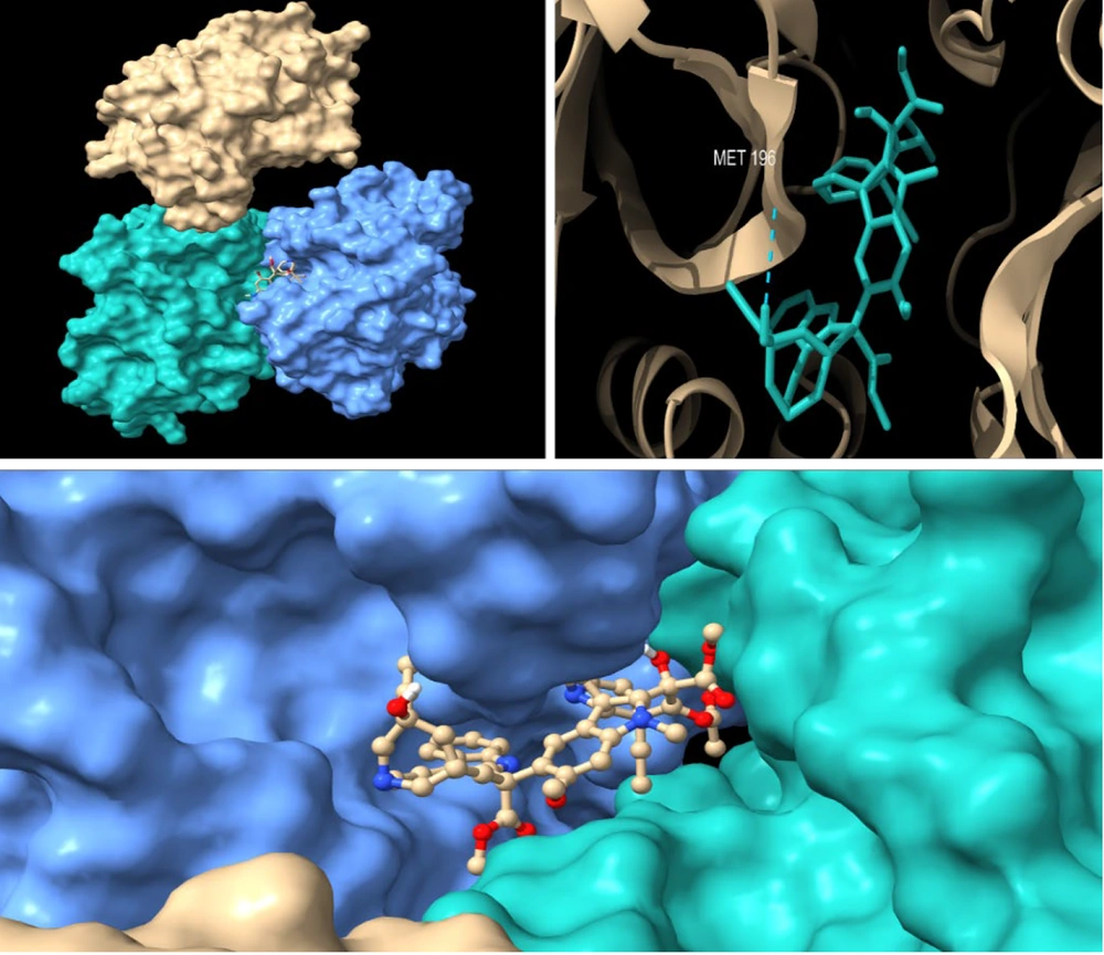
The active site of the CatB enzyme was determined by examining the primary ligands that interact with this enzyme. Among the various sites identified, the cavity situated between the A and B chains was selected as the optimal location for the binding of the vinblastine ligand. The docking analysis performed after the processing phase demonstrated that vinblastine can establish hydrogen bonds with the amino acid Met 196, which is situated on the chain B of the CatB enzyme at this particular location (Figure 2). In addition, the software analysis uncovered that the electrostatic interactions with the residues Glu 255 and Glu 122, also found in chain B, could play a crucial role in significantly enhancing the stability of the binding of this ligand to the enzyme's active site. The total energy was determined to be -218.647 kcal/mol for the MolDock score and -129.661 kcal/mol for the Rerank score. These values indicate a significant affinity of the ligand for binding at this specific location.
Cathepsin B (CatB) enzyme structure and the interaction of vinblastine in the active site with Met 196: Chain A (light sea green), chain B (cornflower blue), and chain C (grey).
Figure 2.

Cathepsin B (CatB) enzyme structure and the interaction of vinblastine in the active site with Met 196: Chain A (light sea green), chain B (cornflower blue), and chain C (grey).

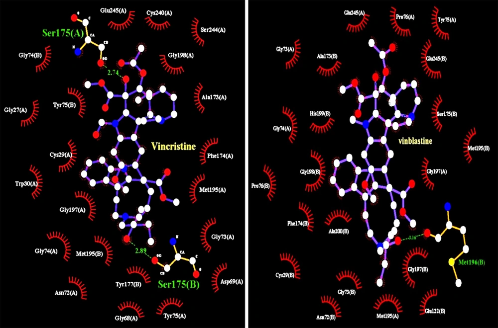
The molecular docking analysis conducted on vincristine with CatB demonstrated that this ligand possesses significant potential for interaction with the enzyme's active site. Specifically, vincristine successfully established hydrogen bonds with Ser 175 located in both chain A and chain B of CatB. Furthermore, the ligand showed the capability to form electrostatic interactions with Glu residues 245 and 122 in chain A. The calculated MolDock score for this interaction was -184.221 kcal/mol, while the Rerank score was -96.239 kcal/mol, indicating a strong binding affinity. Figure 3 provides a detailed representation of the steric interaction bonds that are formed by both the vinblastine and vincristine ligands as they occupy the active site. This illustration highlights the spatial arrangements and interactions that occur between the ligands and the surrounding molecular environment, emphasizing the significance of these steric interactions in the binding process.
The names of the residues that created the steric interaction bonds are displayed in white, while the residues that established the hydrogen bond are shown in green. The estimated hydrogen bond length between vincristine and the Ser 175 residue of the B chain is 2.89 angstroms, whereas the bond length with Ser 175 of the A chain was estimated to be 2.74 angstroms, and the bond length of vinblastine with Met 196 was calculated to be 3.16 angstroms.
Figure 3.

The names of the residues that created the steric interaction bonds are displayed in white, while the residues that established the hydrogen bond are shown in green. The estimated hydrogen bond length between vincristine and the Ser 175 residue of the B chain is 2.89 angstroms, whereas the bond length with Ser 175 of the A chain was estimated to be 2.74 angstroms, and the bond length of vinblastine with Met 196 was calculated to be 3.16 angstroms.

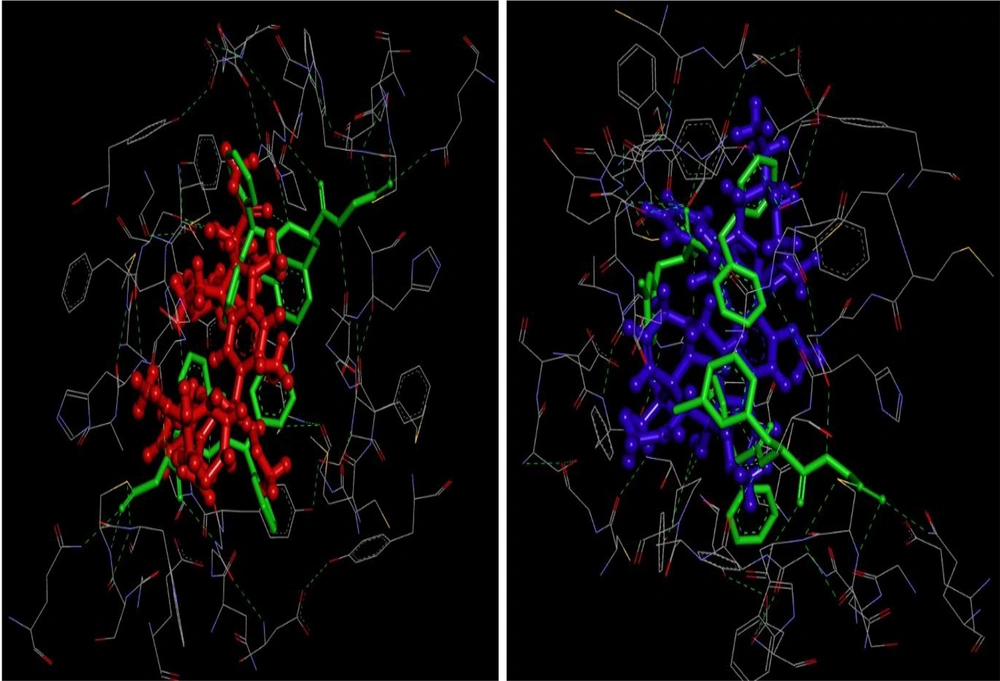
After analyzing the results obtained from BIOVIA Discovery Studio, it was found that both ligands exhibited a strong capacity to bind in an optimal disposition at the specific site where CatB ligands typically attach (Figure 4). This suggests that these ligands have the potential to compete with the primary ligands for binding at this location, possibly occupying it with greater affinity. As a result, this could impede the binding of the main ligands to the site, ultimately disrupting the enzyme's normal function.
The positioning of the ligands vinblastine and vincristine within the active site of the enzyme cathepsin B (CatB) is compared to the primary ligands of this enzyme. The primary ligands of CatB are depicted in green, while vinblastine is represented in red and vincristine in blue.
Figure 4.

The positioning of the ligands vinblastine and vincristine within the active site of the enzyme cathepsin B (CatB) is compared to the primary ligands of this enzyme. The primary ligands of CatB are depicted in green, while vinblastine is represented in red and vincristine in blue.

5. Discussion

The docking process is regarded as an initial step in identifying a specific molecule for the exploration of its potential application in the development of new pharmaceuticals, due to the method's cost-effectiveness, speed, and reliability. Consequently, it is advisable for researchers to first assess the potential of their target ligand utilizing bioinformatics tools; if the ligand demonstrates acceptable potential, further investigation can be conducted in vivo and in vitro in subsequent phases. In 2020, coinciding with the coronavirus pandemic, Singh and Florez extracted 1121 ligands from the ZINK and DrugBank databases, employing bioinformatics tools and molecular docking to evaluate the potential of these ligands in inhibiting the main protease enzyme of the coronavirus. Their findings revealed that Furobinordentatin and Alstiphyllanine F were among the most promising ligands, exhibiting favorable binding energy for the inhibition of this enzyme (25).
Furthermore, in a separate investigation, molecular docking techniques were utilized to discover an appropriate molecule that inhibits the coronavirus protease enzyme, thus identifying a suitable ligand to target this enzyme. In this research, 1600 approved drugs from the ZINC database were examined as potential inhibitors for this enzyme. Ultimately, velpatasvir, molnupiravir, and Ivermectin were chosen for additional studies using virtual screening methods to identify an effective ligand for the treatment of COVID-19 (26).

5.1. Conclusions

This research has demonstrated that the molecules vinblastine and vincristine possess significant potential for targeting the enzyme CatB. Furthermore, these molecules serve as mitotic inhibitors in cancer cells (27-29), making them of considerable interest to researchers in this domain as multi-target drugs. This is particularly valuable for advancing the development of therapies that can simultaneously address multiple targets within cancer cells, thereby reducing the necessity for additional medications that may lead to increased side effects for patients. Consequently, it is advisable for researchers in subsequent phases to explore the stability of the enzyme-ligand complex and to employ molecular dynamics to enhance the precision of experiments conducted on both in vivo and in vitro grounds.
Ultimately, this study has laid the groundwork for acquiring new insights into the application of ligands such as vinblastine and vincristine, which target tubulin in cancer, and also have potential for inhibiting other enzymes with critical roles in cancer progression. The findings indicate that these ligands may hold significant promise for consideration within the pharmaceutical sector.

Footnotes

References

  • 1.
    Xie Z, Zhao M, Yan C, Kong W, Lan F; Narengaowa, et al. Cathepsin B in programmed cell death machinery: mechanisms of execution and regulatory pathways. Cell Death Dis. 2023;14(4):255. [PubMed ID: 37031185]. [PubMed Central ID: PMC10082344]. https://doi.org/10.1038/s41419-023-05786-0.
  • 2.
    Gondi CS, Rao JS. Cathepsin B as a cancer target. Expert Opin Ther Targets. 2013;17(3):281-91. [PubMed ID: 23293836]. [PubMed Central ID: PMC3587140]. https://doi.org/10.1517/14728222.2013.740461.
  • 3.
    Cai Z, Xu S, Liu C. Cathepsin B in cardiovascular disease: Underlying mechanisms and therapeutic strategies. J Cell Mol Med. 2024;28(17). e70064. [PubMed ID: 39248527]. [PubMed Central ID: PMC11382359]. https://doi.org/10.1111/jcmm.70064.
  • 4.
    Frlan R, Gobec S. Inhibitors of cathepsin B. Curr Med Chem. 2006;13(19):2309-27. [PubMed ID: 16918357]. https://doi.org/10.2174/092986706777935122.
  • 5.
    Wang J, Zheng M, Yang X, Zhou X, Zhang S. The Role of Cathepsin B in Pathophysiologies of Non-tumor and Tumor tissues: A Systematic Review. J Cancer. 2023;14(12):2344-58. [PubMed ID: 37576397]. [PubMed Central ID: PMC10414043]. https://doi.org/10.7150/jca.86531.
  • 6.
    Aggarwal N, Sloane BF. Cathepsin B: multiple roles in cancer. Proteomics Clin Appl. 2014;8(5-6):427-37. [PubMed ID: 24677670]. [PubMed Central ID: PMC4205946]. https://doi.org/10.1002/prca.201300105.
  • 7.
    Mort JS, Buttle DJ. Cathepsin B. Int J Biochem Cell Biol. 1997;29(5):715-20. [PubMed ID: 9251238]. https://doi.org/10.1016/s1357-2725(96)00152-5.
  • 8.
    Berquin IM, Sloane BF. Cathepsin B expression in human tumors. Adv Exp Med Biol. 1996;389:281-94. [PubMed ID: 8861022]. https://doi.org/10.1007/978-1-4613-0335-0_35.
  • 9.
    Podgorski I, Sloane BF. Cathepsin B and its role(s) in cancer progression. Biochem Soc Symp. 2003;(70):263-76. [PubMed ID: 14587299]. https://doi.org/10.1042/bss0700263.
  • 10.
    Hoang NH, Huyen NT, Trang DT, Canh NX, Mao CV, Sopjani M, et al. Effects of Vinblastine and Vincristine on the function of chronic myeloid leukemic cells through expression of A20 and CYLD. Cell Mol Biol (Noisy-le-grand). 2022;68(10):47-53. [PubMed ID: 37114273]. https://doi.org/10.14715/cmb/2022.68.10.7.
  • 11.
    Dhyani P, Quispe C, Sharma E, Bahukhandi A, Sati P, Attri DC, et al. Anticancer potential of alkaloids: a key emphasis to colchicine, vinblastine, vincristine, vindesine, vinorelbine and vincamine. Cancer Cell Int. 2022;22(1):206. [PubMed ID: 35655306]. [PubMed Central ID: PMC9161525]. https://doi.org/10.1186/s12935-022-02624-9.
  • 12.
    Jordan MA, Himes RH, Wilson L. Comparison of the effects of vinblastine, vincristine, vindesine, and vinepidine on microtubule dynamics and cell proliferation in vitro. Cancer Res. 1985;45(6):2741-7. [PubMed ID: 3986806].
  • 13.
    Keglevich P, Hazai L, Kalaus G, Szantay C. Modifications on the basic skeletons of vinblastine and vincristine. Molecules. 2012;17(5):5893-914. [PubMed ID: 22609781]. [PubMed Central ID: PMC6268133]. https://doi.org/10.3390/molecules17055893.
  • 14.
    Kumar ASHUTOSH. Vincristine and vinblastine: a review. IJMPS. 2016;6:23-30.
  • 15.
    Ferguson PJ, Phillips JR, Selner M, Cass CE. Differential activity of vincristine and vinblastine against cultured cells. Cancer Res. 1984;44(8):3307-12. [PubMed ID: 6744266].
  • 16.
    Chao MW, Lai MJ, Liou JP, Chang YL, Wang JC, Pan SL, et al. The synergic effect of vincristine and vorinostat in leukemia in vitro and in vivo. J Hematol Oncol. 2015;8:82. [PubMed ID: 26156322]. [PubMed Central ID: PMC4504084]. https://doi.org/10.1186/s13045-015-0176-7.
  • 17.
    Król T, Schmidt M, Witek B, Kołątaj A. The Effect of Vinblastine on the Autophagic Processes in Mouse Hepatocytes. Sci Didactic Yearbook Pedagogical Univ Krakow. 1997.
  • 18.
    Groth-Pedersen L, Ostenfeld MS, Hoyer-Hansen M, Nylandsted J, Jaattela M. Vincristine induces dramatic lysosomal changes and sensitizes cancer cells to lysosome-destabilizing siramesine. Cancer Res. 2007;67(5):2217-25. [PubMed ID: 17332352]. https://doi.org/10.1158/0008-5472.CAN-06-3520.
  • 19.
    Noble CO, Guo Z, Hayes ME, Marks JD, Park JW, Benz CC, et al. Characterization of highly stable liposomal and immunoliposomal formulations of vincristine and vinblastine. Cancer Chemother Pharmacol. 2009;64(4):741-51. [PubMed ID: 19184019]. [PubMed Central ID: PMC2717390]. https://doi.org/10.1007/s00280-008-0923-3.
  • 20.
    Puscasu C, Negres S, Zbarcea CE, Chirita C. Unlocking New Therapeutic Options for Vincristine-Induced Neuropathic Pain: The Impact of Preclinical Research. Life (Basel). 2024;14(11). [PubMed ID: 39598298]. [PubMed Central ID: PMC11595627]. https://doi.org/10.3390/life14111500.
  • 21.
    Yasin YS, Jumaa AH, Jabbar S, Abdulkareem AH. Effect of Laetrile Vinblastine Combination on the Proliferation of the Hela Cancer Cell Line. Asian Pac J Cancer Prev. 2023;24(12):4329-37. [PubMed ID: 38156870]. [PubMed Central ID: PMC10909105]. https://doi.org/10.31557/APJCP.2023.24.12.4329.
  • 22.
    Hafezi A, Khamar Z. Molecular Docking of Silymarin as a Ligand for Aromatase Enzyme and HIV-1 Reverse Transcriptase Using Molegro Virtual Docker. J Microbio. 2025;2(2). https://doi.org/10.5812/jmb-162254.
  • 23.
    Kusumaningrum S, Budianto E, Kosela S, Sumaryono W, Juniarti F. The molecular docking of 1, 4-naphthoquinone derivatives as inhibitors of Polo-like kinase 1 using Molegro Virtual Docker. J App Pharmaceut Sci. 2014;4(11):47-53.
  • 24.
    Naeem S, Hylands P, Barlow D. Docking studies of chlorogenic acid against aldose redutcase by using molgro virtual docker software. J App Pharmaceut Sci. 2013;3(1):13-20.
  • 25.
    Singh S, Florez H. Coronavirus disease 2019 drug discovery through molecular docking. F1000Res. 2020;9:502. [PubMed ID: 32704354]. [PubMed Central ID: PMC7361499]. https://doi.org/10.12688/f1000research.24218.1.
  • 26.
    Manavi MA. In silico study to identification of potential SARS-CoV-2 main protease inhibitors: virtual drug screening and molecular docking with AutoDock Vina and Molegro Virtual Docker. J Cell Molecular Res. 2022;13(2):108-12.
  • 27.
    Ngan VK, Bellman K, Hill BT, Wilson L, Jordan MA. Mechanism of mitotic block and inhibition of cell proliferation by the semisynthetic Vinca alkaloids vinorelbine and its newer derivative vinflunine. Mol Pharmacol. 2001;60(1):225-32. [PubMed ID: 11408618]. https://doi.org/10.1124/mol.60.1.225.
  • 28.
    Jordan MA, Thrower D, Wilson L. Effects of vinblastine, podophyllotoxin and nocodazole on mitotic spindles. Implications for the role of microtubule dynamics in mitosis. J Cell Sci. 1992;102 ( Pt 3):401-16. [PubMed ID: 1506423]. https://doi.org/10.1242/jcs.102.3.401.
  • 29.
    Gigant B, Wang C, Ravelli RB, Roussi F, Steinmetz MO, Curmi PA, et al. Structural basis for the regulation of tubulin by vinblastine. Nature. 2005;435(7041):519-22. [PubMed ID: 15917812]. https://doi.org/10.1038/nature03566.

Similar Articles

31
Jul
2018

Design, Synthesis and Evaluation of Substituted Aryl-2-Nitrovinyl Derivatives as Small Molecules Proteasome Inhibitors

Masoud Faghih Akhlaghi,
Marjan Daeihamed,
Seyed Abdolmajid Ayatollahi,
Farzad Kobarfard,
Athar Ata

Faghih Akhlaghi M, Daeihamed M, Ayatollahi SA, Kobarfard F, Ata A. Design, Synthesis and Evaluation of Substituted Aryl-2-Nitrovinyl Derivatives as Small Molecules Proteasome Inhibitors. Iran J Pharm Res. 2018;17(3):e124795. doi: https://doi.org/10.22037/ijpr.2018.2270

21
Jan
2024
Jundishapur J Microbiol

Identification and Characterization of Indolebutyroyl Aspartic Acid: A Promising Inhibitor of VIM-2 Metallo-β-Lactamase Against Carbapenem-Resistant Pseudomonas aeruginosa Strains

Seyedeh Sara Moosavi,
Mahboobeh Madani,
Sako Mirzaie,
Nima Hosseini Jazani

Moosavi SS, Madani M, Mirzaie S, Hosseini Jazani N. Identification and Characterization of Indolebutyroyl Aspartic Acid: A Promising Inhibitor of VIM-2 Metallo-β-Lactamase Against Carbapenem-Resistant Pseudomonas aeruginosa Strains. Jundishapur J Microbiol. 2023;16(11):e131974. doi: https://doi.org/10.5812/jjm-131974

12
Oct
2025
koomesh

A Computational Study of Some Benzenesulfonamide Derivatives as HIV- 1 Protease Enzyme Inhibitors: Three-Dimensional Quantitative Structure-Activity Relationship and Molecular Docking

Rahman Abdizade,
Tooba Abdizadeh

Abdizade R, Abdizadeh T. A Computational Study of Some Benzenesulfonamide Derivatives as HIV- 1 Protease Enzyme Inhibitors: Three-Dimensional Quantitative Structure-Activity Relationship and Molecular Docking. koomesh. 2025;27(6):e159682. doi: https://doi.org/10.69107/koomesh-159682

11
Aug
2025
J Rep Pharm Sci.

Natural Marine-derived Compounds as Potential Inhibitors of SARS-CoV-2 Main Protease: A Comprehensive Computational Study

Ghazal Nowroozi,
Shokoufeh Rahmani,
Parvin Jalali,
Armin Zareie,
Zahra Asvar,
Amin Nowroozi

Nowroozi G, Rahmani S, Jalali P, Zareie A, Asvar Z, et al. Natural Marine-derived Compounds as Potential Inhibitors of SARS-CoV-2 Main Protease: A Comprehensive Computational Study. J Rep Pharm Sci. 2025;13(1):e159451. doi: https://doi.org/10.5812/jrps-159451

2
Apr
2022
J Health Rep Technol

Discovery of a Potent Inhibitor to Overcome Carbapenem Resistance in Pseudomonas aeruginosa Strains via Inhibition of VIM-2 Metallo-β-lactamases

Seyedeh Sara Moosavi,
Mahboobeh Madani,
Sako Mirzaie,
Nima Hosseini Jazani

Moosavi SS, Madani M, Mirzaie S, Hosseini Jazani N. Discovery of a Potent Inhibitor to Overcome Carbapenem Resistance in Pseudomonas aeruginosa Strains via Inhibition of VIM-2 Metallo-β-lactamases. J Health Rep Technol. 2022;8(2):e121109. doi: https://doi.org/10.5812/ijhls.121109


Crossmark
Crossmark
Checking
Share on
Cited by
Metrics

Ordering Reprints

Articles are published under the Creative Commons license stated on each article. No permission or royalty fee is required for uses permitted by that license. CCC handles optional bulk and customized reprint orders. Any quotation covers production and delivery services only, not copyright permission. > Request Reprints from CCC