Evaluation of Escherichia coli aggR Gene Expression in the Presence of Different Concentrations of Creatinine

Author(s):
Massome AyoubnejadMassome Ayoubnejad1, Mohammadreza MehrabiMohammadreza Mehrabi1,*, Mohsen MirzaeiMohsen Mirzaei1
1Department of Laboratoty Science, BO, C Islamic Azad University, Broujerd, Iran

Jundishapur Journal of Microbiology:Vol. 19, issue 8; e169357
Published online:Jul 18, 2026
Article type:Research Article
Received:Dec 27, 2025
Accepted:May 24, 2026
How to Cite:Ayoubnejad M, Mehrabi M, Mirzaei M. Evaluation of Escherichia coli aggR Gene Expression in the Presence of Different Concentrations of Creatinine. Jundishapur J Microbiol. 2026;19(8):e169357. doi: https://doi.org/10.5812/jjm-169357

Abstract

Background:

Chronic renal failure is an irreversible disease associated with elevated blood creatinine levels and substantial mortality and morbidity in children worldwide. Enteroaggregative Escherichia coli (EAEC) is a diarrheagenic agent that possesses several virulence factors, including aggR.

Objectives:

This study aimed to evaluate aggR gene expression in EAEC under different levels of creatinine exposure.

Methods:

Initially, three biochemically confirmed E. coli isolates obtained from medical laboratories in Borujerd city, western Iran, were molecularly evaluated by 16S ribosomal RNA (16S rRNA) gene amplification. Next, the isolates were assessed for the aggR gene using polymerase chain reaction (PCR). Isolates carrying the aggR gene were cultured in Brain Heart Infusion (BHI) medium supplemented with 1, 3, and 6 mg/dL creatinine (37oC for 24 hours). The expression level of aggR was evaluated in creatinine-exposed isolates using real-time PCR.

Results:

Among 50 clinical E. coli isolates, three were EAEC, and one of these isolates carried the aggR gene. Real-time PCR showed that aggR gene expression decreased significantly in the presence of varying creatinine concentrations (P < 0.0001).

Conclusions:

These findings warrant further investigation to elucidate the role of creatinine in modulating bacterial virulence gene expression in the context of renal disorders.

1. Background

Diarrheal illnesses caused by diverse infectious agents constitute a major public health concern in both adults and children (1, 2). Escherichia coli, a gram-negative, rod-shaped bacterium capable of facultative anaerobiosis, is a prominent diarrhea-causing pathogen among the infectious agents commonly detected in the human intestinal tract (3). Although E. coli is typically part of the normal gut microbiota, certain strains can acquire virulence factors and become pathogenic variants, leading to serious diseases such as diarrhea. These pathogenic strains are classified into 6 distinct pathotypes according to their virulence mechanisms (4). Enteroaggregative E. coli is a major cause of both acute and persistent diarrhea across all age groups, particularly in infants and immunocompromised individuals (5-7). The clinical presentation of Enteroaggregative Escherichia coli (EAEC) infection often includes fever, anorexia, nausea, and bloody-mucosal diarrhea (8). The classic diagnostic method for EAEC involves identifying the characteristic "stacked-bricks" aggregation pattern in HEp-2 cell culture (9). The pAA virulence plasmid is a key molecular marker of EAEC and contains essential pathogenicity genes, including aggR, aatA, and genes encoding aggregative adherence fimbriae (10, 11).
The aggR gene functions as a master regulator of EAEC virulence. Strains carrying this gene are classified as typical EAEC and are generally more virulent, causing more severe disease than atypical strains (12, 13). EAEC pathogenesis is a complex process involving initial adherence to host cells, biofilm formation, and subsequent toxin release that triggers an inflammatory response (14). In addition to diarrheal disease, diarrheagenic E. coli strains, including EAEC, have recently been linked to urinary tract infections (UTIs), which affect more than 150 million people worldwide (15-18). The urinary tract provides an environment in which uropathogenic microorganisms can thrive, and their growth can be influenced by the chemical composition of urine, including the concentrations of urea and creatinine. In patients with renal insufficiency, host-pathogen interactions are markedly altered by the accumulation of various substances, which in turn affects microbial virulence factor expression and host defense mechanisms. Notably, studies have shown that high creatinine levels can inhibit bacterial persistence in the urinary tract (19).

2. Objectives

This study aimed to examine the effect of varying creatinine levels on aggR gene expression in clinically isolated E. coli strains from individuals with urinary tract infections in Boroujerd, western Iran.

3. Methods

3.1. Collection and Maintenance of Clinical Isolates

In total, 50 E. coli strains were obtained from clinical samples from individuals with urinary tract infections at diagnostic medical facilities in Borujerd, Lorestan province. The identity of these isolates had been previously confirmed using biochemical methods. All isolates were preserved at -20°C in Brain Heart Infusion broth supplemented with 15% glycerol for future analysis (20).

3.2. DNA Extraction and Amplification

Genomic DNA was extracted using the boiling method. Briefly, bacterial cultures were grown from glycerol stocks in BHI broth and subcultured on blood agar. Individual colonies were then transferred to BHI broth and incubated for 20 hours. After incubation, cells were pelleted, resuspended in 500 µL of distilled water, and boiled at 95°C for 5 minutes to induce lysis. After centrifugation, the supernatant containing genomic DNA was collected. DNA concentration was measured at 280 nm using a spectrophotometer, and samples were subsequently stored at -20°C (21).

3.3. PCR-Based Identification and Virulence Gene Detection in Clinical Bacterial Isolates

3.3.1. Species Identification via 16S rRNA Gene Amplification

Clinical isolates were identified by targeting the 16S rRNA gene. PCR amplification was performed in a total volume of 25 µL, including 12.5 µL of Ampliqon MasterMix (Ampliqon, Denmark), 1 µL each of forward and reverse primers (5 pmol), 4 µL of purified DNA, and 6.5 µL of sterile distilled water. Amplification was performed using a thermocycler under the following conditions: initial denaturation at 95°C for 1 minute, followed by 45 cycles of denaturation at 95°C for 20 seconds, annealing at 60°C for 25 seconds, and extension at 72°C for 20 seconds, with a final extension at 72°C for 7 minutes (22).

3.3.2. Virulence Gene Screening for aggR

To detect the aggR gene, standard PCR was used. The amplification mixture was prepared as described earlier. Thermal cycling conditions consisted of an initial denaturation step at 94°C for 5 minutes, followed by 45 cycles of denaturation at 94°C for 30 seconds, primer annealing at 58°C for 30 seconds, and elongation at 72°C for 40 seconds, concluding with a final elongation at 72°C for 7 minutes. The resulting amplicons were resolved by electrophoresis on a 1.5% agarose gel using a 50-bp molecular weight marker as a reference. Bands were visualized by UV transillumination. Details of the oligonucleotide primers used to amplify the 16S rRNA and aggR genes are provided in Table 1. Positive amplification results confirmed bacterial strain identity and determined the presence of the aggR virulence factor in the isolates (23, 24).
Table 1.Primer Pairs Used for Amplification of the 16S rRNA and aggR Genes in EAEC Clinical Isolates Involved in Urinary Infections in Medical Laboratories in Boroujerd, Western Iran
GenesF/RPrimer Sequence (5' -> 3')Product LengthReference
aggRFGAATCGTCAGCATCAGCTACA102 bp(39)
R CCTAAAGGATGCCCTGATGA
16S rRNAFAGTTATCCCCCTCCATCAGG99 bp(40)
R TGCAAGTCGAACGGTAACAG

3.4. in vitro Assessment of Creatinine Effects on aggR-Positive E. Coli

To assess the effects of creatinine on aggR-positive E. coli clinical isolates, as confirmed by prior PCR screening, an in vitro assay was performed. The selected creatinine concentrations (1, 3, and 6 mg/dL) were chosen to simulate serum levels commonly observed across the spectrum of renal function: approximately the upper limit of normal in healthy individuals (approximately 1 mg/dL), moderate chronic kidney disease (approximately 3 mg/dL), and severe/advanced chronic kidney disease or pre-dialysis stages (approximately 6 mg/dL) (25, 26). Creatinine was dissolved in sterile distilled water at concentrations of 1, 3, and 6 mg/dL to prepare BHI broth, with the appropriate amount of BHI powder added according to the manufacturer's instructions for the volume of water used. The mixtures were sterilized using 0.22-µm syringe filters (Merck Millipore, Darmstadt, Germany) and inoculated with the isolates. Cultures were incubated at 37°C for 24 hours under aerobic conditions to evaluate the response of the isolates to creatinine.

3.4.1. RNA Extraction and Real-Time PCR

RNA was extracted from 24-hour cultures of aggR-positive E. coli clinical isolates before and after creatinine exposure using a commercial RNA extraction kit (CinnaGen, Iran), according to the manufacturer's protocol. Complementary DNA (cDNA) was synthesized from 25 µL of purified RNA using the CinnaClone kit (CinnaGen, Iran). The cDNA was amplified by conventional PCR in a 25-µL reaction mixture containing 12.5 µL of MasterMix (Ampliqon, Denmark), 3 µL of cDNA, 1 µL each of forward and reverse primers for a housekeeping gene (5 pmol), and 7.5 µL of sterile distilled water, with thermocycling conditions of initial denaturation at 95°C for 1 minute, 45 cycles of denaturation at 95°C for 20 seconds, annealing at 60°C for 25 seconds, and extension at 72°C for 20 seconds, followed by a final extension at 72°C for 1 minute. Amplified cDNA was stored at -20°C for subsequent use. Real-time PCR was performed to quantify aggR gene expression using an ABI StepOne system (Applied Biosystems, USA) in a 15-µL reaction volume comprising 7.5 µL of Real-Time MasterMix, 5.1 µL of DEPC-treated water, 0.7 µL each of specific forward and reverse primers, and 1 µL of cDNA. Amplification included a holding stage at 94°C for 5 minutes, 45 cycles of denaturation at 94°C for 15 seconds, annealing at 58°C for 30 seconds, and extension at 72°C for 40 seconds, followed by a melt-curve stage with steps at 95°C for 15 seconds, 58°C for 30 seconds, and 95°C for 15 seconds. Reactions were performed in duplicate, with a housekeeping gene as a positive control, and data were analyzed using REST software.

4. Results

4.1. PCR Amplification

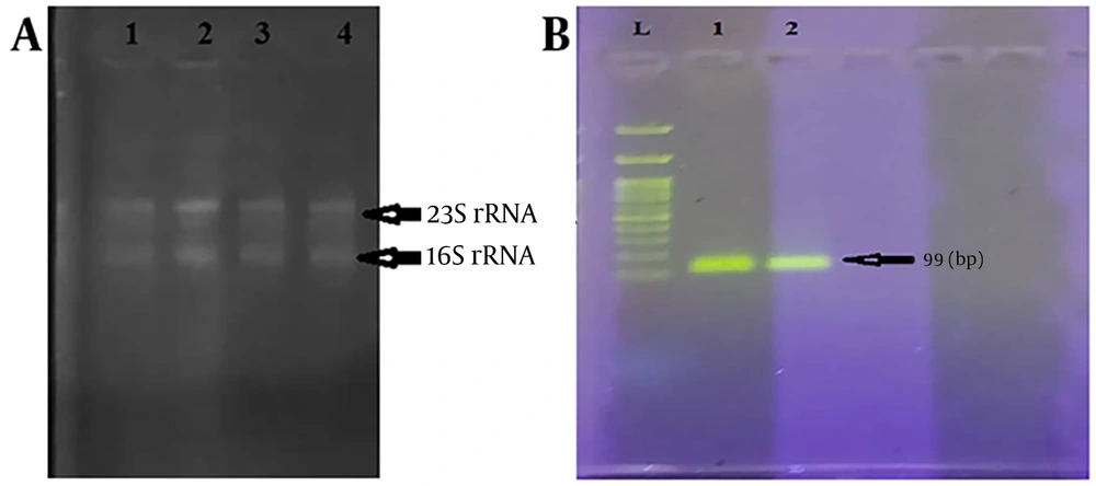
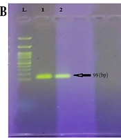
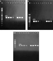
Purified DNA samples from E. coli clinical isolates showed distinct bands on 1% agarose gel electrophoresis, confirming successful extraction (Figure 1A). PCR amplification of the 16S rRNA gene (99 bp) confirmed the molecular identity of the E. coli isolates (Figure 1B). Among the three EAEC isolates, only one tested positive for the aggR gene (102 bp) by PCR (Figure 1C). RNA samples exhibited an A260/A280 absorption ratio of 1.8 - 1.9, indicating high purity, and gel electrophoresis showed distinct 16S and 23S rRNA bands, confirming RNA integrity and purity (Figure 2A).
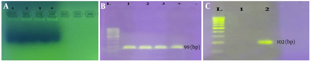
A, Validation of the extracted DNAs from EAEC clinical isolates involved in urinary infections in medical laboratories in Boroujerd, west of Iran. B, Identification of the 16S rRNA gene fragment (length: 99 bp) in EAEC clinical isolates. C, Amplification of the aggR gene (length: 102 bp) in the EAEC clinical isolates. DNA marker: 50 bp.
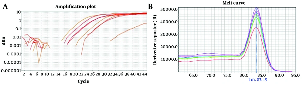
Figure 1.

A, Validation of the extracted DNAs from EAEC clinical isolates involved in urinary infections in medical laboratories in Boroujerd, west of Iran. B, Identification of the 16S rRNA gene fragment (length: 99 bp) in EAEC clinical isolates. C, Amplification of the aggR gene (length: 102 bp) in the EAEC clinical isolates. DNA marker: 50 bp.

A, Quality of the purified RNAs in negative control and samples treated with creatinine concentrations (1, 3, and 6 mg/dL) (lanes 1 - 4); two specific 16S and 23S RNA bands are indicative of the integrity and purity of the extracted RNA. B, cDNA quality control.
Figure 2.

A, Quality of the purified RNAs in negative control and samples treated with creatinine concentrations (1, 3, and 6 mg/dL) (lanes 1 - 4); two specific 16S and 23S RNA bands are indicative of the integrity and purity of the extracted RNA. B, cDNA quality control.

4.2. cDNA Validation and aggR Gene Expression Analysis in Creatinine-Treated E. Coli

Synthesized cDNA from EAEC clinical isolates obtained from patient samples in Borujerd city was validated by agarose gel electrophoresis, which showed clear, distinct bands, confirming successful cDNA synthesis and integrity (Figure 2B). Quantitative reverse transcription PCR results revealed marked and statistically significant downregulation of the aggR gene in bacterial strains after treatment with creatinine at concentrations of 1, 3, and 6 mg/dL (P < 0.0001), indicating a clear dose-responsive pattern (Figure 3A and B). The expression levels of aggR messenger RNA (mRNA) were relatively quantified, showing consistent downregulation across the tested creatinine concentrations compared with untreated controls, with the most pronounced reduction observed at the highest concentration of 6 mg/dL (Figure 3B). These findings are summarized in Table 2, which details the fold changes in aggR mRNA expression for each isolate and concentration, highlighting variability among the isolates and the significant impact of creatinine treatment on virulence gene expression.
Table 2.Relative Quantification Analysis Showing the Expression Level of aggR Gene mRNA in EAEC Clinical Isolates Involved in Urinary Infections in Medical Laboratories in Boroujerd, Western Iran
GenesReaction EfficiencyExpressionResultP-Value
aggR10.9760.000Decrease0.0001
aggR20.9760.002Decrease0.0001
aggR30.9760.009Decrease0.0001
16S0.98871.000--
A, Amplification plot of the aggR gene in the isolated EAEC clinical sample involved in urinary infections in medical laboratories in Boroujerd, west of Iran (ΔRn cycle). B, Melt-curve analysis to ensure the specificity of the amplified aggR gene fragment. The amplification peaks are aligned with each other.
Figure 3.

A, Amplification plot of the aggR gene in the isolated EAEC clinical sample involved in urinary infections in medical laboratories in Boroujerd, west of Iran (ΔRn cycle). B, Melt-curve analysis to ensure the specificity of the amplified aggR gene fragment. The amplification peaks are aligned with each other.

5. Discussion

E. coli, commonly regarded as a harmless resident of the human gut microbiome, can transform into disease-causing forms, including EAEC, a prominent contributor to global diarrheal illness (27, 28). Since the initial identification of bacteria, researchers have sought effective therapies to combat the infections they cause. However, the emergence of antibiotic resistance has created substantial obstacles to treating illnesses caused by diverse microorganisms (29, 30). E. coli is a leading cause of numerous human bacterial infections, and multidrug resistance poses a severe threat, particularly among inpatients (31). This organism is a key factor in urinary tract infections and nosocomial conditions such as sepsis, wound complications, gastrointestinal disorders, and infant meningitis. Moreover, E. coli acts as an opportunistic pathogen in healthcare settings and has developed resistance to beta-lactam drugs through plasmid-mediated extended-spectrum beta-lactamases (32-34).
The present study observed significant dose-dependent downregulation of aggR gene expression in EAEC clinical isolates from Borujerd city, with reductions evident at creatinine concentrations of 1, 3, and 6 mg/dL (P < 0.0001) and most pronounced at 6 mg/dL. These findings highlight creatinine’s potential as a modulator of bacterial virulence, potentially through interference with the transcriptional regulation of the aggR regulon on the pAA plasmid. The creatinine concentration range was selected based on reported serum levels in patients with chronic kidney disease to better reflect the physiological/pathological conditions of patients with chronic kidney failure and to enhance the translational potential of the findings to clinical settings. This observation aligns with prior research demonstrating the antibacterial effects of creatinine, such as that of McDonald et al. (19), who reported that creatinine hydrochloride inhibits the replication of diverse bacteria, including E. coli, by overwhelming proton efflux mechanisms, suggesting a mechanistic basis for the observed gene expression changes beyond mere growth inhibition.
Similarly, Janbakhsh and Janbakhsh et al. observed alterations in rfbE gene expression in E. coli O157:H7 under varying creatinine levels, with initial decreases at low concentrations (1 mg/dL) followed by increases at higher concentrations (3 and 6 mg/dL), indicating that creatinine may exert concentration-specific effects on virulence genes and potentially exacerbate renal pathology in conditions such as hemolytic uremic syndrome through enhanced endotoxin production (35). In contrast, Meiland et al. (36) found no association between E. coli bacteriuria and long-term renal function decline in women, implying that asymptomatic bacteriuria alone may not drive kidney damage. However, our results suggest that elevated creatinine in chronic kidney disease could interact with EAEC virulence in comorbid scenarios, particularly in Iran, where chronic kidney disease prevalence is rising dramatically, from 97,300 cases in 1990 to a projected 423,300 by 2030 (37).
The detection of aggR in only one of three isolates (33% prevalence) is consistent with regional variations in EAEC virulence reported globally, such as 66% in Iranian patients reported by Bouzari et al. (38), underscoring the need for broader molecular surveillance in Borujerd. Clinically, these findings suggest that the modulatory effects of creatinine could mitigate EAEC-driven diarrhea in patients with chronic kidney disease and elevated serum levels, such as 3.8 - 7.2 mg/dL in Iranian chronic kidney disease cohorts reported another study; however, the in vitro design and limited sample size limit extrapolation to in vivo contexts.

5.1. Study Limitations and Conclusions

A major limitation of the present study is the small number of isolates evaluated for aggR gene expression (only one aggR-positive isolate), which primarily resulted from the low prevalence of typical EAEC among the collected clinical strains. Therefore, the results should be considered preliminary and exploratory, and further studies using a larger number of aggR-positive isolates are required to confirm these findings and to investigate potential strain-specific variations in response to creatinine.
This investigation sought to explore the impact of different creatinine levels (1, 3, and 6 mg/dL) on aggR gene expression in clinical E. coli strains isolated from Borujerd, Lorestan province, western Iran. The findings showed that three E. coli strains were confirmed by PCR targeting the 16S rRNA gene, with one carrying the aggR gene. Notably, this work provides initial evidence that aggR gene expression is suppressed in vitro across varying creatinine doses. These observations highlight the need for additional studies to clarify how creatinine influences the regulation of bacterial virulence genes. Future research should prioritize examining the expression of other virulence elements and genes under creatinine exposure, analyzing target gene activity in stool specimens from individuals undergoing dialysis, and expanding the cohort size to improve statistical reliability. These efforts could provide deeper insights into the interplay among creatinine concentrations, microbial pathogenicity, and health implications for individuals with kidney impairment.
Footnotes

References

  • 1.
    Makarova MA. A modern view of diarrheagenic Escherichia coli-a causative agent of acute intestinal infections. Journal of Microbiology, Epidemiology and Immunobiology. 2023;100(4):333-344. https://doi.org/10.36233/0372-9311-410.
  • 2.
    Liang D, Wang L, Liu S, Li S, Zhou X, Xiao Y, et al. Global incidence of diarrheal diseases-an update using an interpretable predictive model based on XGBoost and SHAP: A systematic analysis. Nutrients. 2024;16(18):3217. [PubMed ID: 39339819]. [PubMed Central ID: PMC11434730]. https://doi.org/10.3390/nu16183217.
  • 3.
    Aminian F, Moshirsadri A, Bahrami S, Bahrami S. E. coli bacteria causing diarrhea, threatening travelers in the world, a narrative review. Int J Travel Med Glob Health. 2023;11(1). e170046. https://doi.org/10.30491/ijtmgh.2023.385993.1344.
  • 4.
    Izquierdo-Vega JA, Castillo-Juarez RJ, Sánchez-Gutiérrez M, Ares MA, De La Cruz MA. A mini-review of enteroaggregative Escherichia coli with a specific target on the virulence factors controlled by the AggR master regulator. Polish Journal of Microbiology. 2023;72(4):347-354. [PubMed ID: 37875068]. [PubMed Central ID: PMC10725161]. https://doi.org/10.33073/pjm-2023-037.
  • 5.
    Jenkins C. Enteroaggregative Escherichia coli. Escherichia coli, a Versatile Pathogen. 2018;416:27-50. [PubMed ID: 30232602]. https://doi.org/10.1007/82_2018_105.
  • 6.
    Kaur P, Chakraborti A, Asea A. Enteroaggregative Escherichia coli: an emerging enteric foodborne pathogen. Interdiscip Perspect Infect Dis. 2010;2010(1):254159-10. [PubMed ID: 20300577]. [PubMed Central ID: PMC2837894]. https://doi.org/10.1155/2010/254159.
  • 7.
    Lima AAM, Medeiros PHQS, Havt A. Enteroaggregative Escherichia coli subclinical and clinical infections. Curr Opin Infect Dis. 2018;31(5):433-439. [PubMed ID: 30063473]. https://doi.org/10.1097/QCO.0000000000000477.
  • 8.
    Hebbelstrup Jensen B, Olsen KEP, Struve C, Krogfelt KA, Petersen AM. Epidemiology and clinical manifestations of enteroaggregative Escherichia coli. Clin Microbiol Rev. 2014;27(3):614-630. [PubMed ID: 24982324]. [PubMed Central ID: PMC4135892]. https://doi.org/10.1128/CMR.00112-13.
  • 9.
    Flores J, Okhuysen PC. Enteroaggregative Escherichia coli infection. Curr Opin Gastroenterol. 2009;25(1):8-11. [PubMed ID: 19114769]. https://doi.org/10.1097/MOG.0b013e32831dac5e.
  • 10.
    Vial PA, Robins-Browne R, Lior H, Prado V, Kaper JB, Nataro JP, et al. Characterization of enteroadherent-aggregative Escherichia coli, a putative agent of diarrheal disease. J Infect Dis. 1988;158(1):70-79. [PubMed ID: 2899125]. https://doi.org/10.1093/infdis/158.1.70.
  • 11.
    Boisen N, Struve C, Scheutz F, Krogfelt KA, Nataro JP. New adhesin of enteroaggregative Escherichia coli related to the Afa/Dr/AAF family. Infect Immun. 2008;76(7):3281-3292. [PubMed ID: 18443096]. [PubMed Central ID: PMC2446688]. https://doi.org/10.1128/IAI.01646-07.
  • 12.
    Dias RCB, Tanabe RHS, Vieira MA, Cergole-Novella MC, dos Santos LF, Gomes TAT, et al. Analysis of the virulence profile and phenotypic features of typical and atypical enteroaggregative Escherichia coli (EAEC) isolated from diarrheal patients in Brazil. Front Cell Infect Microbiol. 2020;10. 144. [PubMed ID: 32391284]. [PubMed Central ID: PMC7188757]. https://doi.org/10.3389/fcimb.2020.00144.
  • 13.
    Guerrieri CG, Pereira MF, Galdino ACM, Santos ALSD, Elias WP, Schuenck RP, et al. Typical and atypical enteroaggregative Escherichia coli are both virulent in the Galleria mellonella model. Front Microbiol. 2019;10. 1791. [PubMed ID: 31456762]. [PubMed Central ID: PMC6700222]. https://doi.org/10.3389/fmicb.2019.01791.
  • 14.
    Peng Z, Wang X, Huang J, Li B. Pathogenic Escherichia coli. 11(1). In: Elsevier; 2024. p. 1065-1096. [PubMed ID: 39353923]. [PubMed Central ID: PMC11445235]. https://doi.org/10.1016/B978-0-12-818619-0.00069-1.
  • 15.
    Mancuso G, Midiri A, Gerace E, Marra M, Zummo S, Biondo C. Urinary tract infections: the current scenario and future prospects. Pathogens. 2023;12(4):623. [PubMed ID: 37111509]. [PubMed Central ID: PMC10145414]. https://doi.org/10.3390/pathogens12040623.
  • 16.
    Martino FK, Novara G. Asymptomatic bacteriuria or urinary tract infection? New and old biomarkers. International Journal of Translational Medicine. 2022;2(1):52-65. https://doi.org/10.3390/ijtm2010006.
  • 17.
    Whelan S, Lucey B, Finn K. Uropathogenic Escherichia coli (UPEC)-associated urinary tract infections: the molecular basis for challenges to effective treatment. Microorganisms. 2023;11(9):2169. [PubMed ID: 37764013]. [PubMed Central ID: PMC10537683]. https://doi.org/10.3390/microorganisms11092169.
  • 18.
    Nunes KO, Santos ACP, Bando SY, Silva RM, Gomes TAT, Elias WP. Enteroaggregative Escherichia coli with uropathogenic characteristics are present in feces of diarrheic and healthy children. Pathog Dis. 2017;75(8):ftx106. [PubMed ID: 28961708]. https://doi.org/10.1093/femspd/ftx106.
  • 19.
    McDonald T, Drescher KM, Weber A, Tracy S. Creatinine inhibits bacterial replication. J Antibiot. 2012;65(3):153-156. [PubMed ID: 22293916]. https://doi.org/10.1038/ja.2011.131.
  • 20.
    Murray PR, Rosenthal KS, Pfaller MA. Medical Microbiology. Philadelphia: Elsevier; 2015.
  • 21.
    Ahmed* OB, Dablool AS. Quality improvement of the DNA extracted by boiling method in gram negative bacteria. International Journal of Bioassays. 2017;6(4):5347-5349. https://doi.org/10.21746/ijbio.2017.04.004.
  • 22.
    Moyo SJ, Maselle SY, Matee MI, Langeland N, Mylvaganam H. Identification of diarrheagenic Escherichia coli isolated from infants and children in Dar es Salaam, Tanzania. BMC Infect Dis. 2007;7(1). 92. [PubMed ID: 17688682]. [PubMed Central ID: PMC1976321]. https://doi.org/10.1186/1471-2334-7-92.
  • 23.
    Goldwasser P, Kaldas AI, Barth RH. Rise in serum albumin and creatinine in the first half year on hemodialysis. Kidney Int. 1999;56(6):2260-2268. [PubMed ID: 10594804]. https://doi.org/10.1046/j.1523-1755.1999.00768.x.
  • 24.
    Parker TF, Wingard RL, Husni L, Ikizler TA, Parker RA, Hakim RM. Effect of the membrane biocompatibility on nutritional parameters in chronic hemodialysis patients. Kidney Int. 1996;49(2):551-556. [PubMed ID: 8821843]. https://doi.org/10.1038/ki.1996.78.
  • 25.
    Kashani K, Rosner MH, Ostermann M. Creatinine: from physiology to clinical application. Eur J Intern Med. 2020;72:9-14. [PubMed ID: 31708357]. https://doi.org/10.1016/j.ejim.2019.10.025.
  • 26.
    Levin A, Ahmed SB, Carrero JJ, Foster B, Francis A, Hall RK, et al. Executive summary of the KDIGO 2024 Clinical Practice Guideline for the Evaluation and Management of Chronic Kidney Disease: known knowns and known unknowns. Kidney Int. 2024;105(4):684-701. [PubMed ID: 38519239]. https://doi.org/10.1016/j.kint.2023.10.016.
  • 27.
    Etcheverría AI, Lucchesi PMA, Krüger A, Bentancor AB, Padola NL. Escherichia coli in animals. In: Springer; 2016. p. 149-172. https://doi.org/10.1007/978-3-319-45092-6_7.
  • 28.
    Sandhu KS, Clarke RC, McFadden K, Brouwer A, Louie M, Wilson J, et al. Prevalence of the eaeA gene in verotoxigenic Escherichia coli strains from dairy cattle in Southwest Ontario. Epidemiol Infect. 1996;116(1):1-7. [PubMed ID: 8625998]. [PubMed Central ID: PMC2271236]. https://doi.org/10.1017/S095026880005888X.
  • 29.
    Muteeb G, Rehman MT, Shahwan M, Aatif M. Origin of antibiotics and antibiotic resistance, and their impacts on drug development: a narrative review. Pharmaceuticals (Basel). 2023;16(11):1615. [PubMed ID: 38004480]. [PubMed Central ID: PMC10675245]. https://doi.org/10.3390/ph16111615.
  • 30.
    Naghavi M, Vollset SE, Ikuta KS, Swetschinski LR, Gray AP, Wool EE, et al. Global burden of bacterial antimicrobial resistance 1990 - 2021: a systematic analysis with forecasts to 2050. Lancet. 2024;404(10459):1199-1226. [PubMed ID: 39299261]. [PubMed Central ID: PMC11718157]. https://doi.org/10.1016/S0140-6736(24)01867-1.
  • 31.
    Ikram R, Psutka R, Carter A, Priest P. An outbreak of multidrug-resistant Escherichia coli urinary tract infection in an elderly population: a case-control study of risk factors. BMC Infect Dis. 2015;15(1). 224. [PubMed ID: 26054866]. [PubMed Central ID: PMC4459452]. https://doi.org/10.1186/s12879-015-0974-0.
  • 32.
    Makvana S, Krilov LR. Escherichia coli infections. Pediatr Rev. 2015;36(4):167-170. [PubMed ID: 25834220]. https://doi.org/10.1542/pir.36.4.167.
  • 33.
    Biswas S, Bal M, Pati S, Rana R, Dixit S, Ranjit M. Antibiotic resistance in toxigenic E. coli: a severe threat to global health. Discover Medicine. 2024;1(1). 72. https://doi.org/10.1007/s44337-024-00102-x.
  • 34.
    Aljohni MS, Harun-Ur-Rashid M, Selim S. Emerging threats: antimicrobial resistance in extended-spectrum beta-lactamase and carbapenem-resistant Escherichia coli. Microb Pathog. 2025;200. 107275. [PubMed ID: 39798725]. https://doi.org/10.1016/j.micpath.2024.107275.
  • 35.
    Janbakhsh E, Mehrabi MR. Molecular evaluation of rfbE gene expression changes under different creatinine concentrations in Escherichia coli strains via real-time PCR. Mikrobiolohichnyi Zhurnal. 2023;85(1):26-35. https://doi.org/10.15407/microbiolj85.01.026.
  • 36.
    Meiland R. Association between Escherichia coli bacteriuria and renal function in women: long-term follow-up. Arch Intern Med. 2007;167(3):253-257. [PubMed ID: 17296880]. https://doi.org/10.1001/archinte.167.3.253.
  • 37.
    Shahbazi F, Doosti-Irani A, Soltanian A, Poorolajal J. National trends and projection of chronic kidney disease incidence according to etiology from 1990 to 2030 in Iran: a Bayesian age-period-cohort modeling study. Epidemiol Health. 2023;45. e2023027. [PubMed ID: 36822190]. [PubMed Central ID: PMC10482568]. https://doi.org/10.4178/epih.e2023027.
  • 38.
    Bouzari S, Jafari A, Zarepour M. Distribution of virulence related genes among enteroaggregative Escherichia coli isolates: using multiplex PCR and hybridization. Infect Genet Evol. 2005;5(1):79-83. [PubMed ID: 15567141]. https://doi.org/10.1016/j.meegid.2004.06.005.
  • 39.
    Davoodabadi A, et al. Detection of enteroaggregative Escherichia coli strains from children with diarrhea by use of multiplex PCR and study of their adherence ability to epithelial cells. Iran J Med Microbiol. 2009;2(3):19-24.
  • 40.
    Abbasi P, Kargar M, Doosti A, Mardaneh J, Ghorbani-Dalini S, Anvarinejad M, et al. Enteroaggregative Escherichia coli (EAEC) in South of Iran. Journal of Pediatric Perspectives. 2014;2(2.1):32. https://doi.org/10.22038/ijp.2014.2486.

Similar Articles

26
May
2020
Gene, Cell and Tissue

Prevalence of blaCTX-M, blaSHV, and blaTEM Genes in Escherichia coli Strains Isolated From Urinary Tract Infection Samples of Patients in the Intensive Care Unit in Qom, Iran

Shima Sadat Lesani,
Mohammad Soleimani,
Pegah Shakib,
Mohammad Reza Zolfaghari

Lesani SS, Soleimani M, Shakib P, Zolfaghari MR. Prevalence of blaCTX-M, blaSHV, and blaTEM Genes in Escherichia coli Strains Isolated From Urinary Tract Infection Samples of Patients in the Intensive Care Unit in Qom, Iran. Gene Cell Tissue. 2020;7(2):e102700. doi: https://doi.org/10.5812/gct.102700

5
Jul
2020
Jundishapur Journal of Microbiology

The Frequency of PAI, aer and traT Genes in Escherichia coli Commensal and Urinary Pathogenic E. coli Isolates in Shahrekord and the Relationship Between the Two Groups by Multiplex PCR

Mohamad Hossein Rezaei,
Farshad Kakian,
Abolfazl Gholipour,
Behnam Zamanzad

Rezaei MH, Kakian F, Gholipour A, Zamanzad B. The Frequency of PAI, aer and traT Genes in Escherichia coli Commensal and Urinary Pathogenic E. coli Isolates in Shahrekord and the Relationship Between the Two Groups by Multiplex PCR. Jundishapur J Microbiol. 2020;13(6):e98683. doi: https://doi.org/10.5812/jjm.98683

3
Nov
2019
Jundishapur Journal of Microbiology

Characterization of Uropathogenic Escherichia coli: Distribution of Adhesin-Encoding Genes and O-Serotypes Among Ciprofloxacin Susceptible and Resistant Isolates

Ahmad Rashki,
Masuod Rahdar,
Zahra Rashki Ghalehnoo

Rashki A, Rahdar M, Rashki Ghalehnoo Z. Characterization of Uropathogenic Escherichia coli: Distribution of Adhesin-Encoding Genes and O-Serotypes Among Ciprofloxacin Susceptible and Resistant Isolates. Jundishapur J Microbiol. 2019;12(9):e89179. doi: https://doi.org/10.5812/jjm.89179

6
Aug
2018
Contribution of <i>gyrA</i> and <i>qnrA</i> genes in Ciprofloxacin Resistant <i>Ecsherichia coli</i> Isolates from Patients with Urinary Tract Infections of Imam Khomeini Hospital of Tehran

Contribution of gyrA and qnrA genes in Ciprofloxacin Resistant Ecsherichia coli Isolates from Patients with Urinary Tract Infections of Imam Khomeini Hospital of Tehran

Zahra Zahedi,
Mojdeh Hakemi Vala,
Fatemeh Bagheri Bejestani

Zahedi Z, Hakemi Vala M, Bagheri Bejestani F. Contribution of gyrA and qnrA genes in Ciprofloxacin Resistant Ecsherichia coli Isolates from Patients with Urinary Tract Infections of Imam Khomeini Hospital of Tehran. Arch Pediatr Infect Dis. 2018;6(4):e62129. doi: https://doi.org/10.5812/pedinfect.62129

12
Mar
2018
Antimicrobial Susceptibility Patterns of Enteroaggregative <i>E. coli</i>, as the Most Common Diarrheagenic <i>E. coli</i>, Associated to Gastroenteritis Outbreaks in Iran

Antimicrobial Susceptibility Patterns of Enteroaggregative E. coli, as the Most Common Diarrheagenic E. coli, Associated to Gastroenteritis Outbreaks in Iran

Mohammad Mehdi Soltan-Dallal,
Mohsen Karami-Talab,
Maneli Aminshahidi,
Amir Arastehfar,
Fereshteh Fani

Soltan-Dallal MM, Karami-Talab M, Aminshahidi M, Arastehfar A, Fani F. Antimicrobial Susceptibility Patterns of Enteroaggregative E. coli, as the Most Common Diarrheagenic E. coli, Associated to Gastroenteritis Outbreaks in Iran. Arch Pediatr Infect Dis. 2018;6(2):e11917. doi: https://doi.org/10.5812/pedinfect.11917


Crossmark
Crossmark
Checking
Share on
Metrics

Ordering Reprints

Articles are published under the Creative Commons license stated on each article. No permission or royalty fee is required for uses permitted by that license. CCC handles optional bulk and customized reprint orders. Any quotation covers production and delivery services only, not copyright permission. > Request Reprints from CCC 

Search Relations

Author(s):

Related Articles