
1. Background
Consumption of a Western diet high in fat and/or fructose is associated with increased oxidative stress, insulin resistance, and obesity, which increase the risk of type 2 diabetes (T2D) and nonalcoholic fatty liver disease (NAFLD) and may also contribute to cognitive impairment and neurodegenerative disease (1-3). In mice, we previously showed that chronic intake of an HFD and a high-fructose diet may induce cognitive impairment, characterized by lower discrimination indices and decreased exploration time in recognition-memory tasks, accompanied by lower concentrations of glutamate and glutamine in the hippocampus (4). Moreover, hypercaloric diets can induce obesity, which, together with inflammation and oxidative stress, may impair cognition by disrupting neurotransmitter systems such as glutamate and gamma-aminobutyric acid (GABA) and may also disturb the endocannabinoid system (1, 4).
The endocannabinoid system is a complex network comprising three main components: membrane-bound receptors, their endogenous ligands, and the enzymes involved in the metabolism of these signaling molecules (5). The principal endogenous ligands identified in humans include anandamide (AEA), 2-arachidonoylglycerol (2-AG), and oleamide. Two major cannabinoid receptors have also been described: cannabinoid receptor type 1 (CB1R) and cannabinoid receptor type 2 (CB2R), which mediate many physiological effects associated with these ligands. CB1R is abundant in the hippocampus, cerebellum, and basal ganglia and is also expressed in peripheral tissues such as adipose tissue, skeletal muscle, the gastrointestinal tract, and liver parenchyma. CB2R is widely expressed in the hippocampus, striatum, cortex, nucleus accumbens, thalamus, hypothalamus, and cerebellum, as well as in peripheral tissues such as the immune system (5, 6).
Evidence suggests that activation of hypothalamic CB1R in animal models strongly stimulates preferential intake of high-fat foods, induces hepatic lipogenesis, and increases body weight gain (5). An HFD has also been reported to induce significantly greater food intake in rainbow trout (Oncorhynchus mykiss) than a standard diet, a difference attributed to feed palatability. This increased consumption was associated with elevated AEA and 2-AG concentrations in serum, the hypothalamus, and the telencephalon, a brain region involved in hedonic regulation in fish (5). These findings are consistent with observations in humans, in whom inhibition of CB1R by the antagonist rimonabant reduced appetite and was beneficial for obesity treatment (5-7); however, rimonabant was ultimately withdrawn because of serious mood-related adverse effects. We also previously showed that curcumin restores brain-derived neurotrophic factor (BDNF) levels in the hippocampus of diabetic db/db mice (8) and that a strawberry flavonoid decreased oxidative damage in the hippocampus of obese rats (9).
2. Objectives
This study aimed to evaluate the effects of curcumin on hippocampal CB1R and CB2R expression and endocannabinoid levels (AEA and 2-AG), which were considered the primary outcomes, and to determine their interaction with high-fat and high-fructose diets in C57BL/6 mice. Body weight, body weight gain, and energy intake were assessed as secondary outcomes.
3. Methods
3.1. Ethics Statements and Animal Care
The study was designed to comply with international guidelines applicable to the use of animal models in research and followed the 3Rs principle (refine, reduce, replace) as described in the ARRIVE guidelines (10). Animals were assigned to experimental groups using simple randomization; a block allocation scheme was not used. This decision was based on the homogeneity of the animals at the beginning of the experiment, including the use of the same strain and sex, comparable age, and a narrow range of initial body weight; therefore, no factors requiring additional control through blocking were identified. No animals were lost during the study. All animal procedures were performed in accordance with the National Research Council Guide for the Care and Use of Laboratory Animals and were approved by the Institutional Bioethics Committee of the University of Guanajuato (CIBIUG-P43 - 2020). Animals were housed under standardized conditions on a 12-hour light/dark cycle. No blinding was applied during the experimental procedures, which is acknowledged as a potential source of bias and a study limitation.
3.2. Sample Size Calculation
The primary endpoint used for the a priori sample size calculation was hippocampal AEA concentration. The calculation was based on data reported by Satta et al. (11), who compared hippocampal AEA levels in rats fed a control diet and a HFD, and on the formula for quantitative samples reported by Charan and Biswas (12).
3.3. Experimental Design and Treatment
This comparative experimental study used a hypercaloric diet-induced obese C57BL/6 mouse model to assess the effects of curcumin on hippocampal CB1R and CB2R expression and hippocampal AEA and 2-AG concentrations in mice fed high-fat and high-fructose diets. The mice were treated for 14 weeks as previously described (13): 1) the control group (SD) received a standard diet; 2) the fructose group (SD+Fr) received SD and fructose (30% w/v) in water; 3) the fructose plus curcumin group (SD+C+Fr) received SD supplemented with curcumin (0.75% w/w) and fructose (30% w/v) in water; 4) the HFD group received a high-fat diet; 5) the HFD plus curcumin group (HFD+C) received HFD supplemented with curcumin (0.75% w/w); 6) the HFD plus fructose group (HFD+Fr) received HFD and fructose (30% w/v) in water; and 7) the HFD plus curcumin plus fructose group (HFD+C+Fr) received HFD supplemented with curcumin (0.75% w/w) and fructose (30% w/v) in water.
The SD was purchased as pellets from Purina® Mexico and consisted of 28.5% protein, 13.5% lipids, and 58% carbohydrates (123.3, 58.4, and 250.8 kcal per 100 g of food, respectively). The HFD consisted of 17% protein, 47.5% lipids, and 35.5% carbohydrates (74.6, 332, and 151.6 kcal per 100 g of food, respectively). Curcumin (75% purity) was purchased from Vidanat Mexico (Guadalajara, Jalisco, Mexico); batch information was not recorded and is unavailable. Fructose (30% w/v) in water provided 1.2 kcal/mL. To prepare the experimental diets, standard pellets were ground, and appropriate amounts of lard and/or curcumin were incorporated to achieve the specified concentrations. Fresh batches of diet and fructose solution were prepared every 72 hours and stored at 4 °C to ensure optimal quality for daily administration. Body weight, food intake, and fluid consumption were recorded weekly.
The primary outcomes were hippocampal CB1R and CB2R expression and hippocampal concentrations of AEA and 2-AG. Secondary outcomes included body weight gain and energy intake. The a priori sample size calculation was based on hippocampal AEA levels.
3.4. Euthanasia, Blood Sample Collection, and Hippocampus Dissection
At the end of the treatment period, mice were fasted for 8 hours and then euthanized by isoflurane overdose followed by cervical dislocation. Blood was collected to obtain serum, which was stored at -20 °C until analysis. The hippocampus was then rapidly dissected, washed, and stored at -80 °C in 20 mM Tris-HCl buffer (pH 7.8, 10 mM EDTA, and 2 mM DTT) containing protease inhibitors (Complete, EDTA-free, Roche) for subsequent protein extraction.
3.5. Western Blot Analysis
The hippocampus was homogenized, and proteins were isolated using the phenol extraction method as previously described. Protein precipitates were dried and dissolved in sample buffer (0.5 M Tris-HCl, 10% SDS, 20% glycerol; pH 6.8), followed by determination of protein concentration using the BCA protein assay (Pierce Chemical Co., Rockford, IL, USA), with BSA as the reference standard. Actin, CB1R, and CB2R were determined by western blotting as previously described (13). Briefly, 30 μg of protein was separated on a 10% polyacrylamide gel, and the resolved proteins were transferred to a nitrocellulose membrane. Molecular weights were identified using prestained protein standards (Precision Plus Protein Standards, Bio-Rad Laboratories). Proteins were detected using antibodies against CB1R and CB2R (1:1500; Cayman Chemical) and β-actin (1:2500; Santa Cruz Biotechnology, Inc.). Protein bands were detected using a chemiluminescence kit according to the manufacturer's instructions (Western Lightning Plus-ECL, PerkinElmer), and band optical density was measured using the ChemiDoc XRS system (Bio-Rad, CA, USA). Membranes were stripped to determine cannabinoid receptors and actin on the same membrane. Each experiment was performed in duplicate; therefore, all 3 proteins were determined on the same blot.
3.6. Determination of Endocannabinoids
The endocannabinoids AEA and 2-AG were measured in the hippocampus using ELISA kits for AEA (Cat. MBS1607597; Cayman Chemical, USA) and 2-AG (Cat. MBS1601288; Cayman Chemical, USA), according to the manufacturer's instructions.
3.7. Statistical Analysis
All data were tested for normality using the Shapiro-Wilk test and for homogeneity of variance using Levene's test and are expressed as mean ± SEM. A 3-way analysis of variance (ANOVA) within the general linear model (GLM) was performed to evaluate the main effects of diet (standard vs high-fat), fructose (absence vs presence), and curcumin (absence vs presence), as well as their interactions. When significant effects were detected, Tukey's post hoc test was applied (14). Statistical significance was set at P < 0.05. All analyses were performed using IBM SPSS Statistics version 25.
4. Results
At the beginning of the treatment, the phenotypic characteristics of the mice were similar in terms of strain, sex, age, and body weight, and no between-group differences were observed. At the end of treatment, no atypical results warranted exclusion from the analysis.
4.1. Effect of Hypercaloric Diets and Curcumin on Body Weight Gain
The effects of hypercaloric diets and curcumin on body weight in C57BL/6 mice were first evaluated. As shown in Table 1, initial body weights were similar across all experimental groups. After 14 weeks of treatment, mice in the HFD+Fr group had a significantly higher final body weight than those in the SD group (P = 0.001). Final body weight was lower in the HFD+C+Fr group than in the HFD+Fr group (P = 0.007) and lower in the HFD+C group than in the HFD group (P = 0.039).
Table 1.
Effects of Curcumin on Body Weight and Weight Gain in Male C57BL/6 Mice a
| Groups | Initial Weight (g) | Final Weight (g) | Weight Gain (g) |
|---|---|---|---|
| SD | 20.00 ± 1.035 | 30.03 ± 2.587 | 10.03 ± 2.378 |
| SD+Fr | 19.47 ± 0.856 | 30.52 ± 2.588 | 11.05 ± 2.505 |
| SD+C+Fr | 19.90 ± 0.906 | 27.95 ± 2.660 | 8.05 ± 2.408 |
| HFD | 19.22 ± 1.201 | 33.77 ± 3.775 | 14.55 ± 3.770 b |
| HFD+C | 19.37 ± 1.103 | 29.60 ± 2.792 c | 10.23 ± 2.594 d |
| HFD+Fr | 19.39 ± 1.243 | 35.07 ± 4.590 e | 16.67 ± 4.186 b |
| HFD+C+Fr | 19.50 ± 1.414 | 30.56 ± 3.321 f | 11.06 ± 3.670 g |
a Values are expressed as mean ± SEM (n = 6). Abbreviations: SD, standard diet; SD+Fr, standard diet plus 30% fructose in water; SD+C+Fr, standard diet supplemented with 0.75% curcumin plus 30% fructose in water; HFD, high-fat diet; HFD+C, high-fat diet supplemented with 0.75% curcumin; HFD+Fr, high-fat diet plus 30% fructose in water; HFD+C+Fr, high-fat diet supplemented with 0.75% curcumin plus 30% fructose in water.
b For weight gain: P = 0.014 vs SD.
c For final weight P = 0.039 vs HFD.
d For weight gain P = 0.02 vs HFD.
e For final weight: P = 0.001 vs SD.
f For final weight P = 0.007 vs HFD+Fr.
g For weight gain P = 0.003 vs HFD+Fr.
Body weight gain was calculated by subtracting the initial weight from the final weight (Table 1). Mice fed HFD and HFD+Fr gained more weight (14.55 ± 3.77 and 16.67 ± 4.18 g, respectively) than mice fed SD (10.03 ± 2.37 g; P = 0.014 and P = 0.001, respectively). Curcumin attenuated hypercaloric diet-induced weight gain (HFD+C, 10.23 ± 2.59 g; HFD+C+Fr, 11.06 ± 3.67 g) compared with the respective HFD and HFD+Fr control groups (P = 0.02 and P = 0.003, respectively).
4.2. Effect of Hypercaloric Diets and Curcumin on Energy Consumption
Food and fructose-water intake were recorded separately to determine the effects of hypercaloric diets and curcumin on energy consumption. For energy intake from fructose (Figure 1A), the SD+Fr and SD+C+Fr groups consumed approximately twice as much energy as the HFD+Fr and HFD+C+Fr groups (P < 0.05). Curcumin had no significant effect on energy intake from fructose; no differences were observed between SD+C+Fr and SD+Fr or between HFD+C+Fr and HFD+Fr. For energy intake from food (Figure 1B), the SD group consumed significantly more energy than all other groups (P < 0.001). Curcumin-treated groups did not show a significant change in energy intake from food compared with their respective groups receiving the same diet without curcumin. For total energy consumption (Figure 1C), the SD group consumed more energy than the other 6 groups (P < 0.01), whereas no significant differences were observed between curcumin-treated groups and their respective fructose- or HFD-treated controls. Thus, curcumin did not affect energy intake from either fructose or food.
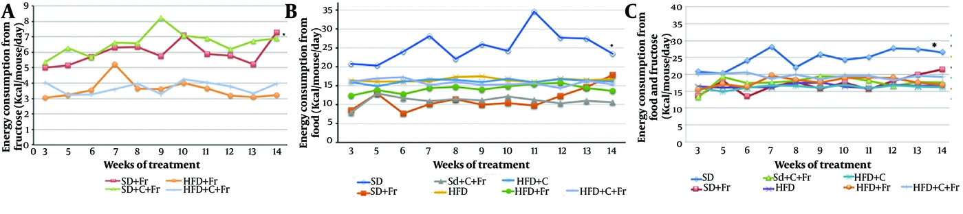
Figure 1.
Effects of curcumin on energy consumption. Data are presented as mean ± SEM (n = 6). SD, standard diet; SD+Fr, standard diet plus 30% fructose in water; SD+C+Fr, standard diet supplemented with 0.75% curcumin plus 30% fructose in water; HFD, high-fat diet; HFD+C, high-fat diet supplemented with 0.75% curcumin; HFD+Fr, high-fat diet plus 30% fructose in water; HFD+C+Fr, high-fat diet supplemented with 0.75% curcumin plus 30% fructose in water. A, Energy consumption from fructose. *P < 0.05 for SD+Fr and SD+C+Fr vs HFD+Fr and HFD+C+Fr. B, Energy consumption from food. *P < 0.001 for SD vs all other groups. C, Total energy consumption. *P < 0.01 for SD vs all other groups.
4.3. Effect of Hypercaloric Diets and Curcumin on Hippocampal Cannabinoid Receptor Type 1 and Type 2 Expression
CB1R and actin (ACT) expression levels were determined on the same nitrocellulose membrane and are expressed as the CB1R/ACT ratio (Figure 2B). As shown in Figure 2A, the hypercaloric diets did not significantly affect CB1R expression in the HFD+Fr group compared with the SD and SD+Fr groups. However, CB1R expression was lower in the HFD+C group than in the HFD group (P = 0.013) and higher in the HFD+C+Fr group than in the HFD+Fr group (P = 0.002). These opposite responses to curcumin suggest that the combination of fructose and fat may have a greater effect on CB1R and may modify the effect of curcumin observed when mice receive fat or fructose alone.
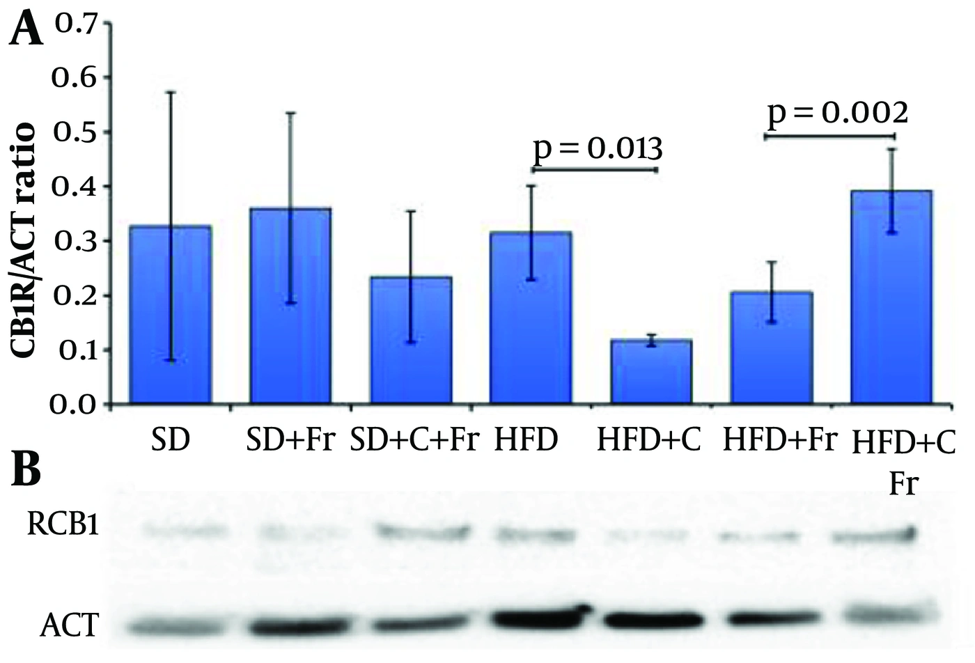
Figure 2.
Effects of hypercaloric diets and curcumin on CB1R expression. A, CB1R/ACT ratio; data are presented as mean ± SEM (n = 6). B, Immunoblotting of CB1R and ACT. SD, standard diet; SD+Fr, standard diet plus 30% fructose in water; SD+C+Fr, standard diet supplemented with 0.75% curcumin plus 30% fructose in water; HFD, high-fat diet; HFD+C, high-fat diet supplemented with 0.75% curcumin; HFD+Fr, high-fat diet plus 30% fructose in water; HFD+C+Fr, high-fat diet supplemented with 0.75% curcumin plus 30% fructose in water.
CB2R and ACT expression levels were also determined on the same nitrocellulose membrane (Figure 3B) and are expressed as the CB2R/ACT ratio (Figure 3A). CB2R expression was lower in the HFD+C group than in the SD (P = 0.006) and HFD (P < 0.001) groups and higher in the HFD+C+Fr group than in the HFD+Fr (P = 0.026) and SD (P = 0.001) groups (Figure 3A).
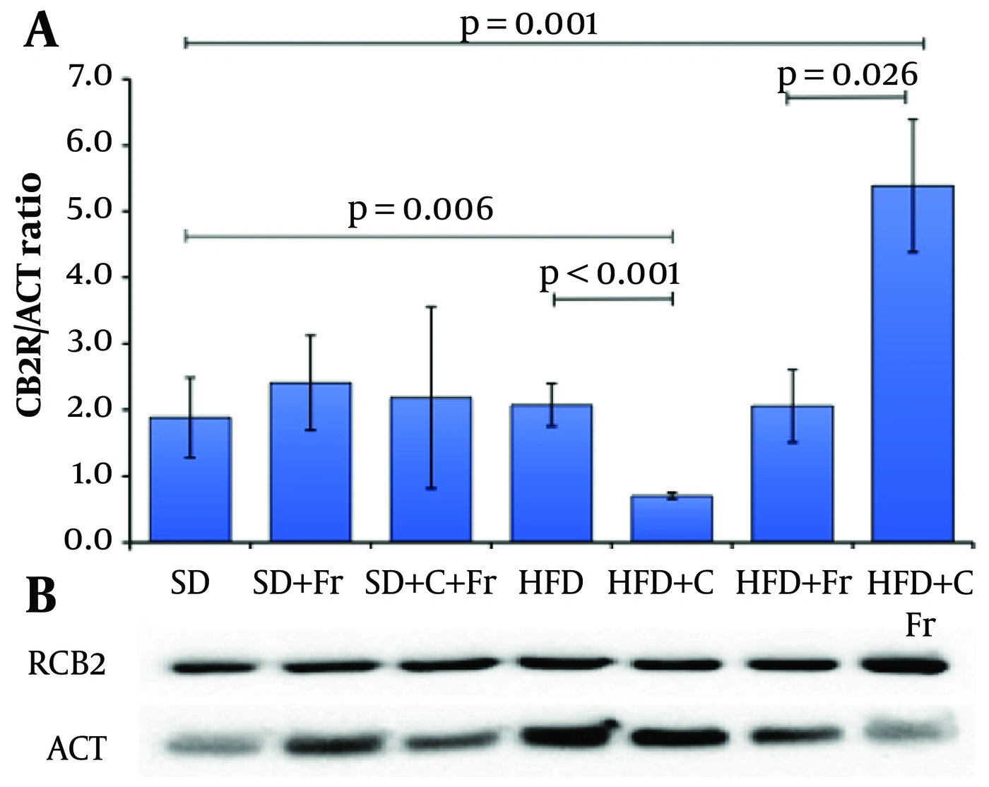
Figure 3.
Effects of hypercaloric diets and curcumin on CB2R expression. A, CB2R/ACT ratio; data are presented as mean ± SEM (n = 6). B, Immunoblotting of CB2R and ACT. SD, standard diet; SD+Fr, standard diet plus 30% fructose in water; SD+C+Fr, standard diet supplemented with 0.75% curcumin plus 30% fructose in water; HFD, high-fat diet; HFD+C, high-fat diet supplemented with 0.75% curcumin; HFD+Fr, high-fat diet plus 30% fructose in water; HFD+C+Fr, high-fat diet supplemented with 0.75% curcumin plus 30% fructose in water.
The CB1R/CB2R expression ratio was subsequently calculated using the CB1R/ACT and CB2R/ACT ratios from Figure 2A and 3A, respectively. No significant differences in the CB1R/CB2R ratio were observed among groups (Figure 4), suggesting that CB1R expression was not proportionally greater than CB2R expression. However, the HFD+Fr group had the highest CB1R/CB2R ratio, approximately twice that of the SD group.
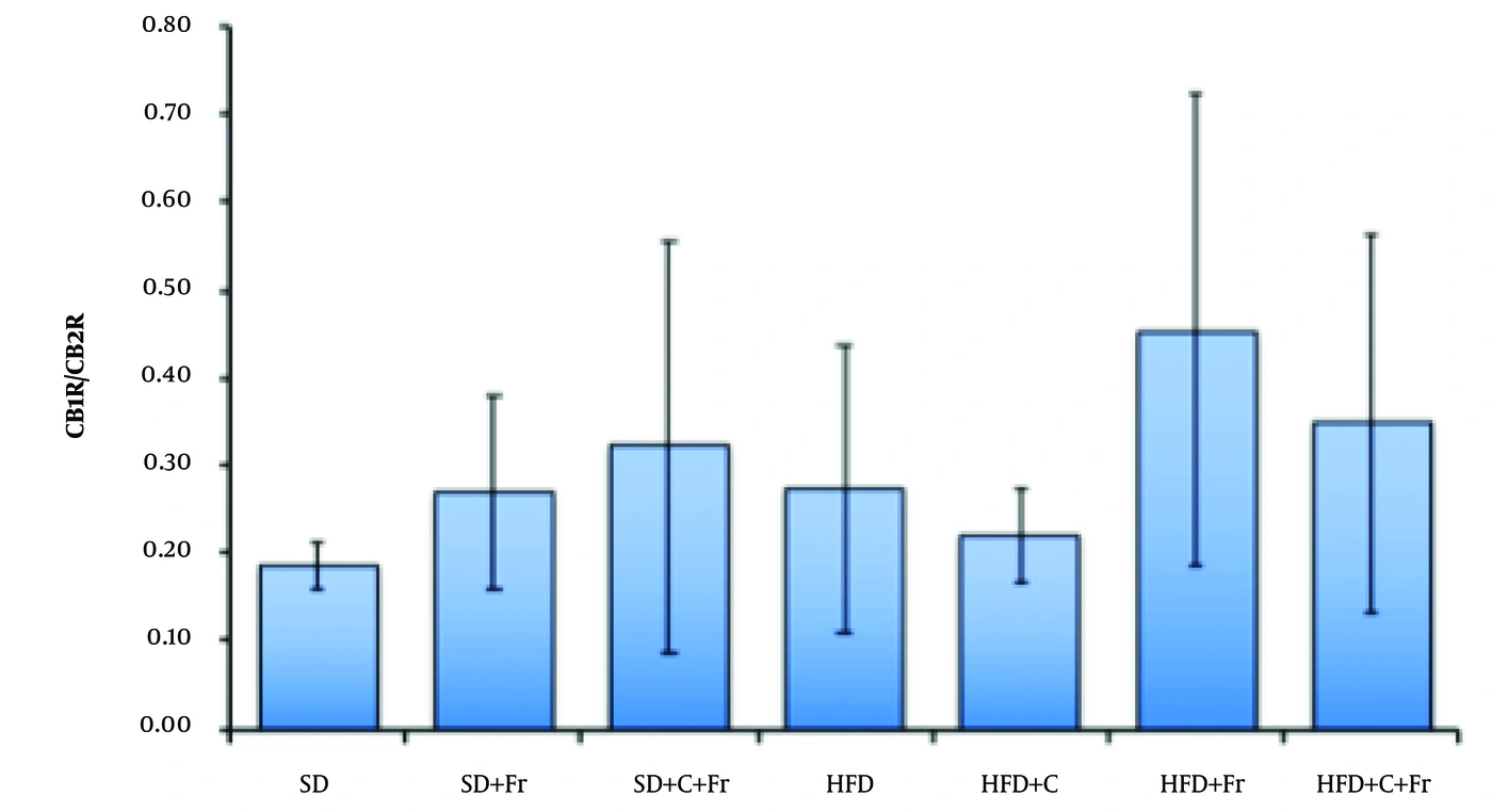
Figure 4.
CB1R/CB2R ratio. Data are presented as mean ± SEM (n = 6) and were obtained from Figures 2A and 3A. SD, standard diet; SD+Fr, standard diet plus 30% fructose in water; SD+C+Fr, standard diet supplemented with 0.75% curcumin plus 30% fructose in water; HFD, high-fat diet; HFD+C, high-fat diet supplemented with 0.75% curcumin; HFD+Fr, high-fat diet plus 30% fructose in water; HFD+C+Fr, high-fat diet supplemented with 0.75% curcumin plus 30% fructose in water.
4.4. Effect of Hypercaloric Diets and Curcumin on Hippocampal Endocannabinoid Concentrations
Hippocampal AEA concentration was significantly lower in the HFD+Fr group than in the SD group (P = 0.049) and in the HFD+C group than in the HFD group (P = 0.04) (Figure 5A). AEA also decreased in the HFD+C+Fr group, but the difference was not statistically significant despite an almost 50% decrease relative to the control. Hippocampal 2-AG concentrations also changed significantly (Figure 5B). The HFD+Fr group had a lower 2-AG concentration than the SD group (P = 0.041). The HFD+C+Fr group showed a slight recovery of this decrease, and no significant difference was observed relative to the SD group, although the concentration remained approximately half that of SD. The HFD+C group had a significantly lower 2-AG concentration than both the HFD group (P = 0.032) and the SD group (P = 0.012).
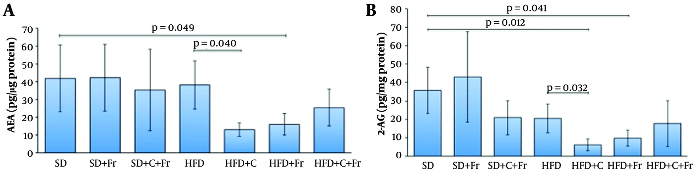
Figure 5.
Effects of hypercaloric diets and curcumin on hippocampal endocannabinoids. A, AEA levels. B, 2-AG levels. Data are presented as mean ± SEM (n = 6). SD, standard diet; SD+Fr, standard diet plus 30% fructose in water; SD+C+Fr, standard diet supplemented with 0.75% curcumin plus 30% fructose in water; HFD, high-fat diet; HFD+C, high-fat diet supplemented with 0.75% curcumin; HFD+Fr, high-fat diet plus 30% fructose in water; HFD+C+Fr, high-fat diet supplemented with 0.75% curcumin plus 30% fructose in water.
4.5. Effect of Diet and Curcumin on Hippocampal Endocannabinoid System Components
To further strengthen interpretation of the findings, a post hoc power analysis was performed. This analysis demonstrated adequate statistical sensitivity for the primary outcomes, with achieved power values of 0.97, 0.99, 0.97, 0.99, and 0.98 for CB1R, CB2R, AEA, 2-AG, and body weight gain, respectively. A GLM analysis was then performed to determine the main effects of diet on endocannabinoid system components and their interactions (Table 2). The multivariate analysis showed significant effects of dietary conditions on CB2R expression (P = 0.005, R2 = 0.556), 2-AG levels (P = 0.004, R2 = 0.562), and AEA levels (P = 0.046, R2 = 0.429). In contrast, CB1R expression (P = 0.090) and the CB1R/CB2R ratio (P = 0.697) were not significantly affected. Further analysis of simple effects showed that curcumin significantly modulated cannabinoid receptor expression in a diet-dependent manner (Table 3). In mice fed a HFD, curcumin supplementation reduced hippocampal CB1R (P = 0.013) and CB2R (P = 0.001) expression. Conversely, in mice fed HFD combined with fructose, curcumin increased CB1R (P = 0.002) and CB2R (P = 0.026) expression.
Table 2.
Main Effects of Diet on Endocannabinoid System Components a
| Dependent Variables | P-Value | Coefficient of Determination (R2) |
|---|---|---|
| CB1R | 0.090 | 0.381 |
| CB2R | 0.005 | 0.556 |
| CB1R/CB2R | 0.697 | 0.174 |
| 2-AG | 0.004 | 0.562 |
| AEA | 0.046 | 0.429 |
a Multivariate ANOVA (GLM) was performed to evaluate the main effects of diet on components of the endocannabinoid system. CB1R, cannabinoid receptor type 1; CB2R, cannabinoid receptor type 2. Statistical significance was set at P < 0.05.
Table 3.
Simple Effects of Curcumin on Cannabinoid Receptor Expression in the Hippocampus a
| Dietary Conditions and Comparisons | Direction of Effect | P-Value |
|---|---|---|
| HFD | ||
| HFD vs HFD+C | ↓ CB1R | 0.013 |
| HFD vs HFD+C | ↓ CB2R | 0.001 |
| HFD+Fr | ||
| HFD+Fr vs HFD+C+Fr | ↑ CB1R | 0.002 |
| HFD+Fr vs HFD+C+Fr | ↑ CB2R | 0.026 |
a Simple effects of curcumin were analyzed using a general linear model (GLM) following significant interactions between factors. Analyses considered diet (standard vs high-fat) and fructose (absence vs presence). HFD, high-fat diet; C, curcumin; Fr, fructose; CB1R, cannabinoid receptor type 1; CB2R, cannabinoid receptor type 2. Statistical significance was defined as P < 0.05.
5. Discussion
The present results show that HFD and HFD+Fr significantly increased body weight gain in C57BL/6 mice, whereas curcumin exerted a protective effect against this phenotype. This effect appeared to be independent of energy intake, suggesting that curcumin may modulate multiple metabolic pathways involved in the development of obesity rather than simply reducing energy consumption. The causal effect of hypercaloric diets on obesity and metabolic syndrome is widely supported in animal models (15-18). Evidence also suggests that the endocannabinoid system may participate in the development of both obesity and cognitive impairment (1, 5, 19), whereas curcumin may exert beneficial effects by reducing oxidative damage in tissues such as the brain (8), liver, heart, kidney, and intestine (9, 17). Ghelani et al. (20) reported a significant reduction in food consumption in rats treated with different concentrations of curcumin (50, 100, and 150 mg/kg). A 2021 study (21) reported that curcumin (100 mg/kg) prevented 53% of body weight gain (g/week) in 5-week-old male C57BL/6J mice fed a HFD for 10 weeks, an effect attributed to decreased food intake. Li et al. (16) demonstrated that supplementation with 0.2% curcumin in a HFD improved metabolic disturbances associated with hepatic steatosis and insulin resistance in obese male C57BL/6 mice. These beneficial effects were linked to alterations in gut microbiota composition and its metabolic products. Similarly, Feng et al. (22) reported that male ApoE (-/-) mice fed a HFD had significantly greater body weight than those receiving the control diet and that curcumin supplementation (0.1% w/w) for 16 weeks significantly prevented body weight gain; no difference in food intake was observed between groups during the experimental period. Therefore, our findings are consistent with these results, showing that curcumin prevents body weight gain without altering energy intake.
Curcumin may also prevent body weight gain by influencing lipid metabolism. A 60% high-fructose diet significantly increased body weight compared with the control diet, and curcumin (200 mg/kg body weight) significantly prevented this increase without affecting food intake; the authors attributed this effect to decreased lipid concentrations through regulation of proteins involved in fatty acid synthesis (20). Maithilikarpagaselvi et al. (23) reported that curcumin reduced body weight gain by preventing hyperlipidemia and hepatic steatosis induced by a high-fructose diet in male Wistar rats. Insulin resistance also plays an important role in body weight regulation. It was previously reported that 23-week-old db/db mice treated with curcumin (0.75% w/w) for 8 weeks had less weight loss than the control group without curcumin. These results suggest that curcumin acts as a metabolic regulator that slows weight loss in older mice with uncontrolled diabetes and hyperphagia, perhaps by decreasing insulin resistance. In addition to influencing lipid metabolism and adipocyte differentiation, curcumin may also influence lean mass gain. Curcumin has previously been reported to increase the expression of proteins involved in protein synthesis (13, 24), supporting the possibility of lean mass gain. However, the percentages of fat and lean mass were not determined in the present study; therefore, it remains unclear whether curcumin’s effect on body weight reflects reductions in fat mass, lean mass, or both.
The endocannabinoid system comprises cannabinoid receptors (CB1R and CB2R), endogenous cannabinoid ligands, and their biosynthetic and degradative enzymes. CB1R and CB2R are expressed in the brain and in some peripheral tissues (5, 25). In the present study, we evaluated the effects of curcumin on CB1R and CB2R expression and on their ligands, AEA and 2-AG. Hippocampal CB1R and CB2R expression decreased significantly in the HFD+C group compared with the HFD and SD groups. This reduction was accompanied by prevention of body weight gain in the HFD+C group compared with the HFD group, suggesting a potentially beneficial effect of curcumin. A similar trend was observed in mice receiving fructose (SD+C+Fr vs SD+Fr), although the difference was not statistically significant.
These findings are of particular interest because CB1R activation has been associated with increased food palatability and a preference for fat- and/or carbohydrate-rich foods, which can increase body weight gain, adipose tissue accumulation, and hepatic lipogenesis in rodents. Evidence also indicates that high-fructose diets enhance central appetite signaling and impair cognitive function (26). Moreover, fructose affects enzymes involved in the synthesis and degradation of endocannabinoids in the hypothalamus. For example, fructose consumption has been shown to increase body weight gain and mRNA expression of fatty acid amide hydrolase while downregulating phospholipase C (27).
In contrast, evidence suggests that hypothalamic stimulation of CB2R increases energy expenditure and promotes normal weight, whereas blocking CB2R with an antagonist increases food consumption, similar to the effect observed when CB1R is stimulated. In humans, administration of the CB1R antagonist rimonabant has been suggested to decrease appetite and body weight, consistent with observations in rodents (7). The crucial role of the endocannabinoid system in body weight control has also been demonstrated in CB1R/CB2R double-knockout mice. These mice do not increase caloric intake or body weight gain on a HFD and are resistant to obesity, possibly because of loss of CB1R or an interactive effect of both receptors (5, 28). This finding supports our results, in which curcumin decreased CB1R expression and body weight. A HFD has been reported not to alter CB1R density in the rat hippocampus but to decrease AEA levels while increasing 2-AG levels (11). These findings indicate that HFD-induced changes in the endocannabinoid system affect brain regions involved in the hedonic aspects of feeding and cognitive processes. In the present study, CB1R and CB2R expression was determined in the whole hippocampus rather than in specific regions, which may have masked increases in the CA1 and CA3 regions induced by HFD that could be physiologically relevant to body weight gain. The curcumin-induced reduction in CB1R and CB2R expression may reflect its effects across the entire hippocampus, consistent with the changes detected by western blotting. Curcumin may also contribute to the effects in the HFD+C group by decreasing CB1R expression or blocking the receptor. Evidence suggests that antioxidant activity activates MEK1/ERK1/2 signaling pathways through CB1R and that administration of curcumin nanoparticles in an animal model of induced depression increases CB1R mRNA and protein and p-MEK1 and p-ERK1/2 in the hippocampus (29). Previous studies have also suggested that curcumin may interact with CB1R and CB2R in a carrageenan-induced pain model in mice (30); however, direct receptor agonism remains to be fully demonstrated.
With respect to CB2R expression, an unexpected finding was that curcumin decreased CB2R expression in mice fed a HFD, whereas it increased CB2R expression in the SD+C+Fr group. Importantly, both groups had lower body weight than groups receiving only hypercaloric diets. These findings suggest that curcumin decreases CB2R expression when mice consume either a high-fat or a high-fructose diet alone; however, when rodents are exposed to a combined high-fat and high-fructose diet, hippocampal CB2R expression increases. One possible explanation is that curcumin may act as a ligand for CB2R, thereby modulating reward-related pathways and contributing to lower body weight. This effect was not accompanied by decreased energy intake, suggesting that curcumin may instead increase energy expenditure and thereby reduce body weight independently of changes in CB2R expression. In this context, bioinformatic molecular analysis has identified curcumin as a potential CB2R agonist (24). Curcumin has also been suggested to bind to both CB1R and CB2R (30). With respect to hippocampal endocannabinoid levels, both AEA and 2-AG were decreased by HFD and HFD+Fr and by curcumin treatment. These reductions could be associated with compensatory changes in hippocampal CB1R and CB2R expression, although this possibility requires further investigation.
Finally, the post hoc power analysis indicated high statistical sensitivity for all evaluated variables, whereas the effect size estimates indicated large to extremely large treatment effects. The largest effects were observed for CB2R and 2-AG, supporting the biological relevance and robustness of the dietary interventions and curcumin-induced changes. Taken together, these findings suggest that the observed effects are robust, biologically meaningful, and unlikely to be attributable to insufficient sample size or random variation.
Collectively, these findings raise new questions regarding the mechanisms underlying the effects of saturated fat and fructose consumption on CB1R and CB2R expression and highlight the need for further studies incorporating multiple metabolic and neurological variables.
5.1. Conclusions
In the hippocampus, intake of HFD and a high-fructose diet, separately or in combination, significantly increased body weight gain. The combined diet had a marked effect on CB1R and CB2R expression and, more importantly, significantly decreased AEA and 2-AG levels. Curcumin supplementation attenuated weight gain and modulated cannabinoid receptor expression in a diet-dependent manner, reducing receptor levels under high-fat conditions while increasing them under combined high-fat and fructose exposure.
5.2. Study Limitations
A key limitation of this study is that protein expression was evaluated in the whole hippocampus rather than in specific hippocampal regions. Analysis of specific hippocampal regions would help clarify the effects of hypercaloric diets and curcumin on cannabinoid receptors. The relatively small sample size is another limitation; therefore, the findings should be confirmed in larger cohorts. In addition, only male animals were included, which limits generalizability to females. Future studies should evaluate both sexes to determine whether sex-specific differences influence the observed effects. Another limitation is that only body weight was determined; the percentages of lean mass and fat were not measured. Cognitive performance was also not assessed. Finally, the lack of blinding in the ELISA and western blot procedures is a limitation and should be considered when interpreting the results. Future research should address these issues and validate the findings in humans.